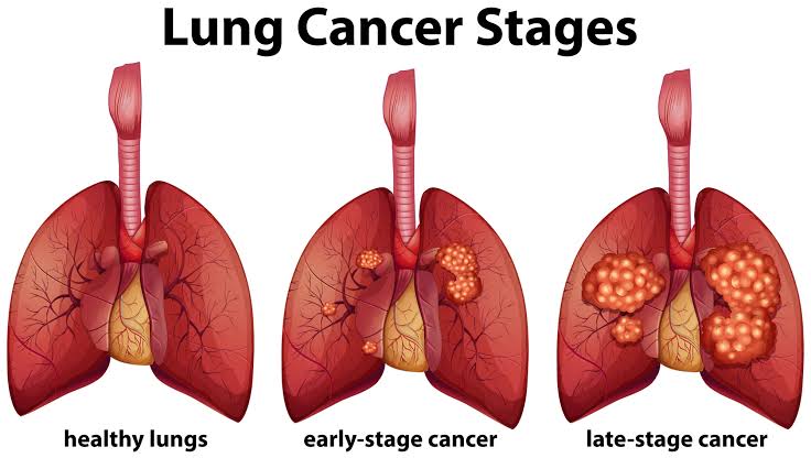
Lung cancer kills more Americans than any other cancer — more than breast, colorectal, and prostate cancer combined. Yet when lung cancer is detected early, outcomes are dramatically better: the five-year survival rate for localized lung cancer is approximately 61%, compared to just 6% for distant-stage disease. The challenge is that lung cancer usually produces no symptoms until it has already spread.
Low dose CT lung cancer screening changed this equation. Two major randomized controlled trials — the U.S. National Lung Screening Trial (NLST, 2011) and the European NELSON trial (2020) — established that annual low dose CT (LDCT) screening reduces lung cancer mortality by 20 to 33% in high-risk individuals. In response, the U.S. Preventive Services Task Force now recommends annual LDCT for adults aged 50 to 80 with a significant smoking history.
This guide goes beyond who qualifies — it explains exactly how low dose CT lung cancer screening works: what makes a CT scan “low dose,” what happens during and after the scan, how radiologists interpret results, and what to do when a nodule is found.
What Makes a CT Scan “Low Dose”?
Standard CT scanning delivers approximately 7 to 8 mSv of radiation — the dose required for high-quality diagnostic images of soft tissue, complex lesions, and cancer staging. Low-dose CT delivers approximately 1.0 to 1.5 mSv — roughly equivalent to six months of natural background radiation. The key is understanding why the lung is uniquely suited to lower-dose imaging: the lung is filled with air, which appears black on CT, while solid nodules appear bright white. This natural contrast means nodules can be detected even in a noisier, lower-dose image.
| Modality | Radiation Dose | Contrast Required | Screening Role |
|---|---|---|---|
| Chest X-ray | ~0.1 mSv | No | NOT recommended for screening (ineffective) |
| Low-Dose CT (LDCT) | ~1.0–1.5 mSv | No | Primary screening tool — USPSTF recommended |
| Standard Chest CT | ~7–8 mSv | Often yes | Diagnostic follow-up for suspicious findings |
| PET/CT | ~14–25 mSv | Yes (tracer) | Evaluation of Lung-RADS 4A/4B findings |
How dose is reduced in LDCT: Three main techniques work together. First, the tube current (mA) is lowered — fewer X-ray photons are produced, creating more image noise but an acceptable image for nodule detection. Second, iterative reconstruction algorithms (vendor-specific names: ASIR from GE, SAFIRE from Siemens, iDose from Philips) mathematically model and reduce image noise, partially compensating for the lower mA. Third, automatic exposure control adjusts mA in real time based on patient body size, so thinner patients receive less radiation and larger patients receive slightly more.
Who Qualifies for Low Dose CT Lung Cancer Screening?
Low dose CT lung cancer screening is recommended only for individuals at high risk — primarily due to heavy smoking history. The current U.S. standard comes from the USPSTF 2021 recommendation.
USPSTF 2021 Eligibility Criteria (all three must be met)
- Age: 50 to 80 years
- Smoking history: 20 or more pack-years
- Smoking status: Currently smoking OR quit within the past 15 years
Medicare (CMS 2022) covers LDCT for beneficiaries aged 50 to 77 who meet the same smoking criteria. Before the first scan, Medicare requires a shared decision-making counseling visit with a clinician to discuss benefits, harms, and the screening process.
Understanding Pack-Years: Pack-Years = (Packs per Day) × (Years Smoked)
Never-smokers, very light smokers (fewer than 20 pack-years), and people who quit more than 15 years ago are not eligible — the evidence does not support screening in lower-risk individuals, and the false positive burden would create more harm than benefit. For deeper coverage of eligibility and trial evidence, see our lung cancer screening guide.
What Happens During the LDCT Scan — Step by Step
One of the reasons low dose CT lung cancer screening has high adherence is that the scan itself is remarkably easy. There is no preparation, no discomfort, and the total visit is usually less than 15 minutes.
- 1 No preparation needed — eat, drink, and take regular medications as usual. No fasting, no contrast dye, no IV line required.
- 2 Check in and change (if needed) — you may be asked to remove clothing with metal (underwire bras, belts, zip-up jackets). A simple T-shirt and loose pants are ideal. The process takes only a few minutes.
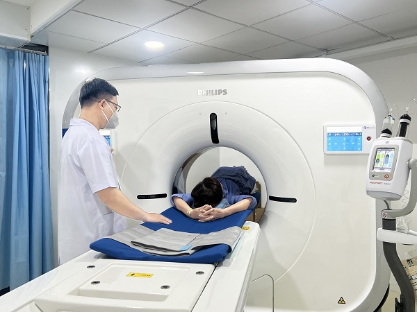
- 3 Lie on the CT table — supine (on your back), arms raised above your head. This position reduces imaging artifact. Unlike an MRI, the CT gantry is short and open — most patients do not experience claustrophobia.
- 4 Hold your breath — the technologist instructs you to take a deep breath and hold it. The actual scan acquisition takes 10 to 15 seconds. This is the only active thing you need to do during the scan.
- 5 Done — leave immediately — no injection, no discomfort from the scanner, no recovery period. Total time from entering the scanner room to leaving is usually 5 to 10 minutes.
- 6 Wait for results — a radiologist reviews images within 1 to 3 business days. Results go to your ordering provider, who contacts you with your Lung-RADS category and next steps.
How Radiologists Read LDCT Scans
Modern LDCT produces thin-slice images — typically 1 to 2.5 millimeters per slice — generating several hundred to over a thousand individual image slices that the radiologist scrolls through on a workstation. Radiologists view images in different window settings: the lung window (optimized for parenchyma and nodule detection) and the mediastinal window (for lymph nodes, heart, and chest wall).
When a nodule is identified, the radiologist measures it. Two methods are used: diameter measurement (longest dimension averaged with the perpendicular) and volume measurement (3D calculation using volumetric software — more reproducible, and the method used in the NELSON trial). Many programs also use computer-aided detection (CAD) software that automatically flags potential nodules — acting as a second reader, though not a replacement for radiologist judgment.
Accredited programs submit data to the ACR Lung Cancer Screening Registry (LCSR), which allows quality benchmarking against national norms for detection rates, positive screen rates, and follow-up compliance.
Understanding Lung-RADS Results
Lung-RADS (Lung Imaging Reporting and Data System) is the ACR standard for LDCT lung cancer screening reports. It replaced the ad-hoc reporting used in the NLST era and substantially reduced false positives by raising size thresholds for follow-up. Every LDCT result at an accredited center includes a Lung-RADS category and a management recommendation.
| Category | Finding | Malignancy Risk | Management |
|---|---|---|---|
| 1 — Negative | No nodules; or benign calcification (central, laminar, popcorn) | <1% | Annual LDCT in 12 months |
| 2 — Benign | Solid nodule <6mm; subsolid <6mm total; benign-appearing ground-glass | <1% | Annual LDCT in 12 months |
| 3 — Probably Benign | Solid 6–7.9mm; part-solid with solid component <6mm; large new ground-glass | 1–2% | 6-month follow-up LDCT |
| 4A — Suspicious | Solid 8–14mm; part-solid with solid component 6–7.9mm | 5–15% | 3-month LDCT or PET/CT |
| 4B — Very Suspicious | Solid ≥15mm; part-solid with solid component ≥8mm | >15% | Contrast CT, PET/CT, tissue sampling |
| 4X — Additional Concern | 4A or 4B with spiculation, growth, or upper lobe location | Higher | PET/CT + tissue sampling |
The NLST used a 4mm threshold for any nodule as “positive,” resulting in ~27% of participants having at least one positive screen per year — and a 96% false positive rate. Lung-RADS raised the threshold to 6mm for solid nodules, reducing the positive screen rate to approximately 10 to 12% while preserving sensitivity for clinically significant cancers.
Nodule Follow-Up — What Happens After a Positive Screen
If your Lung-RADS result is 3 or higher, you will return for additional evaluation. This does not mean you have cancer — most nodules are benign, typically old granulomas from fungal infections (histoplasmosis is common in the Ohio and Mississippi River valleys), scar tissue, or lymph nodes.
Lung-RADS 3 → 6-month repeat LDCT: The radiologist checks whether the nodule has grown. No growth = reassuring, returns to annual screening. Growth = escalation to 4A/4B management. Most Lung-RADS 3 nodules ultimately prove benign.
Lung-RADS 4A → PET/CT or 3-month CT: A PET/CT uses a radioactive glucose tracer — cancer cells metabolize glucose at higher rates than normal tissue, appearing as “hot spots.” Nodules with low metabolic activity on PET are less likely to be malignant. Alternatively, a 3-month repeat CT may be used to assess growth.
Lung-RADS 4B/4X → Tissue sampling: Definitive diagnosis requires biopsy. Options include CT-guided transthoracic needle biopsy (outpatient; small risk of pneumothorax ~10–15%), bronchoscopy (navigational bronchoscopy for peripheral nodules), or VATS surgery (video-assisted thoracoscopic surgery, which allows simultaneous diagnosis and resection). See our lung cancer symptoms guide for context on what symptoms may accompany a positive finding.
Radiation — Is LDCT Safe?
Annual screening for 10 years accumulates approximately 10 to 15 mSv — roughly 3 to 5 additional years of background radiation exposure. The estimated lifetime attributable cancer risk from one LDCT scan is approximately 0.01 to 0.02% — extremely small. When compared to the 20% reduction in lung cancer mortality from the NLST, the benefit of screening clearly outweighs the theoretical radiation risk for eligible high-risk individuals.
This calculation changes for people outside the high-risk category. For a 45-year-old never-smoker, baseline lung cancer risk is very low, and the radiation risk (while still small) is proportionally larger relative to potential benefit. This is why screening is not recommended for lower-risk individuals — the risk/benefit balance does not favor them.
What Else Can LDCT Find? Incidental Findings
Because LDCT images the entire chest, it often detects findings unrelated to lung cancer. These “incidental findings” can be both beneficial and burdensome — they may identify other serious conditions early, but they also generate follow-up testing for some patients.
Coronary Artery Calcium (CAC)
Calcium deposits in coronary artery walls are visible on chest CT. CAC is a validated predictor of cardiovascular risk. Some programs score CAC from LDCT, potentially identifying patients who would benefit from statin therapy or cardiology referral.
Emphysema
CT can characterize and quantify emphysema (air trapping, decreased lung density) — often visible in heavy smokers. May prompt pulmonology referral or COPD management even before respiratory symptoms are significant.
Aortic Aneurysm
Dilation of the thoracic aorta may be detected incidentally. If above threshold diameters, surgical consultation is warranted. This can be a life-saving incidental finding in older smokers.
Interstitial Lung Abnormalities (ILA)
Areas of ground-glass opacity or fibrosis not explained by a known condition. Associated with respiratory outcomes and may warrant specialist evaluation, particularly in older adults.
Vertebral Compression Fractures
Common in older smokers and may indicate osteoporosis. Incidental detection on LDCT can prompt bone density evaluation and treatment before a catastrophic fracture occurs.
How to Choose an LDCT Screening Center
Not all CT scanners or radiology departments are equally suited for low dose CT lung cancer screening. Look for these quality indicators when selecting a center.
What to Look for in an LDCT Screening Program
- ACR accreditation — the American College of Radiology accredits programs meeting quality standards for LDCT protocol, radiologist training, Lung-RADS reporting, and ACR registry participation
- Dedicated screening protocol — a stand-alone LDCT screening order (not a generic chest CT) ensures low-dose protocol and Lung-RADS reporting
- Multidisciplinary follow-up team — pulmonologists, thoracic surgeons, and interventional radiologists available for nodule workup and biopsy
- Smoking cessation resources — both ACR and USPSTF require cessation counseling or referral as part of a complete screening program
- Patient navigation — a nurse navigator or case manager who follows up on abnormal results significantly reduces anxiety and improves follow-up compliance
- ACR Lung Cancer Screening Registry participation — programs that submit data to the LCSR use outcomes for quality improvement and national benchmarking
Frequently Asked Questions
Is low dose CT the same as a regular CT scan?
No. Both use X-rays and produce cross-sectional images, but they differ in radiation dose (~1–1.5 mSv for LDCT vs. ~7–8 mSv standard), imaging protocol, and purpose. LDCT is specifically optimized for lung nodule detection in a screening population; standard chest CT is for diagnostic purposes and requires higher image quality of the mediastinum and soft tissue. They serve different clinical roles and should not be substituted for each other.
Will the LDCT scan find problems other than lung cancer?
Possibly. Low dose CT images the entire chest and can detect coronary artery calcium, emphysema, aortic aneurysm, interstitial lung abnormalities, and other incidental findings. Whether these are reported in detail depends on the program and radiologist. Ask your ordering provider whether your program routinely reports incidental cardiac or vascular findings.
What happens if I can’t hold my breath during the scan?
Tell the technologist before the scan if you have limited breath-holding capacity — for example, due to COPD or severe shortness of breath. The technologist can adapt the instruction. Even a shorter breath hold usually produces adequate images. If needed, the acquisition can be repeated; the additional radiation from one repeat acquisition is small.
How long will it take to get my results?
Most accredited programs report results within one to three business days. Your ordering clinician then contacts you. Some programs have patient portals where results appear within the same timeframe. You should receive a clear Lung-RADS category and any recommended next steps. If you don’t hear within five business days, contact your provider’s office directly.
Does LDCT screening work for non-smokers with other risk factors?
Current USPSTF and NCCN guidelines focus on smoking history as the primary risk factor, because the trial evidence (NLST, NELSON) enrolled primarily heavy smokers. Never-smokers with other risk factors are not covered under standard LDCT screening guidelines — though some clinicians may consider it on an individual basis. Insurance coverage outside standard eligibility criteria is not guaranteed. Understanding the relationship between lung cancer and smoking helps clarify why smoking history is the central eligibility criterion.
Sources
- NLST Research Team — Reduced lung-cancer mortality with low-dose computed tomographic screening; NEJM 2011;365:395–409
- de Koning HJ et al. — Reduced lung-cancer mortality with volume CT screening in a randomized trial (NELSON); NEJM 2020;382:503–513
- USPSTF — Lung cancer screening recommendation; JAMA 2021;325(10):962–970
- American College of Radiology — Lung-RADS Version 2022
- Pinsky PF et al. — Performance of Lung-RADS in the National Lung Screening Trial; Ann Intern Med 2015;163(8):577–583
- Kazerooni EA et al. — ACR practice parameter for the performance and reporting of lung cancer screening CT; 2019
Related reading: Lung cancer screening — who qualifies and the trial evidence | Lung cancer symptoms | Lung cancer and smoking