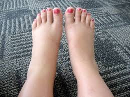
Leg swelling — medically termed peripheral edema — is one of the most visible signs of heart disease and one of the most frequently underestimated. Many patients attribute swollen ankles to long days of standing, warm weather, or the natural effects of aging, without recognizing that bilateral, pitting edema of the lower extremities is a classic manifestation of right heart failure and venous congestion. Understanding why heart disease causes leg swelling, which cardiac conditions are responsible, how cardiac edema is distinguished from other causes, and when swelling demands urgent medical attention can make a meaningful difference in the timely diagnosis and treatment of serious cardiac conditions.
How Heart Disease Causes Leg Swelling
The primary mechanism linking heart disease to leg swelling is right heart failure with venous congestion. When the right ventricle cannot pump blood forward into the pulmonary circulation with adequate force, blood backs up in the systemic venous system. This backed-up blood raises the hydrostatic pressure within the veins and capillaries of the legs. When capillary hydrostatic pressure exceeds the opposing oncotic pressure of plasma proteins, fluid is pushed out of the capillaries into the surrounding tissue — the interstitial space — producing the swelling that is visible and palpable as edema.
Several factors amplify this basic mechanism. Reduced cardiac output from the failing heart activates the renin-angiotensin-aldosterone system (RAAS), a neurohormonal response that signals the kidneys to retain sodium and water. This increases plasma volume, which worsens the venous congestion and adds to the fluid already accumulating in the tissues. Cardiac edema is therefore not simply a matter of fluid leaking out of overpressured capillaries — it reflects a systemic neurohormonal state in which the body is actively retaining more fluid than it can distribute and excrete effectively.
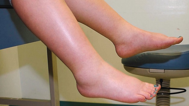
Cardiac edema is gravity-dependent, which accounts for its characteristic distribution. When a patient is upright — standing or sitting — fluid accumulates in the ankles and feet, the lowest points of the body where hydrostatic pressure is greatest. When a patient lies down, the fluid redistributes toward the sacrum and lower back. In severe heart failure, edema can extend up the calves, thighs, genitalia, and abdomen (ascites) as fluid accumulation overwhelms the body’s capacity to redistribute it. This gravity-dependent behavior is one of the clinical features that distinguishes cardiac edema from other causes.
Fluid retention also manifests as rapid weight gain before visible edema appears. The body can accumulate several liters of fluid — representing several kilograms of weight — before swelling becomes visible. This is why daily weight monitoring is a cornerstone of heart failure self-management: a gain of two to three pounds in 24 hours, or five pounds in a week, signals fluid accumulation that typically precedes or accompanies a worsening of leg swelling.
Cardiac Conditions That Cause Leg Swelling
Right heart failure is the most direct cardiac cause of bilateral leg edema, and it can arise from multiple underlying conditions. The most common is left-sided heart failure — when the left ventricle fails, pressure backs up through the pulmonary circulation and eventually overloads the right ventricle, producing biventricular heart failure with both pulmonary congestion (causing dyspnea) and systemic venous congestion (causing leg edema). This is the classic presentation of congestive heart failure.
Pulmonary hypertension from any cause — primary pulmonary arterial hypertension, chronic thromboembolic disease, or secondary pulmonary hypertension from lung disease — raises the resistance against which the right ventricle must pump, eventually producing right ventricular failure and leg edema. Cor pulmonale — right heart failure caused specifically by pulmonary disease — is seen in patients with advanced COPD, pulmonary fibrosis, and severe obstructive sleep apnea, where chronic hypoxia drives pulmonary vasoconstriction and pulmonary hypertension.
Constrictive pericarditis is an underappreciated cause of leg swelling that can mimic liver cirrhosis. When the pericardium becomes fibrotic and rigid following pericarditis, cardiac surgery, or radiation therapy, it constrains the heart’s ability to fill during diastole. The resulting venous congestion produces massive leg edema, ascites, and elevated jugular venous pressure. A distinguishing clinical feature is the Kussmaul sign — a paradoxical rise in jugular venous pressure with inspiration rather than the normal fall — reflecting the pericardial constraint on diastolic filling.
Tricuspid regurgitation — leaking of blood backward through the tricuspid valve from right ventricle to right atrium — raises right atrial and venous pressure, directly congesting the systemic venous circulation and producing prominent leg edema, ascites, and right upper quadrant discomfort from hepatic congestion. Significant tricuspid regurgitation can result from rheumatic disease, right ventricular dilation, or carcinoid syndrome, and generates some of the most severe degrees of venous congestion seen in cardiac practice.
Signs That Leg Swelling May Be Cardiac
Several features distinguish cardiac edema from other causes and should prompt cardiac evaluation. The most important is that cardiac edema is bilateral and symmetric — both legs swell equally, because the underlying problem is systemic venous congestion rather than a local process in one leg. This contrasts with deep vein thrombosis, which characteristically causes unilateral swelling, and with venous insufficiency, which may be asymmetric.
Cardiac edema is pitting — pressing a fingertip into the swollen tissue for a few seconds leaves an indentation that slowly fills back in. This pitting quality reflects the accumulation of free interstitial fluid and is characteristic of the low-protein edema fluid of cardiac and hepatic causes. Dyspnea accompanying leg swelling is a critically important signal: breathlessness with exertion and orthopnea (breathlessness when lying flat) alongside leg swelling make a cardiac diagnosis highly probable. Jugular venous distension — visible engorgement of the neck veins when the patient is at 45 degrees — reflects elevated right heart filling pressures and strongly suggests venous congestion from cardiac or pericardial disease.
Non-Cardiac Causes to Rule Out
Many conditions other than heart disease cause leg swelling, and a systematic evaluation is required before attributing edema to a cardiac cause. Deep vein thrombosis (DVT) is the most urgent alternative diagnosis because it carries the risk of pulmonary embolism. DVT typically causes unilateral leg swelling with calf pain or tenderness, warmth, and sometimes redness. Any patient with unilateral leg swelling should be evaluated urgently with duplex ultrasound to exclude DVT.
Chronic venous insufficiency is perhaps the most common cause of bilateral leg swelling in the general population, particularly in older adults. Incompetent venous valves allow blood to pool in the superficial veins, raising hydrostatic pressure in the capillaries and producing edema. Unlike cardiac edema, venous insufficiency typically causes skin changes — hemosiderin deposits producing brownish discoloration, lipodermatosclerosis (skin hardening), and varicose veins — and is not accompanied by dyspnea or jugular venous distension.
Lymphedema — swelling from impaired lymphatic drainage — develops gradually and becomes non-pitting over time as accumulated protein-rich fluid stimulates fibrosis. Lymphedema characteristically involves the dorsum of the foot (positive Stemmer sign — inability to pinch the skin over the dorsum of the toe), distinguishing it from cardiac edema. Hypoalbuminemia — low serum albumin from liver cirrhosis, nephrotic syndrome, or malnutrition — reduces the plasma oncotic pressure that normally counterbalances capillary hydrostatic pressure, allowing fluid to leak into tissues without elevated venous pressure or dyspnea.
Several medications commonly cause bilateral ankle edema as a side effect. The calcium channel blocker amlodipine is the most frequent offender, producing ankle swelling through capillary dilation rather than systemic fluid retention. This medication-related edema does not respond to diuretics and is not accompanied by dyspnea or weight gain. NSAIDs, steroids, and thiazolidinediones are other common pharmaceutical causes of leg swelling that should be reviewed in any patient presenting with new or worsening edema.
How Cardiac Leg Swelling Is Evaluated
When cardiac leg swelling is suspected, evaluation begins with the history and physical examination. Key historical features include the rate of onset, whether both legs are affected equally, whether dyspnea or orthopnea accompanies the swelling, whether rapid weight gain preceded it, and whether there is known heart disease, atrial fibrillation, or prior cardiac events.
Physical examination assesses the degree and distribution of edema, jugular venous pressure elevation, lung auscultation for crackles or pleural effusion, the presence of an S3 gallop (a low-frequency extra heart sound reflecting increased left ventricular filling pressure), hepatomegaly, and ascites. A BNP or NT-proBNP measurement is one of the most useful initial tests — significantly elevated values confirm ventricular stretch from elevated filling pressures (as seen in heart failure and constrictive pericarditis), while normal values substantially reduce the probability of a cardiac cause.
An echocardiogram provides definitive cardiac characterization: right and left ventricular function, right ventricular size and systolic pressure, pericardial effusion or thickening, valvular abnormalities, and diastolic function. Chest X-ray evaluates heart size, pulmonary vascular pattern, and pleural effusions. An ECG may reveal right heart strain, arrhythmia, or prior MI. Complete blood count, albumin, renal function, and liver function tests assess for non-cardiac contributing causes. Duplex ultrasonography of the lower extremity veins is indicated when unilateral swelling raises concern for DVT.
Treatment of Cardiac Edema
The cornerstone of treatment for cardiac leg swelling is diuresis with loop diuretics. Furosemide, torsemide, and bumetanide block sodium and chloride reabsorption in the loop of Henle, producing a brisk diuresis that rapidly reduces the intravascular volume driving venous congestion and edema. In acute decompensated heart failure, intravenous loop diuretics are preferred because intestinal wall edema from congestion impairs oral absorption. In chronic outpatient management, oral diuretics are titrated to achieve and maintain a dry weight without excessive depletion of volume or potassium.
Sodium restriction is essential for managing cardiac edema. Dietary sodium should be limited to approximately 2 grams per day in patients with heart failure; excess dietary sodium counteracts the effect of diuretics and promotes fluid retention. Guideline-directed medical therapy for the underlying heart failure — ACE inhibitors, beta-blockers, SGLT2 inhibitors, and mineralocorticoid receptor antagonists — addresses the neurohormonal activation driving fluid retention and reduces the need for escalating diuretic doses.
SGLT2 inhibitors (dapagliflozin, empagliflozin) deserve particular mention in cardiac edema management. Through combined glucosuria and natriuresis, they produce a gentle sustained diuretic effect without the electrolyte disturbances of loop diuretics, and clinical trials have shown they reduce heart failure hospitalizations — events largely driven by worsening edema and fluid overload. Elevating the legs above the level of the heart when seated mobilizes dependent edema by reversing the gravitational gradient. Compression stockings can reduce venous pooling, particularly when venous insufficiency coexists with cardiac edema.
Daily weight monitoring is one of the most effective self-management strategies for patients with heart failure and recurrent edema. A weight gain of two to three pounds in 24 hours, or five pounds in a week, reliably identifies early fluid accumulation before severe edema develops. Many heart failure programs provide patients with a written action plan: if a weight threshold is crossed, the patient adjusts their diuretic dose (within a defined range) and contacts their care team promptly.
When to Seek Urgent Care
Certain presentations of leg swelling require urgent or emergency evaluation. New or rapidly worsening bilateral leg swelling accompanied by shortness of breath at rest, orthopnea, or acute respiratory distress may represent acute decompensated heart failure and requires emergency assessment. Unilateral leg swelling with calf pain and tenderness warrants same-day evaluation for DVT, given the risk of pulmonary embolism. Leg swelling with chest pain, rapid heart rate, or profound breathlessness raises concern for pulmonary embolism and requires emergency evaluation. A rapid gain of three or more pounds overnight is a reliable signal of worsening fluid retention in a patient with known heart failure and should trigger prompt contact with a physician or heart failure nurse.
Frequently Asked Questions
Can heart failure cause leg swelling without noticeable shortness of breath?
Yes, though dyspnea commonly accompanies cardiac edema, some patients — particularly those with right-predominant heart failure or cor pulmonale — develop significant leg swelling before dyspnea becomes prominent. Patients who are less physically active may not stress their cardiopulmonary reserve enough to perceive exertional dyspnea, yet still have elevated venous pressure producing edema. However, the absence of dyspnea does not rule out a cardiac cause of bilateral pitting edema, and BNP testing and echocardiography remain appropriate when cardiac edema is clinically suspected.
Does ankle swelling from amlodipine mean I have heart disease?
No. Ankle swelling from amlodipine (and other calcium channel blockers) is a pharmacological side effect of capillary dilation, not a sign of heart failure or fluid retention. It does not respond to diuretics, is not accompanied by weight gain or dyspnea, and is not a marker of cardiac deterioration. It is common, affecting up to 20 percent of patients on high-dose amlodipine. If the swelling is bothersome, switching to a different antihypertensive class (such as an ACE inhibitor or ARB) typically resolves it. Always discuss medication changes with your prescriber before stopping any blood pressure treatment.
Is it safe to reduce fluid intake to manage leg swelling?
Fluid restriction should only be used in specific circumstances and under medical guidance — primarily in heart failure patients with significant hyponatremia (low sodium), where excess fluid intake further dilutes serum sodium. For most heart failure patients with normal or near-normal sodium, the more effective approach is dietary sodium restriction (2g/day) and appropriate diuretic therapy rather than severe fluid restriction, which can cause dehydration, electrolyte imbalance, and worsening of renal function. Always follow your heart failure team’s specific recommendations for fluid management.
Can treating leg swelling reduce my risk of a heart attack?
Treating leg swelling addresses the fluid congestion of heart failure but does not directly reduce the risk of coronary events. However, the underlying guideline-directed medical therapy that manages heart failure — particularly ACE inhibitors, beta-blockers, and SGLT2 inhibitors — reduces cardiovascular mortality and hospitalization beyond their effects on edema. Effective management of heart failure with evidence-based therapy improves overall cardiac prognosis, so treating the cardiac condition driving leg swelling is associated with better long-term outcomes.
For understanding the fatigue that often accompanies leg swelling in heart disease, see our article on fatigue and heart disease. For the breathing symptoms that frequently coexist with cardiac edema, see shortness of breath and heart health. For reference values that help monitor cardiac health, see our article on heart health numbers every adult should know.
The American Heart Association lists leg swelling among the key warning signs of heart failure. The National Heart, Lung, and Blood Institute explains heart failure symptoms including edema and when evaluation is needed. The CDC provides population-level data on heart failure prevalence and outcomes.
Leg swelling is rarely a trivial symptom when it is bilateral, pitting, and accompanied by dyspnea, weight gain, or fatigue. In the context of known heart disease or cardiac risk factors, it is one of the most important signals that the heart is struggling to maintain adequate forward output and that fluid is accumulating in ways that will worsen without targeted treatment. Recognizing cardiac edema early, evaluating it systematically, and treating the underlying cardiac condition with guideline-directed therapy are the steps that convert a visible symptom into an opportunity for meaningful cardiac intervention.
Cardiac Edema in Specific Patient Groups
The presentation and significance of cardiac leg swelling varies across different patient populations. In older adults, bilateral ankle edema is extremely common and frequently attributed to age-related venous insufficiency or medication effects without adequate cardiac evaluation. Yet heart failure prevalence increases sharply with age, and cardiac edema in older adults is often undertreated because the dyspnea component is attributed to deconditioning rather than cardiac congestion. Older patients with new bilateral ankle edema and any degree of exertional breathlessness merit BNP measurement and echocardiographic evaluation rather than empirical diuresis alone.
In patients with chronic kidney disease (CKD), fluid retention and leg edema are common from multiple mechanisms: impaired renal sodium and water excretion, hypoalbuminemia from proteinuria, and the frequent coexistence of heart failure (cardiorenal syndrome). Diuretic resistance — a reduced response to loop diuretics — is common in both advanced CKD and severe heart failure, requiring higher doses, combination diuretic strategies (adding a thiazide-type diuretic to a loop diuretic), or in-hospital IV diuresis. The management of cardiac edema in cardiorenal patients requires close coordination between cardiology and nephrology to avoid diuresis-induced worsening of renal function.
Patients with atrial fibrillation are particularly prone to cardiac edema for several reasons. Atrial fibrillation with rapid ventricular response reduces cardiac output, promotes neurohormonal activation and fluid retention, and is a major cause of de novo heart failure or decompensation of existing heart failure. Rate control (targeting resting heart rates below 80-100 beats per minute) and rhythm control (restoring sinus rhythm through cardioversion or ablation) both reduce the cardiac output impairment driving edema. In patients with atrial fibrillation who develop new leg swelling, evaluation for heart failure and echocardiographic assessment of ventricular function and filling pressures is always warranted.
Long-Term Monitoring of Cardiac Edema
Managing cardiac edema is not a one-time intervention but an ongoing process requiring regular monitoring and adjustment. Heart failure programs typically establish a target “dry weight” — the patient’s weight when adequately diuresed without signs of congestion or dehydration — which becomes the reference point for daily self-monitoring. Patients are instructed to weigh themselves each morning after urinating but before eating, using the same scale and recording the result. Any deviation from the dry weight by more than the action threshold triggers a pre-specified response, ranging from temporarily increasing oral diuretic dose to contacting the heart failure team to presenting for urgent evaluation.
Remote monitoring technologies — including wireless pulmonary artery pressure sensors (CardioMEMS), implantable hemodynamic monitors, and wearable bioimpedance devices — are increasingly used in high-risk heart failure patients to detect early congestion before symptoms and visible edema develop. The CardioMEMS device, implanted in the pulmonary artery via cardiac catheterization, transmits daily pulmonary artery pressures that correlate closely with left-sided filling pressures; in the CHAMPION trial, remote PA pressure monitoring reduced heart failure hospitalizations by 37 percent by enabling earlier medication adjustments before decompensation occurred. These technologies represent the frontier of cardiac edema management, converting reactive treatment of severe decompensation into proactive hemodynamic management.
Regular follow-up with a heart failure specialist or primary care physician familiar with heart failure management is essential for patients with recurrent cardiac edema. Medication adjustments — increasing diuretic dose, adding agents, or introducing SGLT2 inhibitors — require monitoring of renal function and electrolytes, particularly potassium and sodium. Patients should understand that changes in their weight, breathing, or leg swelling between scheduled appointments warrant prompt contact with their medical team rather than waiting for a routine visit, as early intervention prevents the cycle of progressive decompensation that drives hospitalizations.