
What Is a Stroke? Causes, Types, and Brain Effects
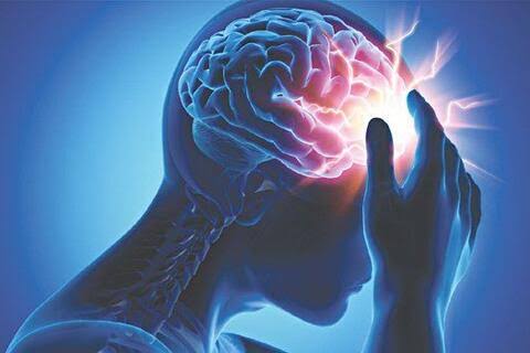
A stroke is a sudden disruption of blood flow to part of the brain that causes brain cells to die within minutes. The brain — despite representing only 2 percent of body weight — receives 15 to 20 percent of cardiac output and consumes 20 percent of the body’s oxygen at rest. Unlike muscle, liver, or fat cells, neurons have virtually no energy stores and cannot function for more than 4 to 5 minutes without continuous oxygen delivery from circulating blood. When a stroke cuts off blood supply to a region of the brain, the affected neurons begin dying almost immediately — at a rate of approximately 1.9 million neurons, 14 billion synapses, and 7.5 miles of myelinated nerve fibers per minute in a large ischemic stroke — producing sudden neurological deficits that reflect precisely which brain region has lost its blood supply.
Stroke is the fifth leading cause of death in the United States and the single leading cause of long-term adult disability, affecting approximately 795,000 Americans annually (610,000 first strokes, 185,000 recurrent strokes) and killing approximately 140,000 each year. Globally, stroke is the second leading cause of death and third leading cause of disability-adjusted life years (DALYs). Despite its enormous burden, stroke is highly preventable — approximately 80 percent of strokes are preventable through risk factor modification — and highly treatable when recognized and addressed immediately. Understanding what a stroke is, why it causes such rapid and severe neurological injury, and what determines outcomes is foundational knowledge for anyone who wants to protect their own or a loved one’s brain health.
The Two Main Types of Stroke — Ischemic and Hemorrhagic
Stroke is divided into two fundamentally different types based on the underlying mechanism — a distinction that is critical for treatment because the therapies for each type are different and, in some cases, directly contraindicated for the other type.
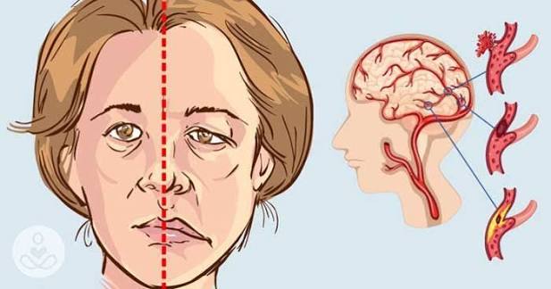
Ischemic stroke accounts for approximately 87 percent of all strokes and results from blockage of a cerebral artery, preventing blood flow to the brain territory supplied by that vessel. The blockage can arise from several mechanisms: thrombosis (clot forming in place on an atherosclerotic plaque within a cerebral or carotid artery), embolism (clot or other material that forms elsewhere — typically in the heart in atrial fibrillation, or in a carotid or vertebral artery plaque — and travels to lodge in a cerebral vessel), or small vessel disease (lipohyalinosis of perforating arteries causing lacunar infarcts in the deep brain structures). The treatment for ischemic stroke is reperfusion — restoring blood flow to the blocked vessel as rapidly as possible through intravenous thrombolysis (IV tPA within 4.5 hours of symptom onset) and/or mechanical thrombectomy (catheter-based removal of the clot, now with evidence supporting treatment up to 24 hours in selected patients with favorable imaging profiles).
Hemorrhagic stroke accounts for approximately 13 percent of strokes but causes a disproportionate share of stroke deaths, representing approximately 40 percent of stroke mortality. Hemorrhagic stroke results from rupture of a blood vessel within or around the brain — either intracerebral hemorrhage (ICH, bleeding directly into the brain parenchyma, most commonly from rupture of a small perforating artery damaged by hypertension or amyloid angiopathy) or subarachnoid hemorrhage (SAH, bleeding into the space surrounding the brain, most commonly from rupture of an intracranial aneurysm). Hemorrhagic stroke treatment focuses on controlling bleeding (reversing anticoagulation, managing blood pressure, surgical or endovascular treatment of the bleeding source), managing intracranial pressure, and preventing secondary brain injury. The thrombolytic drugs used to treat ischemic stroke are contraindicated in hemorrhagic stroke — which is why emergency brain imaging (CT scan) is performed before any stroke treatment, to identify hemorrhage and exclude it from tPA eligibility.
“Time Is Brain” — Why Speed Matters More Than Anything Else
The phrase “time is brain” captures the most fundamental truth about stroke biology and stroke care: every minute of delayed treatment in acute ischemic stroke results in further brain death and reduced prospect of neurological recovery. The calculation — 1.9 million neurons dying per minute during a large artery ischemic stroke — translates clinically into the difference between a patient who walks out of the hospital functionally independent and one who requires nursing home care for the rest of their life, or who dies.
The window for acute ischemic stroke treatment with IV tPA is 4.5 hours from symptom onset. Within this window, earlier treatment produces better outcomes — a patient treated at 60 minutes from symptom onset has substantially better neurological recovery than one treated at 3 hours, who does better than one treated at 4 hours. The benefit of IV tPA is so time-dependent that studies have modeled the value of treatment at different time points in terms of outcomes gained: treating within 90 minutes saves 3 weeks of disability-free life per patient compared to treatment at 90 to 180 minutes.
Mechanical thrombectomy — catheter-based removal of the thrombus from large intracranial arteries — has even more dramatic potential for neurological rescue, with several landmark trials (MR CLEAN, ESCAPE, EXTEND-IA, SWIFT PRIME, DAWN, DEFUSE 3) demonstrating that patients with large vessel occlusion treated with thrombectomy had functional independence rates double or triple those of patients receiving medical management alone. The time window for thrombectomy eligibility has been extended up to 24 hours from last known well in patients with favorable MRI penumbra imaging (demonstrated in the DAWN and DEFUSE 3 trials), but earlier treatment within this extended window still produces better outcomes.
These time dependencies create the clinical imperative for immediate action when stroke symptoms are suspected. Calling 911 immediately — rather than driving to the hospital or waiting to see if symptoms resolve — activates the prehospital stroke protocol that allows hospital notification before arrival, enabling the stroke team to be assembled and the CT scanner to be prepared so that door-to-needle time (time from hospital arrival to IV tPA administration) and door-to-groin time (time from arrival to thrombectomy start) are minimized. Every element of the stroke response chain — recognition, emergency services activation, prehospital notification, emergency department evaluation, imaging, and treatment decision — exists to shorten the total time from symptom onset to reperfusion.
What Happens in the Brain During a Stroke
The brain’s extreme vulnerability to ischemia reflects its unique metabolic profile. Unlike cells that can shift to anaerobic metabolism when oxygen is unavailable, neurons are almost entirely dependent on aerobic (oxygen-dependent) energy production. The brain stores virtually no glycogen for emergency energy use. When blood flow stops, the ATP that maintains ion gradients across neuronal membranes is consumed within seconds, and the pumps that keep sodium and calcium out of the cell fail.
Sodium and calcium flood into neurons, causing them to swell (cytotoxic edema), triggering glutamate release from depolarizing neurons, and activating calcium-dependent enzymes that degrade proteins, membranes, and DNA. This cascade of excitotoxicity, oxidative stress, mitochondrial dysfunction, and inflammation — collectively the “ischemic cascade” — proceeds through two spatially distinct zones: the ischemic core, where blood flow has fallen below the threshold for cellular survival and cells are irreversibly injured within minutes to hours, and the ischemic penumbra, a surrounding zone of marginally reduced blood flow where cells are dysfunctional but still alive — electrically silent but metabolically viable — sustained by residual perfusion through collateral vessels.
The therapeutic opportunity in acute ischemic stroke is the penumbra. Reperfusion that restores flow before the penumbra has converted to irreversible infarction rescues functional neurons that would otherwise die, reducing the final infarct volume and the degree of permanent neurological deficit. The penumbra shrinks over time as the ischemic core expands — driven by failure of collateral perfusion and propagation of the ischemic cascade outward from the core. This dynamic process — the therapeutic window during which penumbra neurons can be rescued — is the biological basis for why every minute matters in stroke treatment.
Risk Factors for Stroke — What Raises the Danger
Stroke shares most major risk factors with coronary artery disease, reflecting the same underlying atherosclerotic and vascular disease process that damages both cerebral and coronary vessels. The most important modifiable risk factors for stroke include:
Hypertension is the single most important stroke risk factor — accounting for approximately 50 percent of stroke risk attributable to all modifiable risk factors. Elevated blood pressure damages cerebral blood vessels through mechanisms including endothelial dysfunction, accelerated atherosclerosis in large arteries, and lipohyalinosis in small perforating arteries that leads to lacunar infarction and small vessel hemorrhage. Each 20 mmHg rise in systolic blood pressure above 115 mmHg approximately doubles stroke risk; conversely, each 10 mmHg reduction in systolic blood pressure reduces stroke risk by approximately 30 percent.
Atrial fibrillation increases stroke risk fivefold by allowing cardioembolic thrombi to form in the left atrial appendage and embolize to the cerebral circulation. AFib-related strokes are typically large and severely disabling, and the stroke risk persists even in paroxysmal AFib. Anticoagulation with DOACs reduces AFib-related stroke risk by 60 to 70 percent and is the highest-priority treatment decision in AFib management.
Diabetes mellitus doubles to quadruples stroke risk through accelerated cerebrovascular atherosclerosis, prothrombotic state, and impaired collateral circulation. Diabetic patients who develop stroke have larger infarcts, worse acute outcomes, and higher recurrence rates than non-diabetic stroke patients, reflecting the additive damage of chronic hyperglycemia on cerebrovascular reserve.
Dyslipidemia — elevated LDL cholesterol — promotes carotid and intracranial atherosclerosis that can either cause ischemic stroke directly (through plaque rupture and arterio-arterial embolism) or serve as a substrate for thrombosis during periods of hemodynamic compromise. High-intensity statin therapy significantly reduces ischemic stroke risk — and paradoxically may slightly increase hemorrhagic stroke risk in some populations, reflecting the complex relationship between cholesterol and small vessel integrity.
Smoking doubles ischemic stroke risk through multiple mechanisms: accelerated carotid atherosclerosis, increased fibrinogen and platelet aggregability, polycythemia (elevated red cell mass that increases blood viscosity), and reduced tissue oxygen-carrying capacity. Smoking cessation reduces stroke risk substantially within 2 to 5 years.
The American Stroke Association’s stroke resources provide comprehensive patient and clinician education on stroke recognition and prevention. The CDC stroke information covers types, risk factors, signs, and prevention. The NHLBI stroke guide addresses causes, diagnosis, treatment, and recovery in detail.
Related reading: Stroke Warning Signs: FAST Explained | Atrial Fibrillation | High Blood Pressure and Stroke | Major Risk Factors for Heart Disease | Ischemic vs Hemorrhagic Stroke
Sources
- Sacco RL, et al. An Updated Definition of Stroke for the 21st Century. Stroke. 2013;44(7):2064-2089.
- Powers WJ, et al. 2019 AHA/ASA Guideline for the Early Management of Patients With Acute Ischemic Stroke. Stroke. 2019;50(12):e344-e418.
- Nogueira RG, et al. Thrombectomy 6 to 24 Hours after Stroke with a Mismatch between Deficit and Infarct (DAWN). N Engl J Med. 2018;378(1):11-21.
- Albers GW, et al. Thrombectomy for Stroke at 6 to 16 Hours with Selection by Perfusion Imaging (DEFUSE 3). N Engl J Med. 2018;378(8):708-718.
- Feigin VL, et al. Global and Regional Burden of Stroke During 1990–2010. Lancet. 2014;383(9913):245-254.
- Siesjö BK. Pathophysiology and Treatment of Focal Cerebral Ischemia. J Neurosurg. 1992;77(2):169-184.
What Brain Area Is Affected? — How Stroke Location Determines Deficits
Because different brain regions control different functions, the neurological deficits produced by a stroke depend entirely on which artery is blocked and which brain territory loses its blood supply. Understanding this anatomy helps explain why stroke symptoms are so variable across patients — a stroke in one patient produces inability to speak, while another patient’s stroke produces paralysis of the right arm, and another causes sudden loss of vision in one eye.
Middle cerebral artery (MCA) strokes are the most common large vessel ischemic strokes and produce the classic stroke presentation: contralateral hemiplegia (weakness of the face, arm, and leg on the opposite side of the body from the stroke), contralateral hemisensory loss, and — when the dominant hemisphere (usually left) is involved — aphasia (language impairment). Aphasia in MCA stroke may involve expressive aphasia (Broca’s area damage — difficulty producing speech while comprehension is preserved), receptive aphasia (Wernicke’s area damage — fluent but nonsensical speech with impaired comprehension), or global aphasia (both areas affected — severe impairment of all language functions). Right MCA strokes produce left hemineglect — a dramatic syndrome in which patients ignore the left side of their body and environment, failing to attend to stimuli on the left despite intact visual and sensory pathways.
Anterior cerebral artery (ACA) strokes produce predominantly leg weakness greater than arm weakness (the opposite of MCA territory, reflecting the somatotopic organization of the motor cortex where the leg representation is on the medial surface supplied by the ACA), along with personality changes, apathy, and frontal lobe behavioral features when the frontal lobe territory is involved.
Posterior cerebral artery (PCA) strokes classically produce contralateral homonymous hemianopia (loss of the same visual field half in both eyes) from damage to the primary visual cortex (occipital lobe). PCA strokes are sometimes mistaken for optic nerve disease or migraine because the visual symptoms may be the predominant or only complaint.
Vertebrobasilar strokes — affecting the posterior circulation (vertebral and basilar arteries supplying the brainstem and cerebellum) — produce a distinctive syndrome of “crossed” deficits: ipsilateral cranial nerve findings (facial weakness, diplopia, dysphagia, dysarthria) with contralateral limb findings (weakness, numbness), along with ataxia (cerebellar incoordination), vertigo, and nausea from cerebellar or vestibular involvement. Basilar artery occlusion — the most severe posterior circulation stroke — can cause locked-in syndrome (quadriplegia and anarthria with preserved consciousness) or death from pontine failure, making it one of the most devastating and time-critical stroke presentations.
Lacunar infarcts — small deep strokes from occlusion of perforating arteries damaged by lipohyalinosis from hypertension or diabetes — produce classic syndromes without cortical features (no aphasia, no hemianopia, no hemineglect): pure motor hemiparesis (weakness without sensory loss), pure sensory stroke (sensory loss without weakness), sensorimotor stroke, ataxic hemiparesis, or clumsy hand-dysarthria syndrome. Lacunar strokes are generally smaller and have better acute outcomes than large vessel strokes, but multiple lacunar infarcts over years (lacunar state, état lacunaire) contribute significantly to vascular cognitive impairment and dementia.
Secondary Stroke Prevention — Reducing the Risk of Recurrence
Stroke recurrence risk is highest in the first days to weeks after an initial stroke — 10 to 15 percent of patients with ischemic stroke have a recurrent stroke within 3 months, with much of this risk concentrated in the first 48 to 72 hours. Secondary prevention — the systematic implementation of evidence-based therapies to prevent recurrence — must begin immediately, even as acute treatment is ongoing.
The specific secondary prevention strategy depends critically on the identified stroke mechanism, because different causes require different preventive approaches:
Cardioembolic stroke from atrial fibrillation: Anticoagulation with a DOAC (apixaban, rivaroxaban, dabigatran, edoxaban) is the cornerstone of secondary prevention — reducing AFib-related stroke recurrence by 60 to 70 percent. The timing of anticoagulation initiation after ischemic stroke in AFib patients involves a risk-benefit judgment between early protection from recurrent embolism and risk of hemorrhagic transformation of the infarct: current practice guidelines suggest anticoagulation within 1 to 14 days depending on stroke size (the “1-3-6-12 day rule” based on stroke severity).
Large artery atherosclerotic stroke (carotid or intracranial atherosclerosis): Antiplatelet therapy (aspirin plus clopidogrel for the first 21 days in high-risk TIA/minor stroke per POINT and CHANCE trials, then single antiplatelet long-term), high-intensity statin therapy (targeting LDL below 70 mg/dL, with evidence for even lower targets in high-risk patients), blood pressure control, and consideration of carotid endarterectomy or stenting for symptomatic high-grade carotid stenosis (benefit strongest for 70 to 99 percent stenosis operated within 2 weeks of the index stroke).
Small vessel/lacunar stroke: Single antiplatelet therapy (aspirin or clopidogrel), aggressive blood pressure control (hypertension is the dominant modifiable risk factor), statin therapy, and diabetes management if present. Dual antiplatelet therapy does not provide incremental benefit over single antiplatelet in lacunar stroke and increases bleeding risk.
Stroke Recovery — What Happens After the Event
Stroke recovery involves a combination of spontaneous neurological improvement (as penumbra neurons that survived the acute event recover function as edema resolves, diaschisis reverses, and collateral circulation improves) and activity-dependent neuroplasticity (in which intensive rehabilitation drives reorganization of surviving brain circuits to compensate for lost functions). The most rapid recovery occurs in the first weeks after stroke, but neurological improvement can continue for months to years — particularly with sustained, intensive rehabilitation and active participation by the patient.
The major rehabilitation modalities for stroke recovery include physical therapy (gait, balance, lower extremity strength and coordination), occupational therapy (upper extremity function, activities of daily living, adaptive strategies and equipment), speech-language therapy (aphasia rehabilitation, dysphagia management, cognitive-communication impairment), cognitive rehabilitation (attention, memory, executive function), and psychological support (depression and anxiety are extremely common post-stroke and require active assessment and treatment). The intensity and duration of rehabilitation is strongly correlated with functional recovery — patients who receive more hours of therapy per day in dedicated stroke rehabilitation units have consistently better outcomes than those in lower-intensity rehabilitation environments.
Post-stroke depression affects approximately 30 to 40 percent of stroke survivors and is the single most important modifiable predictor of functional recovery — depressed stroke patients engage less with rehabilitation, have poorer adherence to secondary prevention medications, and have worse long-term outcomes. Screening for depression at 3, 6, and 12 months post-stroke and providing evidence-based treatment (SSRIs, CBT, or both) is a clinical imperative that is still inconsistently implemented across stroke care systems.
Life expectancy after stroke has improved substantially over the past decades as acute treatment and secondary prevention have advanced. Patients who survive the acute period and complete rehabilitation can achieve meaningful functional recovery — with approximately one-third of stroke survivors experiencing only mild disability, one-third moderate disability, and one-third severe disability requiring significant care. The variables most strongly predicting good functional outcome are younger age at stroke, smaller infarct volume, less severe initial neurological deficit, early and effective reperfusion in eligible patients, and intensive, sustained rehabilitation engagement.
Stroke Prevention — The 80 Percent That Is Preventable
Stroke is among the most preventable of all serious neurological events — population-based modeling and landmark prevention trials consistently estimate that 80 percent of strokes are preventable through systematic management of modifiable risk factors. The five highest-impact prevention targets, ranked by their contribution to population-attributable risk, are hypertension (the single largest contributor), physical inactivity, dyslipidemia, poor diet quality, and obesity — all of which are addressable through a combination of lifestyle modification and pharmacological intervention when indicated.
Blood pressure control deserves particular emphasis because of its outsized contribution to stroke risk and the magnitude of benefit from treatment. The PROGRESS trial demonstrated that blood pressure lowering with perindopril plus indapamide reduced recurrent stroke by 28 percent in patients with prior stroke, regardless of baseline blood pressure level — including in patients already within normal blood pressure ranges. The SPRINT MIND trial showed that intensive blood pressure control (systolic target below 120 mmHg) reduced the composite of MCI and dementia compared to standard treatment (below 140 mmHg), extending the case for intensive BP control beyond cardiovascular events to cognitive preservation. Adults with hypertension who consistently achieve and maintain blood pressure below 130/80 mmHg reduce their lifetime stroke risk by approximately 30 to 40 percent — one of the most impactful risk reduction steps available in all of preventive medicine.
Came across this while researching what is a stroke? causes, types, for a family member. What I liked most was that the article didn’t just say what to avoid — it also gave alternatives. Will definitely be coming back to this site for more health information.
This is one of the clearest explanations of what is a stroke? causes, types, I have found. The article answered questions I didn’t even know I had until I started reading. Exactly the kind of evidence-based information that is hard to find in one place.
As someone dealing with this personally, the what is a stroke? causes, types, section was very helpful. I especially valued the explanation of why these recommendations exist, not just what they are. Exactly the kind of evidence-based information that is hard to find in one place.
Really well-written article on what is a stroke? causes, types,. The article answered questions I didn’t even know I had until I started reading. Appreciate the effort that went into researching and writing this — it shows.
Finally a resource that explains what is a stroke? causes, types, in plain language. I appreciated how the article addressed both the clinical side and the practical adjustments. This gave me real confidence going into my next specialist appointment.
Bookmarked this article on what is a stroke? causes, types, immediately — going to reference it regularly. I have tried following advice from several sources but this is most consistent with what my specialist told me. Keep up this kind of thorough health journalism — it genuinely helps patients like me.