
Pregnancy and Urinary Health: What to Expect and When to Seek Care
Pregnancy and urinary health are intertwined throughout gestation in ways that affect virtually every pregnant person — from the urinary frequency of the first trimester to the stress leakage common in the third, and from the pyelonephritis risk that makes UTI treatment in pregnancy a clinical priority to the pelvic floor changes that influence urinary continence for years after delivery. Understanding why these urinary changes occur, which ones are expected features of a normal pregnancy, and which ones require prompt clinical attention is essential knowledge for pregnant individuals and their care providers. Pregnancy alters the anatomy and physiology of the entire urinary tract: the growing uterus displaces and compresses the bladder; progesterone relaxes the smooth muscle of the ureters; the physiological changes of pregnancy increase renal blood flow by 40–65% and the glomerular filtration rate by 40–50%; and progesterone-induced ureteral relaxation combined with the mechanical ureteral compression of the enlarging uterus predisposes to physiological hydronephrosis (right greater than left) and, importantly, to the urinary stasis that increases UTI risk and its potential to ascend to the kidney. These are not minor incidental changes — they affect clinical decision-making about UTI diagnosis and treatment, the interpretation of urinary symptoms, and the management of pelvic floor health during and after pregnancy.
Urinary tract infections during pregnancy carry risks beyond those in non-pregnant individuals — asymptomatic bacteriuria (bacterial colonization of the urine without symptoms), which in non-pregnant individuals is generally not treated, becomes an indication for antibiotic treatment in pregnancy because the physiological changes of gestation greatly increase the risk of its progression to pyelonephritis (kidney infection), which is associated with preterm labor, low birth weight, maternal sepsis, and fetal and maternal mortality in severe cases. This is one of the most clinically important distinctions between urinary health in pregnancy and in other states, and it underlies the routine urine culture screening performed at the first prenatal visit in most obstetric guidelines. The full spectrum of pregnancy-related urinary changes — physiological, symptomatic, and pathological — requires a differentiated understanding to distinguish normal adaptation from conditions requiring medical intervention.
Urinary Frequency and Urgency During Pregnancy: Why It Happens
Urinary frequency — the need to void more often than usual — is one of the most universal early pregnancy symptoms, often beginning within the first few weeks of gestation and persisting throughout pregnancy, though with different mechanisms at different stages. In the first trimester, frequency is driven primarily by the hormonal effects of human chorionic gonadotropin (hCG) and progesterone on renal blood flow and glomerular filtration rate: the 40–50% increase in GFR that occurs within weeks of conception means the kidneys are producing significantly more urine than before pregnancy, and the bladder must store and empty this increased volume more frequently. In the second trimester, the uterus rises out of the pelvis as it grows, often temporarily reducing bladder compression and providing some relief from first-trimester frequency; many pregnant individuals notice that their urinary frequency improves somewhat in the mid-trimester for this reason. In the third trimester, as the uterus reaches its maximal size and the fetal head descends into the pelvis in preparation for delivery, direct bladder compression returns and worsens — the functional bladder capacity (the volume at which urgency occurs) is reduced by physical compression from both above (the uterine fundus) and below (the presenting fetal part), and frequency and urgency may be more pronounced than at any earlier stage. Urgency — the sudden, compelling need to void — follows a similar pattern, intensifying in the third trimester when bladder capacity is most compromised. These changes are physiological and expected; their clinical importance lies primarily in distinguishing them from the urgency and frequency of urinary tract infection (which typically is accompanied by dysuria, haematuria, or systemic symptoms) or from overactive bladder pathology that is unrelated to pregnancy. For the evaluation framework that helps distinguish physiological urinary frequency from symptomatic conditions requiring investigation, the frequent urination guide on Horizon Health Guide provides the broader differential that applies during and outside pregnancy. For the bladder health context that underlies urinary symptom management generally, the bladder health tips guide covers the evidence-based behavioral strategies applicable across life stages.
UTIs in Pregnancy: Why They Are Treated Differently
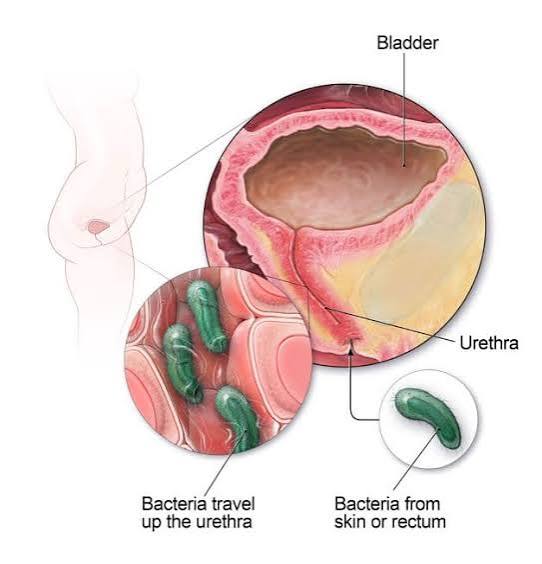
Urinary tract infections during pregnancy are one of the most common medical complications of gestation and one of the most important to diagnose and treat promptly, because the physiological changes of pregnancy dramatically increase the risk of infection ascending from the bladder to the kidney — a progression that in the non-pregnant state is much less likely and in the pregnant state carries serious maternal and fetal consequences. Asymptomatic bacteriuria (ASB) — the presence of bacteria in the urine at clinically significant concentrations (conventionally 100,000 CFU/mL or above of a single organism on a midstream clean-catch specimen) without symptoms of infection — occurs in 2–10% of pregnant individuals and would be managed with watchful waiting in non-pregnant adults. In pregnancy, ASB is treated with antibiotics because the risk of its progression to symptomatic cystitis (20–40% without treatment) and pyelonephritis (20–35%) is dramatically elevated by the physiological hydronephrosis and ureteral stasis of pregnancy. All current obstetric guidelines recommend routine urine culture screening for ASB at the first prenatal visit (typically at 8–12 weeks), with treatment of any positive culture with a pregnancy-safe antibiotic and confirmation of bacterial clearance with a test-of-cure culture 1–2 weeks after completing treatment. The antibiotics appropriate for UTI treatment in pregnancy are limited to those with established safety profiles: nitrofurantoin (avoid near term, at 36+ weeks, as it can cause haemolytic anaemia in the newborn), trimethoprim (avoid in the first trimester due to folate antagonism), amoxicillin (useful when sensitivity confirmed), and cephalexin are commonly used based on culture sensitivity results. Fluoroquinolones (ciprofloxacin, levofloxacin) are generally avoided in pregnancy due to concerns about fetal cartilage development. Symptomatic cystitis in pregnancy — presenting with dysuria, frequency, urgency, and suprapubic discomfort without fever or flank pain — is treated with a pregnancy-safe antibiotic for 3–7 days (longer than the 3-day course for uncomplicated cystitis in non-pregnant individuals, to achieve complete bacterial clearance given the higher risk of relapse and ascent). A test-of-cure urine culture 7–14 days after treatment is recommended to confirm eradication. Pyelonephritis in pregnancy — fever above 38°C with flank pain and costovertebral angle tenderness, often with nausea and vomiting — is a medical emergency in pregnancy, associated with preterm labor (through prostaglandin release from the infected renal parenchyma and endotoxins), maternal sepsis, acute respiratory distress syndrome, and rarely maternal death. Pregnant individuals with clinical pyelonephritis require hospitalization, intravenous antibiotics (typically ceftriaxone or gentamicin until afebrile and clinically improving), intravenous fluid resuscitation, fetal monitoring, and uterine contraction monitoring. After clinical improvement and transition to oral antibiotics, suppressive antibiotic prophylaxis for the remainder of the pregnancy (low-dose nitrofurantoin or cephalexin nightly) is recommended because the recurrence risk after pyelonephritis in pregnancy is approximately 10–15%. The authoritative guidance from the NIDDK UTI resource and from current obstetric guidelines emphasizes the clinical significance of UTI management in pregnancy as distinct from routine adult UTI care.
Stress Urinary Incontinence During and After Pregnancy
Stress urinary incontinence — involuntary leakage of urine with physical effort, coughing, sneezing, or exercise — is extremely common during the third trimester of pregnancy and in the immediate postpartum period, affecting 30–50% of pregnant individuals to some degree during late pregnancy. The mechanisms during pregnancy include the direct pressure of the uterus and fetal weight on the bladder and pelvic floor (reducing the urethral closure pressure reserve against abdominal pressure spikes), progesterone-induced relaxation of periurethral connective tissue, and the gravitational strain on the pelvic floor from the altered centre of gravity of the pregnant abdomen. Vaginal delivery — particularly assisted delivery with forceps or vacuum, delivery of a large fetus, prolonged second stage of labor, and significant perineal tears — causes direct trauma to the pelvic floor muscles (particularly the levator ani complex), pudendal nerve, and periurethral connective tissue that constitutes the primary obstetric risk factor for long-term post-partum SUI. Third trimester SUI that resolves spontaneously within 3–6 months postpartum without treatment is common in women who have had uncomplicated vaginal deliveries; SUI that persists at 6 months postpartum, particularly in women who had prolonged labor or instrumental delivery, is less likely to resolve without intervention and should prompt referral for pelvic floor physiotherapy. Pelvic floor muscle training (PFMT) beginning in pregnancy and continuing postpartum substantially reduces the risk and severity of SUI both during pregnancy and at 3 and 12 months postpartum — multiple randomized trials have confirmed this benefit, and PFMT in pregnancy is recommended by virtually all obstetric guidelines as a preventive intervention for pelvic floor dysfunction. The exercise protocol and evidence base for PFMT applicable to pregnancy and the postpartum period is covered in the pelvic floor exercises and urinary control guide. Women who develop SUI during pregnancy should not assume it will resolve automatically after delivery — seeking pelvic floor physiotherapy postpartum, particularly after vaginal delivery with any of the higher-risk features noted, is the evidence-based approach to maximizing recovery and preventing long-term incontinence. For women with persistent postpartum urinary incontinence beyond 6–12 months, referral to a urogynaecologist for evaluation of surgical options may be appropriate, since obstetric SUI that fails to respond to PFMT can be treated effectively with the midurethral sling with outcomes equivalent to incontinence arising from non-obstetric causes. The full spectrum of urinary health management for women is covered in the urinary health for women guide on Horizon Health Guide. For the postpartum pelvic floor context, the AUA OAB guideline and the StatPearls urinary incontinence review provide the clinical evidence base for postpartum bladder and pelvic floor management.
Sources: NIDDK — UTI · AUA OAB Guidelines · StatPearls — Urinary Incontinence
Overactive Bladder and Urgency Incontinence During Pregnancy
Urgency urinary incontinence — involuntary loss of urine associated with the sudden urge to void — affects a significant minority of pregnant individuals, particularly in the third trimester when bladder capacity is most compressed. The mechanism during pregnancy is the combination of reduced functional bladder capacity from uterine compression (meaning urgency occurs at lower volumes than before pregnancy) and the increased urine production from the elevated GFR, which means the bladder fills to its reduced capacity more quickly. The result can be urgency episodes that produce leakage before the individual can reach the toilet — a distressing complication that is frequently not reported to midwives or obstetricians because of embarrassment. Pelvic floor muscle contraction at the onset of urgency — the urgency suppression technique that is the cornerstone of behavioral management for OAB outside pregnancy — is safe and effective in pregnancy and should be taught to all pregnant individuals experiencing urgency incontinence. The contraction of the pelvic floor at the time of an urgency wave reflexively inhibits the bladder detrusor contraction through sacral afferent-efferent pathways, suppressing the urgency and allowing the individual time to reach the toilet without urgency-driven leakage. Pharmacological treatment for OAB during pregnancy is largely avoided due to limited safety data — antimuscarinics are generally not used in pregnancy, and mirabegron has no established safety profile in gestation. Management is therefore behavioral: urgency suppression, timed voiding (voiding on a schedule every 2–3 hours to prevent the bladder from reaching the volumes at which urgency becomes uncontrollable), pelvic floor strengthening (to improve urgency suppression capacity), and practical adaptations such as close toilet proximity awareness and wearing rapidly removable clothing. Most pregnancy-related urgency incontinence resolves or substantially improves in the weeks after delivery as bladder capacity normalizes and the mechanical compression from the uterus is relieved — women who continue to experience urgency incontinence beyond 6–12 weeks postpartum should raise it at their postnatal appointments, since persistent postpartum urgency incontinence is a treatable condition that responds to behavioral bladder training and, if needed, pharmacological OAB management. The urgency and frequency symptoms of pregnancy should always be distinguished from those of urinary tract infection — if frequency and urgency are accompanied by dysuria, haematuria, cloudy urine, or fever, UTI must be excluded with urinalysis and culture rather than attributed to pregnancy physiology.
Kidney Stones During Pregnancy: Evaluation and Management
Kidney stones present a unique management challenge during pregnancy — symptomatic nephrolithiasis affects approximately 1 in 1,500 pregnancies and is one of the most common non-obstetric reasons for hospitalization during gestation. The physiological changes of pregnancy create a contradictory environment for stone formation: on one hand, the elevated GFR of pregnancy increases urinary calcium and uric acid excretion, raising supersaturation and stone formation risk; on the other, the increased urinary volume from elevated GFR and the elevated urinary citrate and magnesium of pregnancy are inhibitors of crystallization. The net result is that the stone formation rate is not substantially higher in pregnancy than in non-pregnant women of the same age, but when stones form or pre-existing stones become symptomatic, management is complicated by the need to balance diagnostic and therapeutic interventions with fetal safety. The presentation of symptomatic nephrolithiasis in pregnancy — flank pain radiating to the groin with or without haematuria, nausea, and vomiting — overlaps significantly with other causes of acute abdominal pain in pregnancy (round ligament pain, appendicitis, placental abruption, preterm labor), making diagnosis more challenging than in the non-pregnant state. Diagnosis relies primarily on renal ultrasound, which can identify hydronephrosis (though physiological hydronephrosis of pregnancy complicates interpretation) and sometimes visualize large calculi directly; magnetic resonance urography (MR urography without gadolinium) is the gold-standard imaging for suspected urolithiasis in pregnancy when ultrasound is non-diagnostic, avoiding the ionizing radiation of CT urography (which is also used in selected cases when MR is unavailable and clinical need is urgent, using the lowest possible radiation dose). Management of symptomatic nephrolithiasis in pregnancy prioritizes conservative measures: adequate hydration, analgesia (acetaminophen is safe; NSAIDs are avoided in pregnancy particularly after 20 weeks due to renal effects and premature ductus arteriosus closure; opioid analgesia is used short-term for severe pain with appropriate monitoring), and observation for spontaneous passage. The majority of symptomatic stones in pregnancy pass spontaneously, particularly stones below 5 mm. For stones that obstruct the collecting system, cause intractable pain or nausea, or are associated with fever suggesting infection (urinary obstruction with infection is a urological emergency requiring urgent decompression — in pregnancy as in any state), the interventional options are ureteroscopy with laser lithotripsy (performed under general anesthesia with appropriate shielding and fetal monitoring, and considered safe in pregnancy by urological and obstetric guidelines) or percutaneous nephrostomy for decompression when ureteroscopy is not immediately available. Extracorporeal shock wave lithotripsy (ESWL) is contraindicated in pregnancy.
Postpartum Urinary Health: What to Watch For After Delivery
The postpartum period brings its own spectrum of urinary health considerations that extend beyond the immediate resolution of pregnancy-related frequency and the potential persistence of stress incontinence. Postpartum urinary retention — inability to void or severely impaired voiding in the hours after delivery — is a recognized complication of labor and delivery, occurring in approximately 1–4% of women after vaginal delivery and up to 20% after cesarean section. The causes include epidural analgesia impairing detrusor sensation and contractility, pudendal nerve trauma from delivery reducing the afferent limb of the voiding reflex, perineal edema and pain causing voluntary sphincter guarding, and in some cases direct detrusor overdistension from inadequate bladder monitoring during labor. Postpartum retention requires catheterization if the woman cannot void spontaneously within 6 hours of delivery or if the bladder volume exceeds 400–500 mL on bedside ultrasound without successful voiding; most cases resolve within days to weeks as epidural effects wear off and nerve function recovers. Covert postpartum retention — incomplete emptying without overt inability to void — is more common and may go undetected if post-void residual is not routinely checked in the early postpartum period. Postpartum urinary tract infection is common in the puerperium, driven by catheterization during labor or the immediate postpartum period, perineal wound contamination, and incomplete bladder emptying. Symptoms in the early postpartum period may be mistakenly attributed to perineal discomfort from delivery; clinicians should have a low threshold for urine culture in any postpartum woman with urinary symptoms, fever, or non-specific malaise. For women who had pregnancy-related recurrent UTIs or pyelonephritis, a surveillance urine culture at 4–6 weeks postpartum confirms clearance of infection and allows discontinuation of any antibiotic prophylaxis that was used during gestation. Long-term pelvic floor recovery after vaginal delivery — particularly after deliveries associated with higher pelvic floor trauma risk — should be supported by postnatal pelvic floor physiotherapy beginning at 6–8 weeks postpartum. The postnatal assessment of urinary incontinence and pelvic floor function is an opportunity to identify and treat incontinence that, if not addressed in the postpartum period, may persist and worsen through subsequent pregnancies and the postmenopausal years. The pelvic floor exercises guide provides the evidence-based protocol for postpartum pelvic floor rehabilitation. For women with persistent urinary symptoms beyond 3 months postpartum — incontinence, urgency, frequency, or voiding difficulty — seeking evaluation at that point prevents the normalization of what are in fact treatable conditions. The when bladder symptoms need evaluation guide provides the framework for deciding when postpartum urinary symptoms require specialist referral versus self-management with evidence-based behavioral strategies.
Bladder Health Through Pregnancy and Beyond
Maintaining bladder health through pregnancy requires a few practical habits that reduce UTI risk, support pelvic floor function, and prevent the overdistension that can impair bladder recovery postpartum. Adequate hydration — targeting approximately 2–2.5 liters of fluid per day during pregnancy — dilutes urine, reduces crystallization risk (important for stone-prone individuals), and supports the increased renal blood flow of gestation; excessive fluid restriction to manage frequency paradoxically concentrates urine and may increase UTI and stone risk. Avoiding prolonged bladder holding — the tendency of some pregnant individuals to delay voiding because of the inconvenience of frequent toilet visits — risks bladder overdistension and impaired detrusor contractility, particularly as pregnancy progresses; voiding at first urge rather than deferring indefinitely is generally the appropriate approach during pregnancy. Starting pelvic floor exercises in the first trimester — before pelvic floor loading from the growing uterus is maximal and before delivery-related trauma is possible — provides the greatest benefit for both preventing third-trimester incontinence and for postpartum pelvic floor recovery. The behavioral, dietary, and hydration principles that support bladder health during pregnancy are the same as those that support it at any life stage — the bladder health tips guide provides the comprehensive framework applicable to pregnant and non-pregnant individuals alike. Pregnancy is also an opportunity for urinary health awareness that extends beyond the gestational period: women who understand their pelvic floor anatomy, learn proper muscle activation during pregnancy, and receive postnatal physiotherapy are better equipped to maintain urinary continence through subsequent pregnancies and the postmenopausal years — a lifelong investment in pelvic floor health that begins with prenatal education and activation.
I had a urine culture done at my first prenatal appointment at 10 weeks and was told I had asymptomatic bacteriuria — bacteria in my urine with no symptoms whatsoever. My midwife explained that it needed to be treated in pregnancy, which I hadn’t expected since I’d had it before outside pregnancy and been told to just drink plenty of water. The article’s explanation of why ASB is treated differently in pregnancy — the 20-35% risk of ascending to a kidney infection in pregnancy versus the much lower risk outside it — finally explained the clinical decision to me clearly. I had a course of cephalexin, a test-of-cure culture confirmed clearance, and I’ve had no UTI issues since. I’m now 32 weeks and doing pelvic floor exercises daily since my first trimester after reading about the benefit for third-trimester incontinence prevention — something that wasn’t mentioned at my booking appointment and that I only found through my own reading. The point about starting them in the first trimester for best effect is something I wish had been part of my routine prenatal advice.
A clinically accurate and practically important guide that correctly identifies asymptomatic bacteriuria screening and treatment as the most impactful urological intervention in prenatal care. The distinction between the treatment threshold for ASB in pregnancy versus outside it is frequently not well understood by pregnant individuals, and explaining the mechanism — physiological hydronephrosis creating urinary stasis that dramatically increases pyelonephritis risk — is exactly the level of explanation that supports informed consent for antibiotic treatment of a condition with no symptoms. The antibiotic prescribing guidance is correct: nitrofurantoin should be avoided near term (after 36 weeks) due to the haemolysis risk in G6PD-deficient neonates, and fluoroquinolones are contraindicated throughout pregnancy — cephalexin and amoxicillin (when culture-guided sensitivity confirms it) are the most commonly used safe options. The kidney stone section correctly identifies MR urography as the preferred imaging modality over CT when ultrasound is non-diagnostic — the absence of ionizing radiation makes it the preferred second-line investigation for suspected urolithiasis in pregnancy in most centres with adequate MRI access.
Dr. Adeoye, the antibiotic prescribing nuances for UTI in pregnancy are exactly the kind of clinical detail that helps pregnant individuals understand why their prescription looks different from what they might have received outside pregnancy — the safety profile differences between antibiotics at different stages of gestation are real and clinically important, and patients who understand why they are receiving a specific antibiotic are more likely to complete the full course. Natalie, your decision to start pelvic floor exercises in the first trimester on your own initiative is evidence-based — the trials showing the greatest benefit for incontinence prevention do use first-trimester onset, and the postnatal recovery data also favors early onset PFMT. The fact that this wasn’t discussed at your booking appointment reflects a persistent gap in routine prenatal care that physiotherapy associations and obstetric guidelines have been working to close — most first-visit prenatal consultations should now include pelvic floor exercise instruction as standard.