
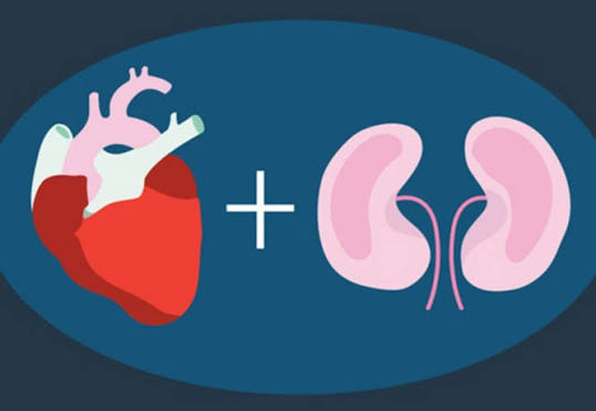
Heart disease and kidney disease are so closely intertwined that kidney specialists have a name for their interaction: cardiorenal syndrome. About half of people with heart failure have chronic kidney disease, and about half of people with chronic kidney disease have cardiovascular disease. Each condition accelerates the other — reduced cardiac output impairs kidney perfusion, and kidney dysfunction drives heart failure, arrhythmias, and accelerated atherosclerosis through mechanisms that are only now being fully understood. Adults with CKD stages 3 to 4 are more likely to die from cardiovascular disease than to ever reach dialysis — a statistic that makes protecting the heart as important as protecting the kidneys in this population. This article covers the biology of the cardiorenal connection, the specific conditions where it plays out, and the treatments that protect both organs simultaneously.
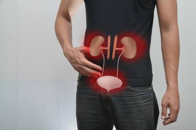
The Cardiorenal Syndrome: A Two-Way Street
Cardiorenal syndrome (CRS) describes the interconnected dysfunction between the heart and kidneys in five distinct patterns, classified by direction and acuity. The most clinically relevant are Type 1 — acute heart failure causing acute kidney injury, as in cardiogenic shock or acute decompensated heart failure — and Type 4 — chronic kidney disease progressively damaging the heart through uremic toxin accumulation, volume overload, anemia, and calcium-phosphate dysregulation.
The concept captures what clinicians observed long before the classification existed: fixing the heart often helps the kidneys, and protecting the kidneys often benefits the heart. The two organs share their blood supply, their hormonal environment (particularly the renin-angiotensin-aldosterone system and the sympathetic nervous system), their inflammatory milieu, and exposure to the same risk factors — diabetes, hypertension, dyslipidemia, smoking, and obesity. Treating either organ without addressing the other is incomplete medicine.
For the full picture of chronic kidney disease — its stages, symptoms, and long-term trajectory — see our article on what is chronic kidney disease.
How Heart Disease Reduces Kidney Function
The kidney receives 20 to 25 percent of cardiac output at rest — making it the most perfusion-dependent organ in the body relative to its size. When the heart’s pumping capacity falls — from heart failure, cardiogenic shock, or a large myocardial infarction — the kidneys feel the consequences almost immediately.
Reduced cardiac output. Falling perfusion pressure at the kidneys triggers the renin-angiotensin-aldosterone system: renin is released, angiotensin II is generated, and aldosterone is secreted. These hormones transiently maintain glomerular filtration by constricting the efferent arteriole, while simultaneously causing sodium and water retention to restore circulating volume. This is adaptive short-term and destructive long-term — sustained angiotensin II drives glomerulosclerosis, and aldosterone promotes renal fibrosis independent of its blood pressure effects.
Neurohormonal activation. Beyond RAAS, heart failure activates the sympathetic nervous system and vasopressin. Sympathetic tone constricts the afferent arteriole, cutting renal blood flow even when cardiac output is only mildly reduced. Vasopressin-driven water retention causes dilutional hyponatremia — a clinical marker of advanced neurohormonal activation and a poor prognostic sign in both conditions.
Inflammation. Chronic heart failure is an inflammatory state. Elevated TNF-α, interleukin-6, and C-reactive protein are consistently found and correlate with outcomes. These cytokines damage the renal endothelium, promote tubular cell apoptosis, and drive renal fibroblast activation — accelerating the fibrotic remodeling that is the final common pathway of CKD progression.
Venous Congestion: The Underappreciated Mechanism
For decades, the dominant clinical picture of cardiorenal deterioration focused on reduced cardiac output — the heart failing to pump forward. Evidence over the past 20 years has established that venous congestion — elevated venous pressure backing up from right heart failure into the renal venous system — is equally or more important in many patients.
Think of the kidney’s filtration pressure as a pressure gradient: what matters is not just the arterial input, but the difference between arterial pressure and venous backpressure. When right-sided heart failure raises central venous pressure from a normal 2 to 8 mmHg to 20 mmHg or more, renal venous pressure rises in parallel. The effective filtration gradient collapses — even if systemic arterial blood pressure looks adequate. Renal interstitial edema develops, compresses the tubules, and paradoxically worsens fluid retention even as the kidneys are bathed in excess fluid.
The ESCAPE trial in acute decompensated heart failure showed this directly: elevated central venous pressure — not low cardiac output — was the strongest predictor of worsening kidney function during hospitalization. The clinical takeaway: decongestion — aggressively removing excess fluid through diuresis — is the priority, even if eGFR dips transiently during treatment. A reversible creatinine rise during effective decongestion is far preferable to the irreversible tubular damage from persistent venous congestion.
How CKD Damages the Heart
CKD exposes the heart to a biological environment that accelerates every form of cardiac disease — atherosclerosis, cardiomyopathy, arrhythmia, and valvular disease.
Uremic cardiomyopathy. Uremic toxins — trimethylamine N-oxide (TMAO), indoxyl sulfate, p-cresyl sulfate — accumulate as kidney function declines and directly damage cardiomyocytes, impairing contractile function and promoting cardiomyocyte apoptosis. The result is uremic cardiomyopathy: left ventricular hypertrophy combined with myocardial fibrosis, reducing both systolic and diastolic function.
Left ventricular hypertrophy (LVH). LVH — pathological thickening of the heart’s main pumping chamber — is present in 40 to 75 percent of CKD stage 3 patients and nearly universal in end-stage renal disease. Its drivers in CKD are multiple and additive: hypertension, volume overload from sodium retention, anemia (forcing the heart to pump harder to deliver adequate oxygen), secondary hyperparathyroidism, and uremic toxin-driven calcium channel dysfunction. LVH substantially increases the risk of sudden cardiac death — the leading cause of mortality in dialysis patients.
Accelerated atherosclerosis. CKD drives coronary artery disease through oxidative stress, endothelial dysfunction, chronic inflammation, dyslipidemia (high triglycerides, low HDL), and uremic toxin exposure. Adults with CKD develop coronary artery disease younger, more extensively, and with worse outcomes after myocardial infarction than matched controls — partly because CKD patients have historically been excluded from cardiovascular trials, resulting in systematic under-treatment.
Vascular Calcification in CKD
One of the most distinctive cardiovascular consequences of advanced CKD is vascular calcification — calcium-phosphate crystal deposition in arterial walls. Unlike the plaque-based intimal calcification of conventional atherosclerosis, CKD primarily drives medial calcification: calcium deposited in the muscular wall of the artery, causing it to stiffen like a pipe.
The mechanism begins with phosphate retention. As eGFR falls, the kidneys can no longer excrete the dietary phosphate load. Rising serum phosphate stimulates fibroblast growth factor-23 (FGF-23) from bone — initially adaptive, but FGF-23 excess directly causes cardiac hypertrophy through cardiomyocyte FGF-23 receptors. Rising phosphate also causes vascular smooth muscle cells to transdifferentiate into osteoblast-like cells that deposit calcium in the vessel wall, stiffening the aorta and coronary arteries. Each heartbeat generates higher pulse pressure against a less elastic aorta, increasing the left ventricle’s afterload — worsening LVH further. Coronary artery calcification in CKD independently predicts cardiovascular events beyond conventional risk scores.
Atrial Fibrillation and Kidney Disease
Atrial fibrillation — the most common sustained cardiac arrhythmia — is two to three times more prevalent in adults with CKD than in the general population, and its prevalence rises progressively with declining eGFR. The shared biology includes chronic inflammation, RAAS overactivation, sympathetic nervous system activation, and fluid-overload-driven atrial remodeling — all promoting the re-entrant electrical circuits that sustain AF.
AF reduces cardiac output by 15 to 25 percent (from loss of the atrial contribution to ventricular filling and the irregular rhythm’s inefficient pumping), cutting renal perfusion. Renal artery embolism from AF-related clot can cause abrupt renal infarction. Stroke risk — already elevated in CKD — is compounded by AF substantially.
Anticoagulation in AF with CKD requires careful selection. Warfarin is now relatively contraindicated in CKD: it inhibits the carboxylation of matrix Gla protein, the molecule that normally keeps calcium out of vessel walls — accelerating the vascular calcification already driven by CKD. This is the “warfarin paradox.” Direct oral anticoagulants are preferred: apixaban has the most favorable safety and pharmacokinetic profile in CKD stages 3 to 4, based on its hepatic-dominant clearance. All DOACs require dose adjustment by eGFR.
Coronary Artery Disease and CKD
Coronary artery disease is two to four times more common in CKD patients than in age-matched controls, and outcomes after myocardial infarction are substantially worse — with dialysis patients experiencing mortality rates four to five times that of the general population after MI. Despite this, CKD patients remain systematically undertreated: less likely to receive statins, dual antiplatelet therapy, or coronary revascularization.
A specific procedural concern is contrast-induced acute kidney injury — the decline in kidney function caused by iodinated contrast dye during coronary angiography. Risk rises sharply at eGFR below 45 mL/min/1.73m², with additional risk from diabetes, volume depletion, and concurrent heart failure. Prevention includes minimizing contrast volume, using low-osmolar or iso-osmolar agents, adequate pre-procedural IV hydration, and avoiding nephrotoxic medications peri-procedure.
Statins reduce major cardiovascular events in CKD stage 1 to 5 (not dialysis). The SHARP trial demonstrated a 17 percent reduction in atherosclerotic events with simvastatin plus ezetimibe in 9,000 CKD patients. Statins are recommended for CKD patients with established cardiovascular disease or high cardiovascular risk. Starting statins de novo in dialysis patients — where the 4D and AURORA trials showed no benefit — is generally not recommended.
Cardiac Biomarkers in Kidney Disease
Interpreting standard cardiac biomarkers is genuinely challenging in CKD — both troponin and BNP are altered by kidney dysfunction in ways that can mislead if not accounted for.
Troponins mark cardiomyocyte injury. In CKD, high-sensitivity troponin T is chronically elevated in most dialysis patients and significantly elevated in advanced CKD — from a combination of reduced renal clearance and subclinical myocardial damage from uremic cardiomyopathy. The diagnostic approach: prefer troponin I over troponin T in CKD (less affected), watch for a serial rise of 20 percent or more over 3 to 6 hours as the signal of acute injury, and integrate all clinical context. Chronically elevated troponin in CKD independently predicts cardiovascular mortality — the organ damage it reflects is real even without an acute MI.
BNP/NT-proBNP reflect ventricular volume stress. In CKD, both are elevated — often two to five times the conventional heart failure threshold — due to reduced renal clearance and volume overload independent of overt cardiac disease. NT-proBNP is more profoundly affected than BNP. Despite the interpretation complexity, BNP in CKD retains prognostic value: higher NT-proBNP levels predict both cardiovascular death and kidney disease progression.
SGLT-2 Inhibitors: Protecting Both Organs at Once
Sodium-glucose cotransporter-2 (SGLT-2) inhibitors — empagliflozin, dapagliflozin, canagliflozin — are the most important pharmacological advance in cardiorenal medicine in a generation. Initially approved as glucose-lowering agents, landmark trials have established organ-protective effects that are largely independent of glucose lowering.
In heart failure: EMPEROR-Reduced, EMPEROR-Preserved, and DAPA-HF showed that empagliflozin and dapagliflozin reduce cardiovascular death and heart failure hospitalization across the ejection fraction spectrum — in people with and without diabetes. SGLT-2 inhibitors are now Class I guideline recommendations for heart failure from the ACC/AHA and ESC.
In kidney disease: DAPA-CKD and EMPA-KIDNEY showed that dapagliflozin and empagliflozin reduce the progression of CKD to kidney failure by approximately 40 percent — and simultaneously reduce cardiovascular events — in adults with CKD with or without diabetes. KDIGO and ADA now recommend SGLT-2 inhibitors for CKD with UACR above 200 mg/g regardless of diabetes status.
The mechanisms explain the simultaneous protection: glycosuria-driven plasma volume reduction (a mild diuretic effect without neurohormonal activation), restoration of tubuloglomerular feedback, direct cardiac energy substrate optimization (shifting myocardial fuel use toward ketone bodies), and anti-inflammatory and anti-fibrotic effects on both cardiac and renal tissue. For the full picture of how diabetes interacts with kidney health, see our article on diabetes and kidney health.
ACE Inhibitors, ARBs, and Heart Failure with CKD
ACE inhibitors and ARBs are cornerstone therapy for heart failure with reduced ejection fraction regardless of kidney function. They reduce ventricular afterload, block the maladaptive RAAS activation of heart failure, and attenuate left ventricular remodeling after myocardial infarction. When heart failure and CKD coexist, these drugs address both: they reduce intraglomerular pressure and proteinuria while simultaneously protecting the failing heart.
Starting RAAS blockade in patients with advanced CKD (eGFR 20 to 30 mL/min/1.73m²) requires close early monitoring — serum creatinine, potassium, and blood pressure within 1 to 2 weeks of initiation. A creatinine rise of up to 30 percent is expected and is not a reason to stop; it indicates the drug is working by reducing intraglomerular pressure. Hyperkalemia above 5.5 mEq/L may require dose reduction, dietary potassium restriction, or potassium binders (patiromer or sodium zirconium cyclosilicate). For the full discussion of RAAS blockade in kidney protection, see our article on high blood pressure and kidney health.
Diuretics in Cardiorenal Syndrome
Loop diuretics — furosemide, torsemide, bumetanide — are the backbone of volume overload management in both heart failure and CKD. In cardiorenal syndrome, decongestion is the priority: removing the fluid driving venous congestion and renal interstitial edema.
In CKD, loop diuretics require higher doses than in normal kidney function: impaired tubular secretion at the organic acid transporter reduces drug delivery to the tubular lumen where it acts. Torsemide’s more predictable oral bioavailability makes it generally preferred in CKD. When high-dose loop diuretics are insufficient, adding a thiazide diuretic for 2 to 3 days — sequential nephron blockade — can dramatically augment the diuretic response.
The guiding clinical principle: a mild, reversible creatinine rise during effective decongestion is preferable to the irreversible tubular damage caused by persistent venous congestion. Achieving a decongested, euvolemic state is the goal — a slightly higher creatinine in a dry patient beats a lower creatinine in a fluid-overloaded one.
Lifestyle Changes for Heart and Kidney Health
The lifestyle interventions that protect the heart and the kidneys overlap almost completely: controlling blood pressure, reducing dietary sodium, reaching a healthy weight, staying physically active, and not smoking are all validated for both organ systems.
Sodium restriction to below 2,300 mg per day — ideally toward 1,500 mg per day in advanced CKD — reduces blood pressure, volume overload, and proteinuria. In combined heart failure and CKD, lower sodium intake also reduces heart failure decompensation hospitalizations.
Physical activity in cardiorenal syndrome is underutilized: patients with both heart failure and CKD are often excluded from exercise programs despite evidence that supervised cardiac rehabilitation reduces sympathetic nervous system overactivation, lowers blood pressure, reduces cardiac fibrosis progression, and improves eGFR. The ACC/AHA heart failure guidelines include a Class I recommendation for cardiac rehabilitation, and no specific CKD contraindication exists.
For the full landscape of risk factors that simultaneously drive both heart disease and kidney disease, see our article on kidney disease risk factors every adult should know.
Anemia in Cardiorenal Syndrome
Anemia is the third leg of the cardiorenal triad — alongside hypertension and diabetes — that links CKD and cardiovascular disease. Anemia in CKD is caused primarily by insufficient erythropoietin (EPO) production from the diseased kidneys, compounded by functional iron deficiency, inflammatory suppression of erythropoiesis, and reduced red blood cell lifespan. By CKD stage 3, approximately 30 percent of adults are anemic; by stage 4 and 5, the majority are affected.
The cardiovascular impact of anemia in CKD is direct and substantial. Lower hemoglobin means lower oxygen-carrying capacity — the heart compensates by increasing its output, generating a high-output state that dilates and hypertrophies the left ventricle. This is one of the drivers of left ventricular hypertrophy in CKD, independent of hypertension. Studies have shown that each 1 g/dL reduction in hemoglobin correlates with an approximately 6 percent increase in LVH prevalence. In patients with existing heart failure, even mild anemia substantially worsens functional capacity and increases hospitalization risk.
Treatment of anemia in CKD involves iron supplementation (intravenous iron is preferred over oral in advanced CKD due to impaired absorption) and erythropoiesis-stimulating agents (ESAs) such as epoetin alfa or darbepoetin. The target hemoglobin is 10 to 11.5 g/dL — not normal range — because the TREAT and CHOIR trials showed that targeting normal hemoglobin with ESAs in CKD increased the risk of stroke and cardiovascular events. More recently, hypoxia-inducible factor prolyl hydroxylase inhibitors (HIF-PHI) — including roxadustat and daprodustat — offer oral alternatives to ESA injection, with different cardiovascular safety profiles currently under regulatory review in different markets.
GLP-1 Receptor Agonists in Cardiorenal Protection
Glucagon-like peptide-1 (GLP-1) receptor agonists — semaglutide, liraglutide, dulaglutide — have emerged as a second class of agents with simultaneous cardiovascular and kidney protective effects, expanding beyond SGLT-2 inhibitors the pharmacological toolkit for cardiorenal syndrome. Originally developed for type 2 diabetes management, their organ protection extends considerably beyond glucose control.
The LEADER trial demonstrated that liraglutide reduced major adverse cardiovascular events (MACE) by 13 percent in adults with type 2 diabetes and high cardiovascular risk. The SUSTAIN-6 trial showed semaglutide reduced MACE by 26 percent. FLOW — the dedicated kidney outcomes trial of semaglutide — was stopped early in 2024 because semaglutide reduced the composite of kidney failure, 50 percent eGFR decline, and kidney or cardiovascular death by 24 percent in adults with type 2 diabetes and CKD. This makes semaglutide the second drug class, after SGLT-2 inhibitors, with proven kidney outcome benefit in a dedicated CKD trial.
The cardiovascular and renal mechanisms of GLP-1 receptor agonists include: reduction in blood pressure (typically 2 to 4 mmHg systolic), weight loss (5 to 15 percent in clinical trials), reduced inflammatory cytokine levels, reduced LDL and triglycerides, reduced proteinuria, and direct cardiac and renal receptor effects beyond metabolic changes. In current KDIGO and ADA guidance, GLP-1 receptor agonists are recommended for adults with CKD and type 2 diabetes who have high cardiovascular risk or existing cardiovascular disease, in combination with SGLT-2 inhibitors when tolerated. Nausea and gastrointestinal side effects are the primary tolerability concern, particularly in the first few weeks of treatment.
The Importance of Multidisciplinary Care in Cardiorenal Disease
The biological complexity of cardiorenal syndrome — involving two organ systems, multiple interacting mechanisms, and a broad pharmacological landscape — requires a care team approach that no single specialty can provide alone. Cardiology manages heart failure, arrhythmias, and coronary disease. Nephrology oversees CKD progression, anemia, metabolic complications, and kidney replacement therapy planning. Endocrinology or primary care optimizes diabetes management. Pharmacy ensures drug dosing is appropriate for eGFR, monitors for drug-drug interactions, and supports medication adherence. A renal dietitian addresses the combined dietary restrictions of heart failure (low sodium, fluid management) and CKD (low potassium, low phosphorus, protein moderation).
Shared decision-making is particularly important in cardiorenal syndrome because the treatment of one condition can adversely affect the other. For example, NSAIDs — commonly used for musculoskeletal pain — are contraindicated in both heart failure and CKD. Contrast-based cardiac imaging requires nephrological input on pre-procedural hydration and post-procedure creatinine monitoring. Potassium-sparing diuretics for heart failure require close nephrological monitoring in CKD. These decisions benefit from proactive communication between specialists, supported by a primary care clinician who synthesizes the overall picture.
For adults with CKD who want to understand their complete risk profile — including the cardiovascular risks that come with declining kidney function — our article on kidney disease risk factors every adult should know provides a comprehensive starting point for that conversation with a care team.
Sources: American Heart Association, heart.org; National Institute of Diabetes and Digestive and Kidney Diseases, niddk.nih.gov; National Kidney Foundation, kidney.org. KDIGO Cardiorenal Syndrome Guidelines; EMPEROR-Reduced/Preserved Trials; DAPA-HF; EMPA-KIDNEY; SHARP Trial; ESCAPE Trial.
My husband has both heart failure and stage 3 CKD. His cardiologist and his nephrologist seem to give him different advice about his fluid intake. It’s very confusing for us. Is this normal?
Carol, what your husband is experiencing is one of the most common practical challenges in cardiorenal medicine, and you’re absolutely right that it can feel contradictory. The cardiologist is typically focused on preventing fluid overload (which stresses the heart), while the nephrologist monitors kidney function closely during diuresis (which can temporarily raise creatinine). Both are correct from their specialist perspective — the complication is that the right balance requires both specialists talking to each other, not just to the patient separately. One practical step: ask whether both doctors can communicate directly about his fluid targets, or whether a shared care coordinator or his primary care physician can synthesize the plan into one set of instructions. At the center of the answer for most cardiorenal patients is: gentle but sustained decongestion (reaching a dry weight target) is usually the priority, even if creatinine bumps up a little.
I had a heart attack last year and my kidney function dropped during the hospital stay. It came back to normal after discharge. Should I still be worried about my kidneys long term?
Tom, the episode you’re describing — acute kidney injury during a heart attack hospitalization, with recovery afterward — is actually quite common and your question is an important one. Even when eGFR returns to pre-event levels, a hospitalization AKI leaves behind a slightly higher long-term CKD risk, particularly if the drop was significant or lasted more than a few days. The general recommendation is to have your eGFR and UACR checked at 3 months, 12 months, and annually going forward — and to make sure you’re on an ACE inhibitor or ARB both for post-MI cardiac protection and for the kidney-protective benefit. If those numbers stay stable over two or three years of monitoring, the long-term concern diminishes substantially. We’d encourage you to make sure your cardiologist has kidney function monitoring in your follow-up plan.
Thank you for covering heart disease and kidney health so thoroughly without being overly technical. I have tried following advice from several sources but this is most consistent with what my specialist told me. Thank you for making complex medical information accessible without dumbing it down.
Bookmarked this article on heart disease and kidney health immediately — going to reference it regularly. The section on managing this condition day-to-day was especially useful for planning. Will definitely be coming back to this site for more health information.
Thank you for covering heart disease and kidney health so thoroughly without being overly technical. The article answered questions I didn’t even know I had until I started reading. This is exactly why I prefer this website over generic health platforms.