
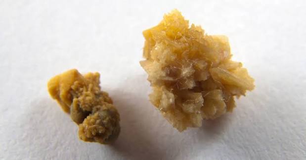
Uric Acid Stones: Causes, Diagnosis, and Treatment
Uric acid stones are the second most common type of kidney stone after calcium oxalate stones, accounting for approximately 5 to 10% of all kidney stones in the United States — though rates are rising in parallel with the increasing prevalence of obesity, type 2 diabetes, and metabolic syndrome, conditions that strongly predispose to uric acid stone formation. What makes uric acid stones clinically distinctive is not their frequency but their characteristics: they are radiolucent (invisible on plain X-ray), they form in persistently acidic urine rather than as a result of excess mineral excretion, and — critically — they are the only common kidney stone type that can be completely dissolved by medical treatment alone, without any surgical or urological procedure. This last feature makes correct identification of uric acid stones both clinically important and potentially highly consequential for patients who might otherwise undergo unnecessary lithotripsy or ureteroscopy for a stone that would respond to oral urinary alkalinization.
Understanding the pathophysiology of uric acid stone formation, the conditions that predispose to it, and the specific management approach required for this stone type is essential for patients who form these stones and for clinicians managing them. The management of uric acid stones differs fundamentally from that of calcium stones — the same dietary advice and the same medications are not interchangeable across stone types, and a treatment plan built for calcium stones will be ineffective, and potentially counterproductive, for a patient with uric acid stones.
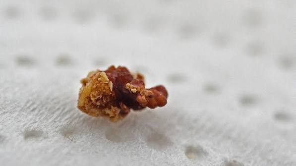
What Causes Uric Acid Stones?
Uric acid is the end product of purine metabolism in the human body. Purines are nitrogen-containing compounds found in many foods (particularly animal proteins, organ meats, and shellfish) and are also generated internally by the normal turnover of cells. Uric acid is excreted primarily through the kidneys — approximately 70% of daily uric acid excretion occurs in the urine, with the remainder excreted in the gastrointestinal tract. When urinary uric acid concentration exceeds its solubility, uric acid crystallizes and, if crystals aggregate and grow, forms a uric acid stone.
The critical biochemical feature of uric acid that distinguishes its stone formation from calcium stone formation is its strong pH dependence. Uric acid (pKa = 5.35) exists in two forms in solution: the ionized urate form (dominant above pH 5.35, highly soluble) and the un-ionized uric acid form (dominant below pH 5.35, poorly soluble). The solubility of uric acid in urine changes dramatically with small pH changes: at urinary pH 5.0, the maximum solubility of uric acid is approximately 60 mg/L; at pH 6.0 it is 220 mg/L; at pH 7.0 it exceeds 1,500 mg/L. This 25-fold difference in solubility across a 2-unit pH range means that persistently acidic urine is a more potent driver of uric acid crystallization than the total amount of uric acid produced or excreted. Raising urinary pH — not reducing uric acid production — is the primary therapeutic target for dissolving existing uric acid stones and preventing new ones.
Three key metabolic conditions drive uric acid stone formation and often coexist in the same patient: persistently low urinary pH (below 5.5), elevated urinary uric acid (hyperuricosuria), and low urine volume. The relative contribution of each varies between patients, but low urinary pH is the dominant factor — some patients form uric acid stones despite normal uric acid excretion because their urine is so persistently acidic that even normal uric acid concentrations exceed the pH-dependent solubility threshold.
Who Is at Risk for Uric Acid Stones?
Type 2 diabetes and insulin resistance are among the strongest risk factors for uric acid stone formation. Insulin resistance impairs the kidney’s ability to excrete ammonium (NH₄⁺) in the urine — ammonium is the primary urinary buffer that neutralizes the protons generated by daily metabolism, and when ammonium excretion is deficient, urinary pH falls. Patients with type 2 diabetes or metabolic syndrome have been shown in large epidemiological studies to have consistently more acidic urine than metabolically healthy controls, and their uric acid stone rate is correspondingly elevated. The prevalence of uric acid stones among diabetic patients is approximately three times that in the general population, and some series report that uric acid stones account for up to 35 to 40% of kidney stones in patients with type 2 diabetes.
Gout and hyperuricemia are the conditions most classically associated with uric acid stones in the public mind. Hyperuricemia — elevated serum uric acid — is present in approximately 25% of patients with uric acid kidney stones but is not universally required for uric acid stone formation. Gout patients have a 10 to 25% prevalence of kidney stones, of which a majority are uric acid stones. The mechanism in gout is dual: elevated uric acid production generates more uric acid for renal excretion (hyperuricosuria), and many gout patients also have the insulin resistance–related defect in urinary ammonium excretion that further reduces urinary pH.
High animal protein diets contribute to uric acid stone risk through two mechanisms. First, dietary purines from animal protein — especially organ meats (liver, kidney), shellfish (anchovies, sardines, mussels), red meat, and game meat — are metabolized to uric acid, increasing urinary uric acid excretion. Second, the high sulfur amino acid content of animal protein generates a metabolic acid load that reduces urinary pH and urinary ammonium excretion. Plant-based diets, by contrast, produce lower uric acid generation and a more alkaline urinary pH — explaining the consistently lower kidney stone rates in vegetarians and near-vegetarians in epidemiological studies.
Chronic diarrheal states — including inflammatory bowel disease (Crohn’s disease more than ulcerative colitis), short bowel syndrome after intestinal resection, and chronic laxative use — cause uric acid stones through dehydration and bicarbonate loss from stool. Profuse diarrhea depletes body bicarbonate, producing a metabolic acidosis that acidifies the urine, and the resulting low urine volume concentrates all urinary solutes including uric acid. Patients with Crohn’s disease have a significantly elevated stone rate, and uric acid stones account for a larger fraction of their stones than in the general population (though enteric hyperoxaluria from fat malabsorption also contributes to calcium oxalate stone formation in this population).
Obesity independently predicts uric acid stone formation beyond its association with insulin resistance and type 2 diabetes. Obese individuals have lower urinary pH, higher urinary uric acid excretion, and lower urinary citrate than normal-weight individuals. The mechanism links to adipose tissue–mediated insulin resistance, altered purine metabolism, and dietary patterns. Population-level data confirm that the incidence of uric acid stones tracks with body mass index — as average BMI has increased in Western populations over the past three decades, uric acid stone rates have increased proportionally.
Certain medications and medical conditions also predispose to uric acid stone formation. Uricosuric agents — drugs that lower serum uric acid by increasing renal uric acid excretion (probenecid, losartan at high doses) — raise urinary uric acid and can promote uric acid stone formation. Rapid cell turnover from chemotherapy for leukemia, lymphoma, or other malignancies releases large quantities of purines from dying cells, generating a surge in uric acid production. Radiographic contrast agents transiently acidify the urine in susceptible patients. Genetic variants in uric acid transport genes (SLC2A9, ABCG2) alter renal uric acid handling and explain some of the familial clustering of uric acid stone disease.
Symptoms and How Uric Acid Stones Are Identified
The symptoms of uric acid stone passage — flank pain, hematuria, nausea, urinary urgency — are identical to those of calcium stones; the stone type does not alter the clinical presentation of acute renal colic. What does differ is the imaging. Uric acid stones are radiolucent — composed entirely of uric acid with no calcium content — and therefore invisible on plain abdominal X-ray (KUB) or standard KUB films. A patient with classic renal colic symptoms, hematuria on urinalysis, and a negative plain X-ray has a uric acid stone (or other radiolucent stone) until proven otherwise. Non-contrast CT of the abdomen and pelvis is the definitive diagnostic imaging: it detects uric acid stones reliably but typically shows a relatively lower Hounsfield unit density (100 to 450 HU) compared to calcium oxalate stones (600 to 1,200 HU), and this lower density is a clue to the stone’s composition even before formal analysis.
Urinary pH is a critical diagnostic clue. A spot urine pH below 5.5 — measured with pH paper or electronic pH meter — in a patient with flank pain and a negative plain X-ray strongly suggests a uric acid stone. A 24-hour urine collection measuring urinary pH across multiple time points (ideally with sequential collections to capture day-to-day variation) provides the most accurate assessment of chronic urinary pH and is standard in the metabolic evaluation of uric acid stone formers. Urinary pH consistently below 5.5 in a patient with a history of radiolucent stones is essentially diagnostic of uric acid stone disease even without stone analysis, and alkalinization treatment can begin immediately on this presumptive diagnosis.
For additional context on how uric acid stones compare to the other kidney stone types — calcium oxalate, calcium phosphate, struvite, and cystine — see the types of kidney stones guide on Horizon Health Guide. Early recognition of the stone type — and awareness of the imaging finding that uric acid stones are invisible on X-ray — can prevent unnecessary repeat X-rays and delay in appropriate treatment for patients whose urologist or emergency physician is expecting to see a stone on plain film.
Dissolving Uric Acid Stones: The Unique Treatment Advantage
The defining therapeutic feature of uric acid stones — and the reason that stone type identification is so clinically important — is that existing uric acid stones can be completely dissolved by urinary alkalinization. This is a treatment option unavailable for any other common kidney stone type: calcium oxalate, calcium phosphate, struvite, and cystine stones do not dissolve in response to urinary pH manipulation. For uric acid stones, dissolution therapy is standard of care for stones that are not causing acute obstruction requiring urgent urological intervention, and even for stones that have caused acute obstruction, alkalinization therapy can be initiated after relief of obstruction to dissolve residual stone fragments.
The dissolution protocol involves raising urinary pH to 6.5 to 7.0 using oral potassium citrate. At pH 7.0, uric acid solubility is approximately 1,500 mg/L — far exceeding the typical urinary uric acid concentration — allowing existing crystalline uric acid to dissolve back into solution over days to weeks, depending on stone size. Small stones (under 5 mm) often dissolve within 2 to 4 weeks; larger stones (10 to 20 mm) may require 4 to 12 weeks of sustained alkalinization. The target is a urinary pH of 6.5 to 7.0, not above 7.0 — excessive alkalinization above pH 7.5 can promote calcium phosphate crystallization and should be avoided. Patients monitor their urinary pH at home 2 to 3 times daily using pH paper (urine pH strips) and adjust their potassium citrate dosing in response to pH readings, with guidance from their urologist or nephrologist.
Sodium bicarbonate is an alternative alkalinizing agent for patients who cannot tolerate potassium citrate (due to gastrointestinal symptoms or hyperkalemia risk), though it carries additional sodium load that may worsen hypercalciuria in patients with mixed stone history and requires attention to dietary sodium management. In patients with significant hyperuricemia or hyperuricosuria, allopurinol (a xanthine oxidase inhibitor that reduces uric acid synthesis) is added to alkalinization therapy to further reduce the uric acid concentration driving crystallization. CT imaging is repeated at 4 to 6 week intervals to monitor stone dissolution and adjust therapy. Patients should be informed that the dissolution process requires compliance with medication and frequent pH monitoring — inconsistent alkalinization, with pH falling below 5.5 at any point, allows crystallization to resume and negates the dissolution effect.
Diet and Lifestyle Changes for Uric Acid Stone Prevention
Dietary modification is an essential component of uric acid stone prevention alongside pharmacological alkalinization. The dietary targets for uric acid stone prevention differ from those for calcium stones, and the advice must be tailored to the stone type.
Reduce high-purine foods. The highest-purine foods — organ meats (liver, sweetbreads, kidneys), sardines, anchovies, mussels, scallops, herring, red meat (particularly red meat consumed in large portions), and concentrated meat broths — generate the most uric acid on metabolism and most directly elevate urinary uric acid. Limiting these foods, especially the organ meats and oily fish with very high purine density, reduces the urinary uric acid load and lowers the amount of alkalinization required to achieve dissolution. This does not require eliminating all animal protein — moderate portions of poultry and lean fish are acceptable — but the dietary pattern should shift toward plant proteins, which do not raise urinary uric acid to the same degree.
Limit fructose and sugar-sweetened beverages. Fructose independently raises serum and urinary uric acid by accelerating adenosine monophosphate (AMP) catabolism in the liver, increasing purine degradation and uric acid production. High-fructose corn syrup in sweetened beverages, fruit juices, and ultra-processed foods is the largest dietary fructose source in Western diets. Epidemiological studies consistently show that sweetened beverage consumption strongly predicts both gout and kidney stone risk — one large prospective cohort study found that women consuming one or more sugar-sweetened cola drinks per day had double the kidney stone risk of non-consumers. Alcohol — particularly beer, which contains both purines and yeast fermentation products that raise uric acid — should be minimized or eliminated in patients with recurrent uric acid stones.
High fluid intake to achieve urine output above 2 liters per day dilutes uric acid concentration and reduces stone formation risk independent of pH. Water is the preferred fluid; lemon juice in water contributes citrate and may modestly raise urinary pH. Fluid intake is particularly important for patients with chronic diarrheal conditions contributing to their uric acid stone risk — these patients have increased insensible losses and require higher total intake to achieve adequate urine volume.
Manage underlying metabolic conditions. For patients in whom uric acid stones are driven by insulin resistance or type 2 diabetes, improvements in metabolic health — through weight loss, increased physical activity, dietary quality improvements, and appropriate pharmacological management of diabetes — can meaningfully improve the urinary pH defect that underlies stone formation. Insulin sensitization (with metformin or weight loss) has been shown to improve renal ammonium excretion and raise urinary pH in patients with type 2 diabetes and uric acid stone disease, adding a mechanistic basis for the clinical observation that metabolic improvement reduces stone recurrence in this patient population.
When to See a Doctor About Uric Acid Stones
Any patient who has passed a kidney stone — or has been told they have a stone on imaging — should be evaluated by a urologist or nephrologist for stone type identification and metabolic risk assessment. For uric acid stones specifically, the stakes of correct identification are high: a patient whose stone is incorrectly assumed to be a calcium stone and is managed accordingly will receive ineffective treatment; a patient correctly identified as a uric acid stone former can achieve complete dissolution of existing stones and reliable prevention of new ones with relatively simple, non-invasive medical management.
Urgent evaluation is required for any of the following: acute flank pain with fever or chills (suggesting infection above an obstructing stone — a urological emergency); bilateral obstruction; obstruction of a solitary kidney; or flank pain with hemodynamic instability. For stable patients with uric acid stones identified on non-contrast CT, the acute management is pain control and, once obstruction status is determined, initiation of alkalinization therapy. For detailed information on kidney stone symptoms and the pain pattern of renal colic, see the kidney stone pain guide. For an overview of all kidney stone treatment options — from oral dissolution to shock wave lithotripsy and ureteroscopy — see the complete kidney stones overview on Horizon Health Guide. For preventive dietary guidance specific to all stone types, see the diet and kidney stone prevention guide. Clinical guidelines are available from the American Urological Association and the National Institute of Diabetes and Digestive and Kidney Diseases.
Sources: AUA Kidney Stone Guidelines · NIDDK — Kidney Stones · StatPearls — Nephrolithiasis
Monitoring and Long-Term Management of Uric Acid Stones
Long-term management of uric acid stone disease requires ongoing monitoring of urinary pH, periodic imaging to assess stone burden, and consistent attention to the underlying metabolic conditions driving stone formation. Unlike calcium stone prevention — where the emphasis is primarily on dietary modification and, when needed, pharmacological adjustments to urinary chemistry — uric acid stone management centers on maintaining urinary pH in the therapeutic range of 6.5 to 7.0 continuously, not just during treatment periods. The pH dependence of uric acid solubility is so steep that even periodic dips in urinary pH to below 5.5 — caused by dietary lapses, medication non-adherence, or intercurrent illnesses — can allow crystallization to resume and small stones to begin growing again.
Home urinary pH monitoring is an essential component of uric acid stone management in a way that it is not for calcium stone prevention. Patients should test their urine pH 2 to 3 times daily — typically on waking (when urine is most acidic after overnight fasting and concentrated), after the largest meal of the day, and at bedtime. Urine pH test strips (available over the counter) are adequate for this purpose; they measure pH in 0.5-unit increments and are sufficient for titrating potassium citrate dosing. The goal is to maintain pH readings consistently between 6.0 and 7.0 across all daily measurements — a reading below 5.5 on any measurement indicates under-treatment, and a reading consistently above 7.0 (especially with phosphaturia) indicates potential over-alkalinization with risk of calcium phosphate precipitation. Patients should bring their pH monitoring log to follow-up appointments so their urologist or nephrologist can adjust dosing precisely.
Follow-up imaging at 4 to 6 week intervals is standard for patients undergoing active dissolution therapy, to document stone shrinkage and adjust the treatment timeline. Once dissolution is confirmed on CT, imaging intervals can be extended to 6 to 12 months for surveillance, returning to more frequent imaging if symptoms recur or 24-hour urine parameters worsen. Annual 24-hour urine collections remain the gold standard for ongoing metabolic monitoring — the results confirm that urinary pH, uric acid, and citrate levels remain in the target range and allow detection of any new metabolic abnormalities (such as emerging hypercalciuria from thiazide side effects or dietary shifts) before they contribute to recurrent stones.
For patients whose uric acid stones are driven primarily by metabolic syndrome or type 2 diabetes, improvement in the underlying condition produces measurable improvements in urinary pH and stone-forming risk. Weight loss of 5 to 10% of body weight has been shown in clinical studies to raise urinary pH in obese patients with uric acid stone disease, and this improvement is independent of and additive to pharmacological alkalinization. Physical activity improvements, dietary quality upgrades (reducing processed foods, sweetened beverages, and organ meats while increasing vegetables, whole grains, and legumes), and better glycemic control from diabetes management all contribute to a more favorable urinary environment for stone prevention. These lifestyle changes are not a substitute for potassium citrate therapy but reduce the dose required for adequate alkalinization and reduce the risk of recurrence if pharmacological management is ever interrupted.
Uric Acid Stones in Special Populations
Children and adolescents with uric acid stones represent a small but important subset of the pediatric stone population. In children, uric acid stones are more likely to be associated with inherited disorders of purine metabolism — Lesch-Nyhan syndrome (complete hypoxanthine-guanine phosphoribosyltransferase deficiency), partial HPRT deficiency, or phosphoribosyl pyrophosphate synthetase superactivity — that cause massive uric acid overproduction from early life. These genetic conditions present with extreme hyperuricemia, hyperuricosuria, and severe stone disease beginning in infancy or childhood, and require specialized management by metabolic geneticists and pediatric nephrologists alongside standard urological stone treatment. In older adolescents, uric acid stones increasingly resemble the adult phenotype of metabolic syndrome–associated stone disease, tracking the rising rates of obesity and insulin resistance in this age group.
Patients undergoing chemotherapy for hematological malignancies — particularly leukemia, lymphoma, and myeloproliferative disorders — face dramatically elevated uric acid stone risk during and after treatment, from the massive purine release associated with tumor lysis syndrome. Tumor lysis syndrome can raise serum uric acid to extraordinarily high levels within 24 to 72 hours of cytotoxic treatment initiation, overwhelming the kidney’s capacity for uric acid excretion. Standard supportive care for tumor lysis risk includes vigorous intravenous hydration, urinary alkalinization, and in high-risk patients, prophylactic rasburicase (a recombinant uricase enzyme that converts uric acid to the more soluble allantoin). Nephrolithiasis is one of several potential complications of tumor lysis syndrome, and oncology teams routinely incorporate uric acid monitoring and alkalinization into the supportive care protocol for at-risk patients.
Patients with gout who are started on uricosuric therapy — agents that increase renal uric acid excretion to lower serum uric acid levels — face an initial period of elevated uric acid stone risk as urinary uric acid rises. Urinary alkalinization should be initiated concomitantly with uricosuric therapy in these patients, and high fluid intake (targeting urine output above 2 liters per day) reduces the concentration-related crystallization risk during the uricosuric phase. This is a well-recognized clinical consideration in gout management and illustrates the general principle that any intervention raising urinary uric acid excretion — whether dietary purine loading, uricosuric medication, or cytotoxic chemotherapy — increases uric acid stone risk and should be accompanied by appropriate urinary pH management in stone-prone patients.
I have gout and recently passed what my urologist confirmed was a uric acid stone. What struck me reading this article is that I had no idea the stone was invisible on plain X-ray — the ER told me my X-ray was ‘clear’ and seemed confused about why I was in so much pain. It was only after a CT scan that they saw it. The information about dissolution therapy is also something I wasn’t told: my stone was 7mm and my urologist wanted to do a procedure immediately, but another opinion suggested trying potassium citrate first. Three weeks later it had dissolved completely on repeat CT. I wish I had found information like this before my ER visit.
Excellent review of uric acid stone pathophysiology and management. The section on insulin resistance and urinary pH is particularly well done — the impaired ammonium excretion mechanism is often omitted from patient-facing resources but is clinically important for understanding why diabetic patients have such a high uric acid stone prevalence even when their serum uric acid is not dramatically elevated. The dissolution therapy section accurately sets the target pH at 6.5–7.0 and correctly cautions against over-alkalinization above 7.5, which is a real clinical issue we see when patients try to push pH higher thinking more alkaline is always better.
Thank you for that clinical perspective, Dr. Okafor — the over-alkalinization caution is one we emphasized because it is a practical pitfall in self-managed dissolution therapy. Patients monitoring their own pH at home sometimes assume that higher is better, but pushing urinary pH above 7.5 shifts the crystallization risk toward calcium phosphate, which is not soluble at high pH and can begin precipitating in the collecting system. The 6.5–7.0 target is narrow but achievable with consistent potassium citrate dosing and frequent pH monitoring. Daniel, your experience — a negative plain X-ray in a patient with clear renal colic symptoms — is one of the most common clinical scenarios where uric acid stone disease is initially missed. The lesson is that a negative KUB in the setting of classic colic symptoms should trigger CT, not reassurance that there is no stone.