Inflammation is the body’s first-line defense against infection and injury — a precisely coordinated mobilization of immune cells, signaling molecules, and repair mechanisms designed to eliminate threats and restore normal tissue. In its acute form, inflammation is protective, time-limited, and essential for survival.
But chronic inflammation is something fundamentally different. When inflammatory signals persist for months or years — driven by chronic infection, autoimmune disease, obesity, or environmental exposures — the same biological machinery that normally protects tissue begins to damage it. Sustained oxidative stress, pro-proliferative cytokine signaling, impaired immune surveillance, and continuous cycles of cell damage and repair create a biological environment in which cancer thrives.
The magnitude of this connection is significant: approximately 20% of cancers worldwide are attributable to chronic infection-related inflammation alone. The 2011 revision of the “Hallmarks of Cancer” framework explicitly lists “tumor-promoting inflammation” as a recognized hallmark — acknowledging that inflammation is not merely an upstream risk factor but is actively co-opted and maintained by established tumors to support their own survival, growth, and spread.
Acute vs Chronic Inflammation — A Fundamental Distinction
Acute inflammation is the familiar response to a cut, an infection, or a wound. Blood vessels dilate, immune cells flood the affected tissue, pathogens and debris are cleared, and within days to weeks the threat is eliminated, the response resolves, and tissue repairs itself.
Chronic inflammation occurs when this resolution fails — when a persistent stimulus maintains inflammatory signaling indefinitely. What was designed as a temporary defensive posture becomes a permanent microenvironment.
The pathologist Harold Dvorak described tumors as “wounds that won’t heal.” Tumors recruit the same inflammatory cells, cytokines, and remodeling factors that wounds use to regenerate — but without the resolution phase. The result is a self-reinforcing cycle: the tumor promotes inflammation to access nutrients and evade immunity; inflammation accelerates tumor growth and invasion.
In chronic inflammation, three interconnected processes damage DNA and enable cancer:
- Sustained ROS and RNS production by macrophages and neutrophils, directly mutating oncogenes and tumor suppressors
- Persistent pro-proliferative signaling via NF-κB, STAT3, and MAP kinase pathways, accelerating cell division in a mutation-prone environment
- Suppressed immune surveillance by immunosuppressive cytokines and myeloid suppressor cells that prevent normal immune elimination of pre-cancerous cells
The NF-κB Pathway — Inflammation’s Master Switch
At the molecular core of inflammation-driven carcinogenesis sits NF-κB — the transcription factor that functions as the master regulator of inflammatory gene expression. In chronic inflammation, it remains constitutively active — turning on a gene program that cancer cells exploit.
What NF-κB activates:
- Inflammatory cytokines (TNF-α, IL-1β, IL-6, IL-8) — creating a tumor-supportive cytokine milieu
- COX-2: converts arachidonic acid to prostaglandin E2 (PGE2) — a potent mediator of tumor-promoting inflammation
- iNOS: produces nitric oxide and reactive nitrogen species → DNA damage
- VEGF: drives angiogenesis — new blood vessel formation that tumors require to grow
- Anti-apoptotic genes BCL-2 and BCL-XL: protect cancer cells from programmed cell death
- Matrix metalloproteinases (MMPs): degrade the extracellular matrix, enabling tumor invasion
Constitutively active NF-κB is found in the majority of pancreatic cancers, many colorectal cancers, and a substantial proportion of breast and ovarian cancers. Cancer cells co-opt NF-κB to resist chemotherapy-induced apoptosis.
COX-2 and prostaglandin E2 deserve particular emphasis. COX-2 is overexpressed in more than 70% of colorectal cancers and at elevated levels in breast, lung, gastric, and esophageal cancers. PGE2 acts on tumor cells to stimulate proliferation, inhibit apoptosis, induce angiogenesis, and suppress NK cell and T cell cytotoxic activity — allowing tumor cells to escape immune destruction. This explains why COX-2 inhibitors reduce cancer risk in multiple clinical studies.
Oxidative Stress and DNA Damage
Activated macrophages and neutrophils generate reactive oxygen species (ROS) — superoxide, hydrogen peroxide, hydroxyl radical — and reactive nitrogen species (RNS) including nitric oxide and peroxynitrite. In chronic inflammation, persistent ROS/RNS damages the host’s own DNA:
- 8-oxoguanine: the most common oxidative DNA lesion; misread by DNA polymerases → G:C→T:A transversion mutations in KRAS and TP53
- Strand breaks: single- and double-strand breaks leading to chromosomal rearrangements
- Nitrosative damage: deamination of cytosine → C→T transitions; DNA crosslinks
Critically, ROS also activate NF-κB, creating a positive feedback loop: inflammation → ROS → NF-κB → more inflammation → more DNA damage. Epigenetic consequences compound this: inflammatory cytokines and ROS alter DNA methylation and histone modifications, silencing tumor suppressor genes without changing the underlying DNA sequence — changes that propagate through cell divisions.
The Tumor Microenvironment — Inflammation from Within
Once established, a tumor does not merely tolerate the inflammatory microenvironment — it actively recruits and maintains it.
Tumor-associated macrophages (TAMs) are among the most abundant immune cells in solid tumors. Polarized toward the M2 (pro-tumor) phenotype under tumor-derived signals, M2 TAMs produce IL-10 and TGF-β (suppressing anti-tumor immunity), VEGF (stimulating angiogenesis), and MMPs (enabling invasion). High TAM infiltration predicts poor prognosis in breast cancer, ovarian cancer, gastric cancer, and glioblastoma.
Myeloid-derived suppressor cells (MDSCs) — immature myeloid cells arrested in chronic inflammation — suppress T cell and NK cell cytotoxic activity through arginine depletion and TGF-β secretion, disabling anti-tumor immune responses.
Cancer-associated fibroblasts (CAFs) activated by TGF-β produce pro-inflammatory signaling molecules and extracellular matrix proteins that scaffold tumor invasion.
The IL-6/STAT3 axis is one of the most consequential oncogenic signaling cascades in this network. IL-6 — produced by TAMs, CAFs, and cancer cells — activates STAT3 in cancer cells, driving accelerated proliferation, resistance to apoptosis, cancer stem cell renewal, and immune evasion. Constitutively activated STAT3 is found in approximately 70% of human tumors.
Infection-Driven Inflammation and Cancer
H. pylori and Gastric Cancer
Helicobacter pylori colonizes the gastric mucosa of roughly half the world’s population and is the dominant cause of non-cardia gastric cancer — the third-leading cause of cancer mortality globally.
H. pylori triggers chronic gastritis that persists for decades. Virulent strains carrying the CagA gene inject the CagA protein into gastric epithelial cells, disrupting cell polarity and activating NF-κB and ERK oncogenic signaling. H. pylori also stimulates IL-8, TNF-α, and ROS from gastric immune cells, causing direct DNA damage.
The sequence from infection to cancer follows the Correa cascade: H. pylori gastritis → chronic atrophic gastritis → intestinal metaplasia → dysplasia → adenocarcinoma — a progression taking 20–40 years. Eradicating H. pylori with antibiotics reduces gastric cancer risk by approximately 35%. H. pylori is classified as an IARC Group 1 carcinogen.
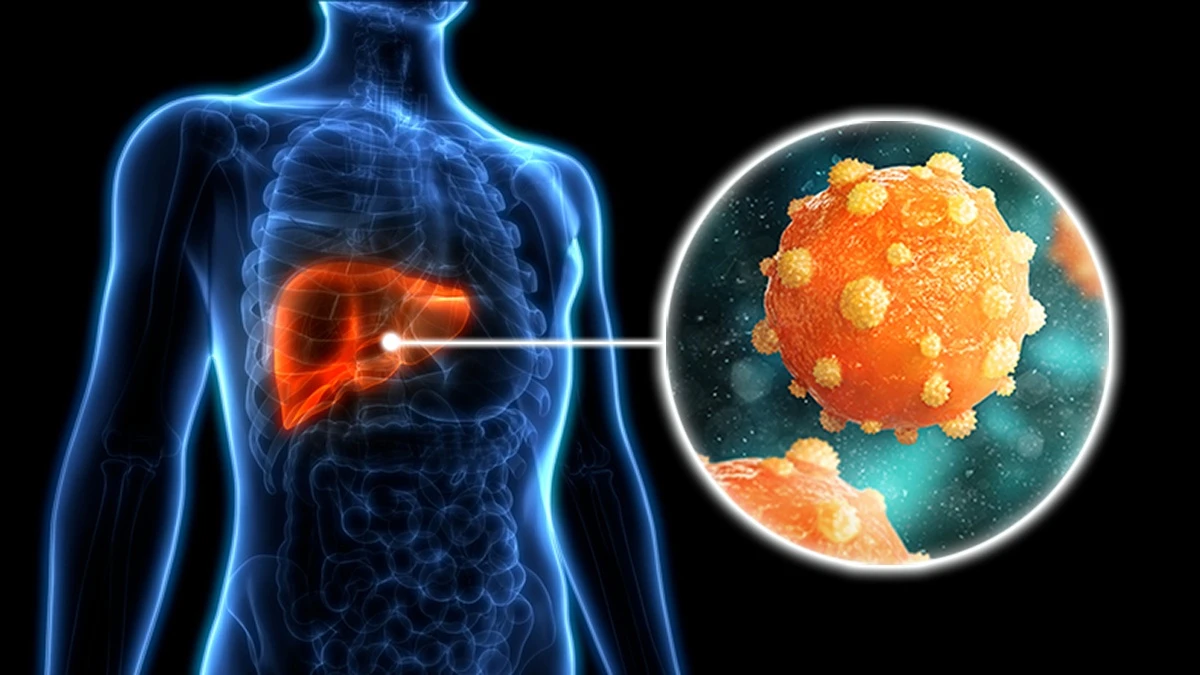
HBV/HCV and Hepatocellular Carcinoma
Approximately 80% of HCCs worldwide are associated with chronic viral hepatitis.
HBV: Creates persistent hepatic inflammation; its HBx protein activates NF-κB and promotes epigenetic silencing of tumor suppressors. Chronic inflammation → cirrhosis → HCC over decades. HBV vaccination is one of the most effective cancer prevention interventions — Taiwan’s universal vaccination program (1984) reduced HCC incidence in vaccinated cohorts by over 70%.
HCV: NS5A protein activates pro-inflammatory pathways. Curative antiviral therapy with DAAs reduces HCC risk by 60–70% but does not eliminate it in patients who already have cirrhosis — structural damage persists even after viral clearance.
HPV and Cervical/Oropharyngeal Cancer
HPV 16 and 18 drive cervical and oropharyngeal cancer through direct oncogene activation combined with local inflammation. HPV E6 degrades p53 and E7 degrades pRb — disabling the two most critical cell cycle checkpoints. HPV vaccination prevents approximately 70% of cervical cancers.
Other Infection-Associated Cancers
- EBV: Burkitt lymphoma, Hodgkin lymphoma, nasopharyngeal carcinoma — via LMP1-mediated NF-κB activation
- Schistosoma haematobium: bladder cancer through chronic local inflammation (IARC Group 1)
- H. pylori and gastric MALT lymphoma: antibiotic eradication cures early-stage gastric MALT lymphoma in ~75% of cases — treating an infection to cure a cancer
Chronic Disease–Driven Inflammation
Inflammatory Bowel Disease
Ulcerative colitis (UC) and Crohn’s disease generate chronic mucosal inflammation with repeated cycles of epithelial damage and repair. UC carries approximately 2× colorectal cancer risk after 10 years of disease, rising to ~4× risk after 20 years of extensive, poorly controlled colitis. Surveillance colonoscopy every 1–2 years in patients with long-standing extensive colitis is standard of care. Mesalamine (5-ASA) maintenance therapy is associated with approximately 25–50% reduced CRC risk in observational studies, through COX-2 inhibition and NF-κB suppression in colonic epithelium.
Obesity and Adipose Tissue Inflammation
Excess visceral fat is not inert storage — it is metabolically active inflammatory tissue that chronically secretes TNF-α, IL-6, and leptin (pro-proliferative) while producing reduced anti-inflammatory adiponectin. Concurrent chronic hyperinsulinemia and elevated IGF-1 activate PI3K/AKT and RAS/MAPK → cell proliferation and anti-apoptosis. Aromatase in adipose tissue converts androgens to estrogens, driving ER-positive breast cancer and endometrial cancer. Obesity is recognized as a cause of at least 13 cancer types, with adipose tissue inflammation as a central thread.
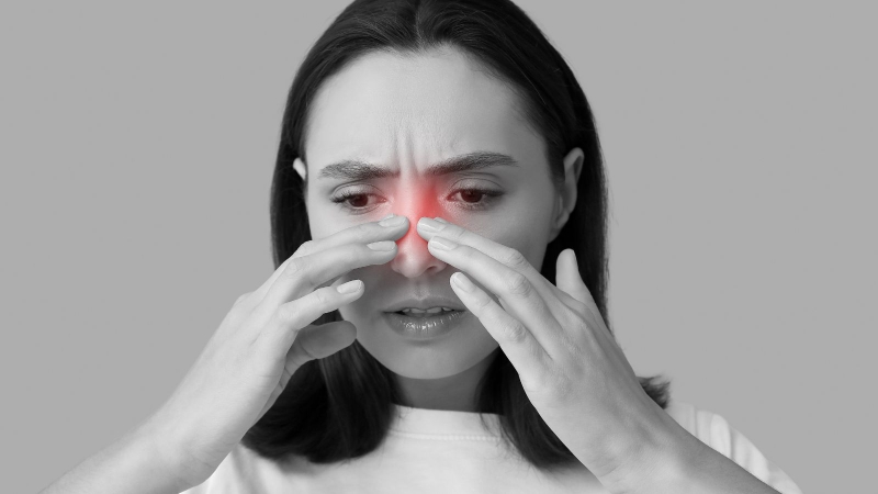
GERD and Barrett’s Esophagus
Chronic acid and bile reflux triggers NF-κB activation and COX-2 upregulation in esophageal epithelium, driving metaplastic change (Barrett’s esophagus) that can progress to esophageal adenocarcinoma — one of the fastest-rising cancers in Western countries.
Anti-Inflammatory Strategies with Cancer Prevention Evidence
Aspirin and NSAIDs
Daily aspirin shows the strongest anti-inflammatory cancer prevention evidence. Meta-analyses (Rothwell PM, Lancet 2010) demonstrate 20–30% reduction in colorectal cancer incidence with regular low-dose aspirin (100–300mg daily) over 5+ years. Selective COX-2 inhibitor celecoxib reduced colorectal adenoma recurrence by 25–45% in clinical trials. Important caveat: regular aspirin carries real gastrointestinal bleeding risk; current guidelines do not recommend it for general population cancer prevention. It is most clearly supported in specific high-risk groups such as Lynch syndrome.
Infection Eradication and Vaccination
- H. pylori eradication: ~35% reduction in gastric cancer risk; cures early MALT lymphoma in ~75%
- HBV vaccination: >70% reduction in HCC in vaccinated cohorts
- HPV vaccination: prevents ~70% of cervical cancers
Mediterranean Diet
Associated with 10–20% reduced overall cancer incidence in prospective cohort studies. Anti-inflammatory mechanisms include omega-3 fatty acids shifting eicosanoid balance away from pro-inflammatory prostaglandins; dietary fiber fermenting to butyrate, which inhibits NF-κB in colonic epithelium; and olive oil polyphenols suppressing COX-2 and NF-κB in experimental systems.
Weight Loss and Exercise
Both reduce adipose tissue inflammatory burden, lower circulating TNF-α and IL-6, improve insulin sensitivity, and reduce IGF-1 — directly addressing the pro-inflammatory, pro-proliferative hormonal environment linking obesity to cancer.
Frequently Asked Questions
Does inflammation cause cancer?
Chronic inflammation is a major contributor to cancer — not a direct cause in isolation, but a sustained biological environment that enables and accelerates cancer in susceptible tissues. It promotes DNA damage via ROS, provides pro-proliferative cytokine signals, suppresses immune surveillance, and facilitates the angiogenesis tumors need to grow. The clearest evidence involves infection-driven inflammation (H. pylori, HBV/HCV, HPV) and disease-related inflammation (IBD, obesity). Acute inflammation from a wound or common cold does not cause cancer.
How does H. pylori cause stomach cancer?
H. pylori colonizes the stomach lining and triggers chronic inflammation that persists for decades. Its CagA virulence protein — injected directly into gastric epithelial cells — activates NF-κB and ERK oncogenic signaling while disrupting cell polarity. Inflammatory cells recruited to the stomach produce ROS that directly damage DNA. Over 20–40 years, this damage follows the Correa cascade — gastritis → atrophic gastritis → intestinal metaplasia → dysplasia → cancer — in susceptible individuals. Antibiotic eradication of H. pylori reduces gastric cancer risk by approximately 35%.
Can aspirin reduce cancer risk?
Regular low-dose aspirin (100–300mg/day) reduces colorectal cancer incidence by approximately 20–30% in people who take it for 5+ years, primarily through COX-2 inhibition reducing prostaglandin E2. However, aspirin also carries real gastrointestinal bleeding risks, and current guidelines do not recommend it for general population cancer prevention. People at high CRC risk — particularly those with Lynch syndrome — may benefit most. This is a decision to make with a physician who can weigh individual benefits and risks.
What is the tumor microenvironment?
The tumor microenvironment (TME) is the ecosystem surrounding and infiltrating a tumor — cancer cells, immune cells (particularly tumor-associated macrophages and myeloid-derived suppressor cells), cancer-associated fibroblasts, blood vessels, and extracellular matrix. Far from being passive bystanders, these cells are recruited and reprogrammed by tumors to support their growth: macrophages become immunosuppressive, fibroblasts remodel the matrix for invasion, blood vessels proliferate to supply nutrients. Inflammation is the dominant feature of most tumor microenvironments and a central target for immunotherapy development.
Does IBD increase cancer risk?
Yes. Ulcerative colitis increases colorectal cancer risk approximately 2× after 10 years and ~4× after 20 years of extensive, active disease. Chronic mucosal inflammation with DNA damage and pro-proliferative NF-κB signaling drives colorectal carcinogenesis over time. Management includes surveillance colonoscopy every 1–2 years and anti-inflammatory maintenance therapy with mesalamine, which is associated with reduced CRC risk in observational studies. Crohn’s disease also carries elevated CRC risk, though somewhat lower than UC.
How does obesity cause inflammation that promotes cancer?
Excess visceral adipose tissue chronically secretes TNF-α, IL-6, and leptin while producing reduced anti-inflammatory adiponectin, creating systemic low-grade inflammation that activates NF-κB and STAT3 signaling throughout the body. Concurrent chronic hyperinsulinemia activates IGF-1R → PI3K/AKT → cell proliferation and anti-apoptosis. Aromatase in adipose tissue also produces excess estrogen, driving ER-positive breast cancer and endometrial cancer. These interconnected inflammatory and hormonal mechanisms explain why obesity is linked to at least 13 cancer types.
Can a Mediterranean diet reduce cancer risk through anti-inflammatory effects?
Adherence to the Mediterranean diet is associated with 10–20% reduced overall cancer incidence in large prospective cohort studies. The anti-inflammatory mechanisms are real: omega-3 fatty acids shift eicosanoid balance away from pro-inflammatory prostaglandins; dietary fiber ferments to butyrate, which inhibits NF-κB in colonic epithelium; olive oil polyphenols suppress COX-2 and NF-κB in experimental systems. However, dietary association studies cannot establish causation, and no single dietary intervention has been proven to prevent cancer in randomized controlled trials. The Mediterranean diet remains the best-supported dietary pattern for overall chronic disease prevention.
- Hanahan D, Weinberg RA. Hallmarks of Cancer: The Next Generation. Cell. 2011;144:646–674.
- Parkin DM. The global health burden of infection-associated cancers in the year 2002. Int J Cancer. 2006;118:3030.
- Rothwell PM, et al. Short-term effects of daily aspirin on cancer incidence, mortality, and non-vascular death. Lancet. 2010;376:1741.
- Ma JL, et al. Fifteen-year effects of Helicobacter pylori, garlic, and vitamin treatments on gastric cancer incidence and mortality. J Natl Cancer Inst. 2012;104:488.
- Eberhart CE, et al. Up-regulation of cyclooxygenase 2 gene expression in human colorectal adenomas and adenocarcinomas. Gastroenterology. 1994;107:1183.
- Eaden JA, et al. The risk of colorectal cancer in ulcerative colitis: a meta-analysis. Gut. 2001;48:526.
- Chang MH, et al. Universal hepatitis B vaccination in Taiwan and the incidence of hepatocellular carcinoma in children. N Engl J Med. 1997;336:1855.
- Yu H, Bhatt DL, Mazzone P. Signal transducers and activators of transcription 3 (STAT3) — a tumor promoter. Nat Rev Cancer. 2004;4:457.
- Hanahan D. Hallmarks of Cancer: New Dimensions. Cancer Discov. 2022;12:31–46.
- Balkwill FR, Mantovani A. Cancer-related inflammation: Common themes and therapeutic opportunities. Semin Cancer Biol. 2012;22:33–40.