
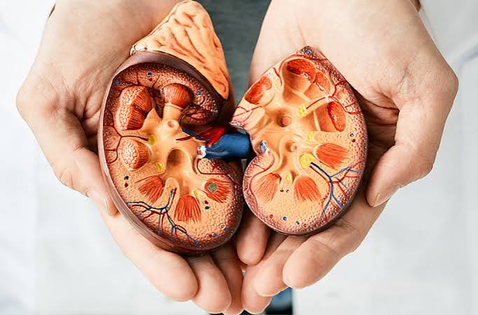
Kidney disease has identifiable causes in the vast majority of cases — and knowing the cause is not just a matter of medical curiosity. It determines the treatment approach, the expected rate of progression, the specific monitoring schedule, and whether the cause itself can be treated to slow or halt the damage. Approximately 90 percent of chronic kidney disease in the United States is attributable to diabetes, high blood pressure, or a combination of both. But the full range of causes extends well beyond these two — from immune diseases attacking the kidney’s filtration units, to inherited conditions where cysts replace healthy tissue, to medications used for years without awareness of their nephrotoxic effect. This article covers the major causes of kidney disease in detail, explains the mechanism by which each damages kidney tissue, and identifies what can be done — early — to interrupt each cause before irreversible damage accumulates.
Diabetes — The Number One Cause of Kidney Disease
Diabetes mellitus is responsible for approximately 44 percent of new cases of end-stage kidney disease in the United States. Sustained high blood glucose damages the glomeruli through thickening of the glomerular basement membrane, expansion of mesangial cells, and injury to podocytes — the specialized cells whose foot processes create the filtration barrier. As podocytes are lost, the filtration barrier becomes leaky, allowing albumin to spill into the urine — appearing first as microalbuminuria (UACR 30–300 mg/g). Left untreated, diabetic nephropathy advances to macroalbuminuria (UACR above 300 mg/g), followed by progressive eGFR decline and eventually end-stage kidney disease over 10 to 20 years.
Critical protective interventions include maintaining HbA1c below 7% (ADA 2024 target), blood pressure control below 130/80 mmHg, an ACE inhibitor or ARB for any adult with diabetes and UACR above 30 mg/g, and an SGLT-2 inhibitor (dapagliflozin, empagliflozin, canagliflozin) for adults with type 2 diabetes and CKD. SGLT-2 inhibitors reduce both proteinuria and intraglomerular pressure — an effect independent of blood glucose lowering, which explains why they benefit non-diabetic CKD patients as well.
High Blood Pressure — The Second Leading Cause
Hypertension accounts for approximately 28 percent of new end-stage kidney disease cases. The mechanism is hypertensive nephrosclerosis: chronically elevated systemic blood pressure transmits into the glomerular capillaries, causing progressive glomerulosclerosis and tubular atrophy. The relationship is bidirectional — high blood pressure damages kidneys; damaged kidneys worsen hypertension by impairing sodium excretion, activating the RAAS, and reducing vasodilatory prostaglandin production. RAAS blockade (ACE inhibitors or ARBs) interrupts this cycle at the pharmacological level. The blood pressure target for all adults with CKD is below 130/80 mmHg (KDIGO 2021).
Renovascular hypertension — caused by renal artery stenosis reducing perfusion to one or both kidneys — triggers aggressive RAAS activation and ischemic nephropathy. Treating the stenosis by angioplasty or stenting may partially restore kidney function if fibrosis has not yet become irreversible.
Glomerular Diseases — When the Filtration Barrier Is Attacked
IgA nephropathy (Berger’s disease) is the most common primary glomerular disease worldwide — IgA immune complex deposits in mesangial cells cause hematuria and proteinuria. Approximately 30 to 40 percent of patients progress to ESRD over 20 years if untreated. SGLT-2 inhibitors have shown significant proteinuria reduction in IgA nephropathy; sparsentan was approved for it in 2023.
Focal segmental glomerulosclerosis (FSGS) produces scarring in glomerular segments. Primary FSGS causes heavy nephrotic proteinuria with poor prognosis. Secondary FSGS results from obesity, HIV, heroin, or vesicoureteral reflux. Membranous nephropathy is the most common nephrotic syndrome cause in adults — anti-PLA2R antibodies drive 70 to 80 percent of primary cases; rituximab has replaced cyclophosphamide as preferred treatment (MENTOR trial 2019). Lupus nephritis complicates 50 percent of SLE cases and is a leading CKD cause in young women; voclosporin and belimumab have improved remission rates. ANCA-associated vasculitis and anti-GBM disease (Goodpasture syndrome) are rapidly progressive forms requiring emergency immunosuppression and plasma exchange.
Polycystic Kidney Disease — The Genetic Cause
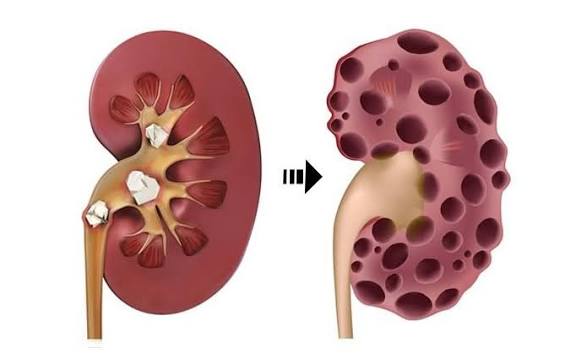
Polycystic kidney disease (PKD) affects approximately 1 in 400 to 1,000 adults. The autosomal dominant form (ADPKD) is caused by PKD1 mutations (85%, faster progression) or PKD2 mutations (15%). Fluid-filled cysts proliferate in both kidneys from birth, progressively replacing nephron mass — kidneys can become massively enlarged while eGFR remains normal, because remaining nephrons compensate. More than 50 percent of patients with PKD1 mutations reach kidney failure by age 60 to 70. Tolvaptan (vasopressin V2 receptor antagonist) slows cyst growth in rapidly progressing ADPKD but requires monthly liver enzyme monitoring. Extra-renal manifestations include liver cysts and intracranial aneurysms in approximately 10 percent of patients — justifying screening in those with a family history of aneurysm.
Medications and Nephrotoxins
NSAIDs are the most common preventable medication cause of CKD. Chronic use reduces prostaglandin-mediated renal vasodilation, causing tubular ischemia and, long-term, tubulointerstitial fibrosis. Lithium (bipolar disorder) causes nephrogenic diabetes insipidus and, after 10 to 20 years, focal tubular atrophy and CKD — requiring creatinine monitoring every 6 months. Calcineurin inhibitors (cyclosporine, tacrolimus) cause afferent arteriolar vasoconstriction and direct tubular toxicity in transplant patients. Aristolochic acid, found in some traditional herbal preparations, causes rapidly progressive tubulointerstitial fibrosis — among the most nephrotoxic substances known. Heavy metals (lead, cadmium) from occupational or environmental exposure cause tubular toxicity and CKD.
Obstructive Nephropathy
Long-standing urinary obstruction causes progressive kidney damage through elevated hydrostatic pressure in the renal pelvis and tubules, driving tubular atrophy and interstitial fibrosis. The most common cause in men over 60 is benign prostatic hyperplasia (BPH), often developing silently over years until substantially elevated creatinine is discovered. Prompt treatment (alpha-blockers, 5-alpha reductase inhibitors, or surgery) can stabilize or partially recover kidney function. Bilateral ureteral obstruction from pelvic malignancy and bilateral kidney stones (or unilateral stones in a single functioning kidney) cause rapidly progressing CKD requiring urgent decompression.
Infections That Damage the Kidneys
HIV-associated nephropathy (HIVAN) presents as collapsing FSGS with heavy proteinuria — predominantly in adults of African ancestry with APOL1 G1/G2 high-risk variants, typically with advanced HIV. Effective antiretroviral therapy substantially reduces HIVAN risk. Hepatitis B causes membranous nephropathy through immune complex deposition; antiviral therapy (tenofovir, entecavir) treats the kidney disease by treating the virus. Hepatitis C causes cryoglobulinemic vasculitis affecting glomeruli; direct-acting antivirals now cure HCV in over 95 percent of patients, with kidney disease typically remitting with viral clearance. Chronic pyelonephritis from recurrent urinary tract infections causes renal scarring — more clinically relevant in adults with structural urinary tract abnormalities or vesicoureteral reflux.
Hereditary and Genetic Kidney Diseases
Alport syndrome is caused by mutations in COL4A3, COL4A4, or COL4A5 genes encoding type IV collagen — a structural component of the glomerular basement membrane. Defective collagen leads to progressive glomerulonephritis with sensorineural hearing loss and ocular abnormalities; RAAS blockade slows progression. Fabry disease is an X-linked lysosomal storage disorder with alpha-galactosidase A deficiency leading to progressive kidney disease, treated with enzyme replacement therapy (agalsidase alfa or beta). CAKUT (congenital anomalies of the kidney and urinary tract) — including solitary kidney, horseshoe kidney, and posterior urethral valves — reduce nephron endowment from birth, predisposing to CKD through reduced renal reserve.
How Kidney Disease Causes Converge on a Common Final Pathway
Despite the diversity of causes, most forms of CKD converge on the same final histological pattern: interstitial fibrosis and tubular atrophy (IFTA). Activated fibroblasts, driven by TGF-beta and other pro-fibrotic signals, replace functional nephron tissue with collagen-rich scar. This is why advanced CKD looks similar on biopsy regardless of whether it started as diabetic nephropathy, hypertensive nephrosclerosis, or IgA nephropathy — and why treatments targeting common mechanisms (intraglomerular hypertension, oxidative stress, inflammatory signaling) can benefit patients across multiple underlying causes.
What You Can Do Based on Your Cause
For modifiable causes — diabetes and hypertension, which together account for most CKD — the protective actions are known and accessible: control blood glucose and blood pressure, use RAAS blockade if albuminuria is present, add an SGLT-2 inhibitor if eligible, avoid NSAIDs chronically, and undergo annual kidney screening. For glomerular diseases, early diagnosis by urine testing and timely biopsy allows specific treatment before irreversible fibrosis develops. For hereditary causes, genetic counseling and family screening allow at-risk relatives to begin monitoring before damage is advanced. For medication-related causes, awareness and regular creatinine monitoring are the primary protective tools. For more context on the early warning signs of kidney damage, see our article on early signs of kidney problems. For what to measure to track kidney health, see our guide on kidney health numbers every adult should know. For a broad overview of the conditions these causes produce, see our guide on common kidney problems in adults. For the key distinction between chronic and acute kidney conditions, see our article on what is chronic kidney disease.
Diabetic Kidney Disease — The Full Mechanism
Understanding exactly how diabetes damages the kidneys helps explain why intervention must begin early — before the damage becomes structural and irreversible. In normal kidneys, the glomerular filtration rate is regulated partly by the tone of the afferent and efferent arterioles surrounding each glomerulus. In diabetes, chronic hyperglycemia activates several damaging pathways simultaneously: the polyol pathway (which accumulates sorbitol in glomerular cells), the hexosamine pathway, advanced glycation end products (AGEs) that cross-link proteins in the glomerular basement membrane, and activation of protein kinase C, which promotes vascular permeability and inflammatory cytokine release. Together, these processes produce glomerular hyperfiltration in the early stages — actually an elevated GFR as the damaged glomeruli try to compensate — followed by basement membrane thickening, mesangial expansion, and the characteristic Kimmelstiel-Wilson nodular glomerulosclerosis visible on kidney biopsy in advanced diabetic nephropathy.
Podocyte loss is the most sensitive early marker of diabetic nephropathy — podocytes can be detected in the urine before albuminuria becomes measurable on standard tests. Once the filtration barrier is disrupted and albumin begins leaking, the clinical trajectory toward ESRD is set in motion unless reversed. The CREDENCE trial (2019) and DAPA-CKD trial (2020) demonstrated that SGLT-2 inhibitors slow this trajectory significantly — not just by lowering glucose, but by reducing the afferent arteriolar tone and intraglomerular pressure that drives the hyperfiltration injury at the heart of diabetic nephropathy. GLP-1 receptor agonists (semaglutide, liraglutide) have also shown kidney protective benefits in trials, and their combination with SGLT-2 inhibitors represents the current frontier of diabetic kidney disease management.
Renovascular Disease and Ischemic Nephropathy
Renal artery stenosis — narrowing of one or both renal arteries — is an underrecognized cause of CKD and treatment-resistant hypertension. The most common cause in adults over 55 is atherosclerosis involving the ostia of the renal arteries. In younger adults, fibromuscular dysplasia — a non-atherosclerotic, non-inflammatory arteriopathy — is the more common cause and predominantly affects women. When renal artery stenosis reduces perfusion pressure to one or both kidneys, the affected kidney perceives chronic ischemia and activates the RAAS maximally to restore blood flow — driving severe hypertension through angiotensin II-mediated vasoconstriction and aldosterone-mediated sodium retention. Over time, the ischemic kidney’s tubules and interstitium undergo progressive atrophy — a condition called ischemic nephropathy. If both kidneys are affected (bilateral renal artery stenosis) or only one kidney is functioning, the result is progressive CKD driven by ischemia rather than the usual diabetic or hypertensive mechanisms.
A key clinical clue to renovascular disease is a sharp decline in eGFR after starting an ACE inhibitor or ARB — these drugs, by blocking the efferent arteriolar constriction that the ischemic kidney depends on to maintain filtration pressure, reveal the underlying stenosis by causing GFR to fall. This “ACE inhibitor-induced AKI” in the setting of bilateral renal artery stenosis is a diagnostic opportunity as much as a complication. Doppler ultrasound, CT angiography, or MR angiography can detect renal artery stenosis; treatment by balloon angioplasty and stenting can restore perfusion and stabilize kidney function if performed before irreversible cortical scarring has occurred.
Analgesic Nephropathy and Chronic NSAID Use
Analgesic nephropathy — chronic kidney disease from long-term analgesic overuse — was originally described in the mid-twentieth century from mixed analgesic products containing phenacetin (now banned), aspirin, and caffeine. In contemporary practice, chronic NSAID use is the most relevant contributor. NSAIDs inhibit cyclooxygenase (COX) enzymes, blocking the synthesis of prostaglandins that maintain renal vasodilation under low-perfusion states (dehydration, heart failure, sodium depletion). In people with normal kidneys and adequate hydration, short-term NSAID use carries minimal kidney risk because prostaglandins are less critical to maintaining perfusion under normal conditions. In people with CKD, heart failure, cirrhosis, or any state of volume depletion, NSAID use removes the vasodilatory protection that the kidney depends on — causing acute reduction in GFR that, with repeated exposures, may become chronic.
The characteristic pathological finding of analgesic nephropathy is renal papillary necrosis — ischemic death of the papillary tips caused by disruption of the juxtamedullary capillary blood flow that depends on prostaglandins for its maintenance. Papillary necrosis can cause pieces of papillary tissue to slough off into the urinary collecting system, producing hematuria and, if the sloughed papilla causes obstruction, acute flank pain resembling kidney stone episodes. Adults with chronic joint pain who take NSAIDs daily over years should have annual creatinine monitoring and should be counseled to use the lowest effective dose, use alternative analgesics (acetaminophen, topical NSAIDs, duloxetine, physical therapy) wherever possible, and avoid NSAIDs entirely during any period of dehydration or acute illness.
Reflux Nephropathy — A Cause That Begins in Childhood
Vesicoureteral reflux (VUR) is a condition in which urine flows backward from the bladder into the ureters and renal pelvis during voiding — the opposite of the normal one-way flow direction. In mild cases (Grade I–II), reflux is confined to the ureter and rarely causes kidney damage. In severe cases (Grade III–V), reflux carries infected urine directly into the renal parenchyma during each voiding episode, causing recurrent pyelonephritis and progressive renal scarring. The combination of high-grade reflux and urinary tract infection — each episode capable of leaving a permanent scar on the growing kidney — is the mechanism of reflux nephropathy. By the time the affected child reaches adulthood, the kidney may have lost 30 to 50 percent of its cortical mass from cumulative scarring, leaving a reduced nephron endowment that predisposes to CKD from early adulthood onward through glomerular hyperfiltration in the remaining nephrons.
Many adults with reflux nephropathy do not know their diagnosis — they may have had recurrent UTIs as children, or may have been told they had “kidney infections” without a formal diagnosis of VUR. Adults with a history of frequent childhood UTIs or “kidney problems” as children should undergo kidney function screening (eGFR and UACR) as part of their routine adult health care, because the structural damage from childhood reflux nephropathy manifests as CKD in adulthood — often without any ongoing active process, simply from the reduced nephron mass that has been present since childhood.
Sickle Cell Nephropathy — A Cause Often Overlooked Outside Hematology
Sickle cell disease causes kidney damage through repeated episodes of sickling in the renal microvasculature — particularly in the hypoxic, hyperosmotic, acidic environment of the renal medulla, which is the most favorable environment in the body for hemoglobin S polymerization and sickling. The result is medullary ischemia, papillary necrosis, and progressive loss of tubular concentrating ability — manifesting clinically as inability to concentrate urine (isosthenuria), leading to high daily urine volumes and polyuria that require high fluid intake to maintain hydration. Renal tubular acidosis, proteinuria, and hematuria are also common manifestations of sickle cell nephropathy in adults with HbSS disease. Over time, glomerular hyperfiltration (from chronic anemia-driven increased cardiac output and renal blood flow) causes focal segmental glomerulosclerosis that progresses to CKD. Hydroxyurea, which increases fetal hemoglobin production and reduces sickling frequency, is the most important disease-modifying therapy for sickle cell disease and is associated with reduced kidney disease progression. Adults with sickle cell disease should have annual kidney monitoring including eGFR, UACR, and urinalysis as part of their comprehensive sickle cell care.
Obesity and Kidney Disease — A Growing Cause
Obesity is an increasingly recognized independent cause of CKD, separate from its role as a risk factor for diabetes and hypertension. The mechanism is obesity-related glomerulopathy — the combination of elevated renal blood flow and increased glomerular filtration rate caused by obesity-driven hyperdynamic circulation causes glomerular hypertension and eventually focal segmental glomerulosclerosis. Adults with a body mass index above 35 kg/m² have substantially elevated risk of developing proteinuria and CKD even in the absence of diabetes or hypertension, and the degree of proteinuria is proportional to the degree of obesity. Weight loss — whether through diet, exercise, bariatric surgery, or GLP-1 receptor agonist therapy — reduces proteinuria in proportion to the degree of weight reduction. This makes obesity-related nephropathy one of the few forms of CKD where the primary cause is directly addressable through weight management.
Metabolic syndrome — the cluster of central obesity, insulin resistance, dyslipidemia, and hypertension — compounds kidney risk beyond any individual component. Adults with metabolic syndrome have approximately double the CKD risk of those without it, through the combined effects of glomerular hyperfiltration from obesity, intraglomerular hypertension from hypertension, hyperuricemia-related tubular injury, and chronic low-grade inflammation. Recognition of metabolic syndrome as a kidney risk factor — rather than only a cardiovascular risk factor — allows for kidney function monitoring in adults whose diabetes and hypertension risk profiles do not yet cross the standard diagnostic thresholds but whose cumulative metabolic burden is already injuring nephrons. For context on the full picture of conditions that emerge from these causes, see our overview of common kidney problems in adults.
Knowing Your Cause Shapes Your Protection
The most important take-away from understanding what causes kidney disease is that the cause is rarely unknown — it can be identified by a focused clinical evaluation combining medical history, lab testing, urine sediment analysis, kidney imaging, and sometimes biopsy. And identified causes are treatable causes. A diabetic nephropathy caught at the microalbuminuria stage responds to SGLT-2 inhibitors and RAAS blockade. A membranous nephropathy caught before significant GFR decline responds to rituximab. An obstructive nephropathy caught before irreversible fibrosis responds to decompression surgery. An analgesic nephropathy stopped at its source — the NSAID prescription — may plateau and stabilize rather than progress. The window of opportunity for each cause is different, but for all of them, the window is widest early. This is why the answer to “what causes kidney disease?” is not just a scientific question — it is a clinical question with practical implications for every adult who carries a risk factor or receives an abnormal kidney lab result. Knowing the cause is the first step toward interrupting it before it reaches the irreversible fibrosis that ends the opportunity for meaningful intervention.
Sources: National Institute of Diabetes and Digestive and Kidney Diseases (NIDDK), niddk.nih.gov; National Kidney Foundation, kidney.org; American Kidney Fund, kidneyfund.org. USRDS Annual Data Report 2022; KDIGO CKD Guidelines 2012/2024; ADA Standards of Care 2024.
Pingback: Kidney Disease Risk Factors Every Adult Should Know
Pingback: Diabetes and Kidney Health
I shared this article on what causes kidney disease? with my doctor and they appreciated the level of detail. The practical tips made this immediately actionable, not just theoretical. Thank you for making complex medical information accessible without dumbing it down.
My doctor recommended I look into what causes kidney disease? and this article covered it perfectly. I have tried following advice from several sources but this is most consistent with what my specialist told me. This is exactly why I prefer this website over generic health platforms.
Bookmarked this article on what causes kidney disease? immediately — going to reference it regularly. The practical tips made this immediately actionable, not just theoretical. Exactly the kind of evidence-based information that is hard to find in one place.