
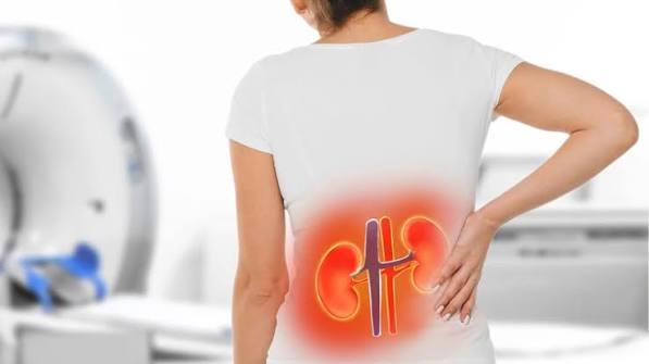
Flank pain — pain in the lateral region of the body between the lower ribcage and the hip — is one of the signature presentations of kidney disease. It is also one of the most frequently misidentified symptoms, often attributed to a muscle strain when the kidney is actually the source. The distinction matters because kidney conditions causing flank pain range from a small stone passing on its own to a life-threatening infected obstruction requiring emergency drainage. This article covers the anatomy of flank pain, the main kidney-related causes, non-kidney conditions to exclude, and when flank pain constitutes a medical emergency.
What Is the Flank and Why Does the Kidney Cause Pain There?
The flank is the lateral region of the torso between the lowest rib and the upper edge of the iliac crest (hip bone) on each side — more lateral than the back, typically felt between the side and the posterior waist, and usually unilateral.
The kidneys sit directly in this region. Each kidney occupies the retroperitoneal space at approximately T12 to L3, enclosed in a fibrous capsule. When the kidney swells, becomes infected, obstructs, or is invaded by tumor, the stretching renal capsule generates pain felt in the ipsilateral flank. The costovertebral angle (CVA) — formed by the junction of the 12th rib and the lateral border of the erector spinae muscle — is the anatomical reference point for kidney pain; CVA tenderness on percussion is the classic physical examination finding, covered in detail in the article on lower back pain and kidney health.
When a kidney stone enters the ureter, ureteral spasm generates colicky flank pain that radiates from the flank downward — to the groin, inner thigh, and genitalia — in a pattern unlike any muscular or spinal pathology. This radiation pattern is one of the most specific clinical features distinguishing ureteral from other causes of flank pain.
Kidney-Related Causes of Flank Pain
Kidney Stones — The Most Common Cause
Kidney stones cause the majority of acute severe flank pain emergency presentations. When a stone obstructs the ureter, ureteral spasm produces the characteristic renal colic: sudden, severe, colicky flank pain with waves of intensity, radiating toward the groin and genitalia, accompanied by nausea, vomiting, and an inability to find a comfortable position. Blood in the urine (hematuria) is present in approximately 90 percent of cases. The article on blood in urine covers hematuria evaluation in detail.
Stone passage depends on size: stones under 5 mm pass spontaneously approximately 68 percent of the time; stones between 5 and 7 mm pass approximately 47 percent; stones above 7 mm pass in fewer than 25 percent of cases. NSAIDs (ketorolac, ibuprofen) are the preferred first-line analgesics for renal colic — superior to opioids for this condition. Tamsulosin (alpha-blocker) improves spontaneous passage by relaxing ureteral smooth muscle. (NIDDK, 2024)
The most serious complication: an infected, obstructed kidney — blocked ureter plus infected urine above the obstruction — produces a clinical picture of sepsis (fever, hypotension, rigors) with flank pain. This is a urological emergency requiring emergent decompression via nephrostomy tube or ureteral stent, plus IV antibiotics, regardless of stone size. Delaying drainage in an infected obstructed kidney can be fatal.
Pyelonephritis — Kidney Infection
Pyelonephritis produces flank pain as part of the classic clinical triad: flank pain, fever, and dysuria. CVA tenderness is the hallmark. Rigors signal bacteremia and hospitalization. Approximately 80 percent of cases are caused by E. coli; the remainder include Klebsiella, Pseudomonas, and Proteus. Urinalysis shows WBCs, bacteria, nitrites, and often RBCs. Cloudy urine with foul odor often accompanies infection — covered in the article on cloudy urine. (NIDDK, 2024)
Emphysematous pyelonephritis is a gas-forming infection of the renal parenchyma, seen almost exclusively in diabetics, caused by organisms (E. coli, Klebsiella) that ferment glucose to produce gas visible on CT. Mortality exceeds 20 percent even with treatment; it requires emergent percutaneous drainage or nephrectomy alongside IV antibiotics — oral antibiotics are insufficient.
Perinephric abscess presents with persistent fever despite antibiotic therapy plus flank pain. CT is diagnostic; CT-guided percutaneous drainage alongside prolonged antibiotics is required.
Renal Infarction
Renal infarction — ischemia from arterial occlusion — is frequently misdiagnosed as kidney stone. Most common cause: cardiac embolism in atrial fibrillation. Clinically: sudden severe flank pain, hematuria, nausea — identical to renal colic on history alone. Distinguishing features: no stone on CT, and markedly elevated LDH disproportionate to other liver enzymes. CT angiography is diagnostic. Treatment: anticoagulation (A-fib); thrombolysis for acute arterial thrombosis; management of the underlying cause.
Renal Vein Thrombosis
Renal vein thrombosis (RVT) is most commonly a complication of nephrotic syndrome, in which severe urinary protein loss creates a hypercoagulable state. Acute RVT presents with sudden flank pain, gross hematuria, and worsening edema in a patient with known nephrotic syndrome. Doppler ultrasound is the first-line diagnostic test; CT venography confirms when ultrasound is inconclusive. Treatment: anticoagulation and management of the underlying condition.
Renal Cell Carcinoma
RCC producing symptomatic flank pain generally indicates advanced disease. The classic triad — flank pain, gross hematuria, and a palpable mass — is present in only approximately 10 percent of patients and usually indicates locally advanced or metastatic disease. When flank pain from RCC is present, it is typically a persistent, dull, unilateral ache from tumor capsule distension or local invasion, accompanied by intermittent hematuria, constitutional symptoms (weight loss, fatigue, night sweats), and occasionally a palpable mass. Any persistent dull unilateral flank pain with hematuria or constitutional symptoms in an adult over 50 warrants CT imaging. (ACS, 2024) Monitoring kidney health numbers including urinalysis for hematuria is the most accessible early detection tool.
Polycystic Kidney Disease
ADPKD produces three patterns of flank pain. Most common: chronic bilateral aching from progressive kidney enlargement. Most acute: cyst hemorrhage — sudden severe unilateral flank pain with dark cola-colored or pink urine, typically self-limited over 2 to 7 days (NSAIDs should be avoided in PKD; acetaminophen is the safer analgesic). Cyst infection presents with fever above 38°C and flank pain without a stone on CT; requires prolonged fluoroquinolone or TMP-SMX antibiotics (4 to 6 weeks) — cephalosporins do not adequately penetrate cyst fluid.
Hydronephrosis and Ureteropelvic Junction Obstruction
Hydronephrosis — collecting system distension from urinary tract obstruction — can cause dull unilateral flank ache (chronic, gradual) or acute severe pain (acute obstruction). Causes include kidney stones (most common), ureteral stricture, external compression from tumor or retroperitoneal fibrosis, and BPH (bilateral outflow obstruction). Ureteropelvic junction obstruction (UPJO) produces intermittent flank pain exacerbated by high fluid intake — pain peaks 30 to 90 minutes after drinking large volumes (Dietl’s crisis). Diagnosis: CT with contrast and MAG3 renography. Treatment: pyeloplasty.
Non-Kidney Causes of Flank Pain to Rule Out
Musculoskeletal: Quadratus lumborum muscle strain, rib fracture — movement worsens the pain; no urinary symptoms.
Gastrointestinal: Left-sided diverticulitis (left flank + fever + bowel changes); retrocecal appendicitis (right flank rather than classic RLQ); colitis from any cause.
Pulmonary: Right lower lobe pneumonia or pleurisy; pulmonary embolism with infarction.
Vascular: Abdominal aortic aneurysm (AAA) rupture — sudden severe bilateral back-flank pain in an older man with hemodynamic instability; immediately life-threatening; do not delay for imaging if the patient is unstable.
Herpes zoster: Dermatomal burning or stabbing flank pain 1 to 3 days before the characteristic vesicular rash appears — suspect in older or immunocompromised adults with unilateral burning flank pain without urinary symptoms, no stone on CT.
When Flank Pain Is a Medical Emergency
Go to the emergency department immediately:
- Flank pain + fever + hypotension/rapid heart rate + rigors — infected obstructed kidney (urosepsis); emergent urological drainage required
- Sudden severe bilateral back-flank pain in an older adult with hemodynamic instability — AAA rupture; surgical emergency
- Flank pain + anuria or markedly reduced urine output — bilateral obstruction causing acute kidney injury
- Sudden severe flank pain in an A-fib patient + no stone on CT — renal infarction; CT angiography urgently needed
See a doctor the same day:
- Flank pain + visible blood in urine (pink, red, brown)
- Flank pain + fever without hypotension — possible pyelonephritis
- Flank pain + dysuria in a pregnant woman
Schedule within 1–2 weeks:
- Recurrent intermittent unilateral flank pain without urinary symptoms or fever
- Persistent dull flank ache with known family history of PKD or history of kidney stones
How Flank Pain Is Evaluated
Urinalysis with microscopy: first test for all unexplained flank pain — blood, WBCs, nitrites, protein.
CT without contrast: gold standard for kidney stones (99% sensitivity); also identifies hydronephrosis.
CT with contrast (CT urogram): renal masses, infection complications, ureteral anatomy.
CT angiography: diagnostic for renal infarction (A-fib + flank pain + no stone + elevated LDH); also diagnoses AAA.
Doppler ultrasound: first-line for renal vein thrombosis and hydronephrosis; preferred in pregnancy.
Blood tests: CBC (leukocytosis for infection), BMP (creatinine/BUN for kidney function), LDH (markedly elevated in renal infarction), blood cultures (if sepsis suspected).
Frequently Asked Questions
What does flank pain feel like?
Flank pain is typically felt as a dull ache or sharp, colicky discomfort in the lateral region between the lower rib and the hip, usually on one side. Kidney stone pain comes in waves of intense, sharp pain radiating from the flank toward the groin. Kidney infection produces a constant, deep ache with fever. Both can be severe enough to cause nausea and vomiting. (Mayo Clinic, 2024)
Is flank pain always a kidney problem?
No, though kidney causes are the most common explanation. Gastrointestinal causes (diverticulitis, retrocecal appendicitis, colitis), musculoskeletal causes (muscle strain, rib fracture), pulmonary causes (lower lobe pneumonia, pleurisy), and vascular causes (aortic aneurysm) can all produce flank pain. Herpes zoster produces dermatomal flank pain before the rash appears. Associated symptoms usually guide the distinction.
What causes sudden severe flank pain?
Four primary causes. Kidney stone (renal colic): the most common; colicky waves radiating to the groin, with hematuria and nausea. Renal infarction: sudden sustained severe flank pain in a patient with A-fib; no stone on CT, markedly elevated LDH. Cyst hemorrhage in PKD: sudden localized flank pain with hematuria. AAA rupture: sudden severe bilateral flank-back pain in an older man with hemodynamic instability — life-threatening emergency.
When should I go to the ER for flank pain?
Immediately for: flank pain + fever + rapid heart rate or low blood pressure (infected obstructed kidney); sudden severe bilateral flank-back pain in an older man with hemodynamic instability (AAA rupture); flank pain + anuria. Same day for: visible blood in urine with flank pain; flank pain + fever without low blood pressure; flank pain during pregnancy with any urinary symptoms.
Can flank pain go away on its own?
Kidney stone pain may resolve if the stone passes — more likely with smaller stones (under 5 mm). Pyelonephritis does not resolve without antibiotics. Renal infarction requires treatment of the underlying cause. Any flank pain lasting more than a few hours, accompanied by fever, or recurring warrants evaluation. Understanding kidney disease risk factors and monitoring kidney health numbers helps contextualize recurrent flank pain episodes.
Flank pain is the kidney’s most consistent way of announcing a problem. Most flank pain from kidney causes is a kidney stone — sudden, severe, colicky, and radiating toward the groin — which either passes on its own or is managed with urological intervention. But the critical skill is recognizing the small fraction of presentations that require emergency response: the infected obstructed kidney producing sepsis, the sudden flank pain in an A-fib patient without a stone, and the older man with sudden severe back-flank pain whose aortic aneurysm has ruptured. In each of these cases, the response time directly affects the outcome.
Flank Pain in Special Groups
Pregnant Women
Flank pain during pregnancy presents a specific diagnostic challenge. Physiological hydronephrosis of pregnancy — partial obstruction of the right ureter by the gravid uterus as it crosses the iliac vessels — is present in approximately 80 percent of pregnancies by the third trimester and can produce mild right-sided flank discomfort. This is usually intermittent, positional, and not accompanied by fever or hematuria.
However, pregnancy also substantially increases the risk of symptomatic kidney stones (urinary stasis and hypercalciuria from increased intestinal calcium absorption), pyelonephritis (UTIs ascend more readily during pregnancy due to ureteral dilation and urinary stasis), and bilateral hydronephrosis from progressive uterine pressure. Any pregnant woman with flank pain and fever requires same-day evaluation and likely hospitalization — pyelonephritis in pregnancy carries risk of preterm labor and sepsis that makes home management inappropriate. Ultrasound is the preferred imaging modality in pregnancy to avoid ionizing radiation; if a stone must be confirmed, low-dose CT or MRI is used when the diagnosis is clinically critical and ultrasound is inconclusive.
Older Adults
In adults over 60, the differential diagnosis for flank pain broadens considerably. Kidney stones remain common, but renal cell carcinoma — which peaks in incidence between 60 and 74 years — becomes a meaningful consideration for any persistent dull unilateral flank pain, particularly with hematuria or constitutional symptoms. Abdominal aortic aneurysm (AAA) is predominantly a disease of older men; any older adult with sudden severe bilateral back-flank pain and hemodynamic instability should be treated as AAA rupture until proven otherwise — not as a kidney stone, not as musculoskeletal pain.
Herpes zoster reactivation is more common with age and immunosenescence. The prodromal burning or aching flank pain of zoster — 1 to 3 days before the rash — is frequently evaluated as renal colic or musculoskeletal pain. In an older adult with unilateral burning flank pain, no stone on CT, and no urinary symptoms or fever, examining the skin of the corresponding dermatome for early vesicles is a critical step before concluding the evaluation is negative.
Patients With Known Kidney Disease
For patients already diagnosed with a kidney condition — CKD, PKD, recurrent kidney stones, or prior pyelonephritis — new flank pain requires interpretation in the context of the established diagnosis. For PKD patients, new acute unilateral flank pain most commonly signals cyst hemorrhage or a new kidney stone; if fever is also present, cyst infection is the priority diagnosis. For patients with a history of recurrent stones, new flank pain with the same character as prior stone episodes should still prompt urinalysis and imaging to confirm — a new stone may be larger than the prior ones, and an infected obstructed stone is dangerous regardless of prior stone history. For patients with advanced CKD, even mild flank pain may signal an acute complication — obstruction from stones in a patient with reduced kidney reserve can precipitate rapid deterioration of function — and should prompt earlier evaluation than it would in someone with normal baseline kidney function.
Long-term management of kidney health — tracking eGFR, monitoring for proteinuria, and maintaining blood pressure control — is the foundation of preventing both recurrent acute episodes and progressive kidney function decline. The articles on chronic kidney disease and kidney health numbers provide the practical framework for this long-term monitoring.
Preventing Recurrent Flank Pain: What You Can Do
For most people, a single episode of flank pain — resolved with treatment — is a one-time event. But for others, the underlying condition creates risk for recurrence: recurrent kidney stones affect roughly 50 percent of patients within 10 years of a first stone; recurrent UTIs and pyelonephritis are particularly common in women with anatomical or immune factors; and PKD, by its nature, is lifelong.
Taking targeted steps based on the underlying cause significantly reduces the chance of repeat episodes.
Stone Formers
Adequate hydration is the most important lifestyle intervention for kidney stone prevention. The goal is urine output of 2 to 2.5 liters per day, which means drinking enough fluid — primarily water — to keep urine pale yellow throughout the day. Concentrated, dark yellow urine is a signal that stone-forming minerals are accumulating at higher levels than the urine can safely carry.
Beyond hydration, dietary adjustments depend on stone type. Calcium oxalate stones — by far the most common — are paradoxically worsened by a low-calcium diet, because dietary calcium normally binds oxalate in the gut before it reaches the kidney. The correct approach is adequate dietary calcium (around 1000 to 1200 mg per day from food, not supplements) paired with limiting high-oxalate foods such as spinach, nuts, and beets. Sodium restriction (below 2300 mg per day) also reduces urinary calcium excretion. If metabolic evaluation reveals elevated urinary calcium or uric acid, medications such as thiazide diuretics or allopurinol are added. Following up with a urologist who specializes in stone disease after a first stone is an important step that many patients skip — a 24-hour urine collection identifies specific metabolic risk factors and allows targeted prevention rather than generic advice.
Recurrent Pyelonephritis
Women who experience two or more episodes of pyelonephritis in a year often benefit from low-dose prophylactic antibiotics or post-coital antibiotics if episodes cluster around sexual activity. Urological evaluation is warranted to rule out structural abnormalities — vesicoureteral reflux (VUR), an incomplete bladder or ureteral obstruction — that predispose to ascending infection. Adequate hydration and prompt treatment of lower UTI symptoms before they ascend are practical strategies that reduce the risk of repeat kidney involvement.
Managing Flank Symptoms in PKD
For patients with polycystic kidney disease, flank pain is a near-universal long-term companion: cyst enlargement, hemorrhage, and infection each produce episodic flank discomfort throughout the course of the disease. Blood pressure control — typically with ACE inhibitors or ARBs — remains the cornerstone of slowing cyst-driven kidney enlargement and preserving function. For patients with ADPKD and rapidly enlarging kidneys or rapidly declining eGFR, tolvaptan (a V2 receptor antagonist) has been shown to slow cyst growth and eGFR decline, though it requires careful liver monitoring. Infection episodes should prompt prompt medical attention given the difficulty of antibiotic penetration and the risk of sepsis from poorly treated cyst infections.
The connection between flank pain and long-term kidney health makes this symptom one that deserves more than symptomatic treatment alone. Understanding the cause, addressing the root condition, and following up with a nephrologist or urologist after a significant episode gives patients the best chance of avoiding both recurrence and progressive kidney function loss. For a full picture of how to monitor kidney health over time, the guide on kidney health numbers every adult should know provides the essential framework.
When to Go Back to the Emergency Room
After being evaluated and sent home for flank pain, certain developments warrant an immediate return — regardless of how recently you were seen or what diagnosis was given. Return to the ER without delay if you develop a fever above 38.5°C (101.3°F) after a diagnosis of kidney stone, because fever combined with an obstructed ureter means infected urine cannot drain and urosepsis can develop within hours. Return immediately if the pain becomes severe and unrelenting rather than intermittent and colicky — this shift in pain character can signal stone migration into a position where it is no longer expected to pass spontaneously, or escalation to a complication such as hydronephrosis under pressure. Return if you develop inability to keep fluids down for more than 12 hours, because pain control and passage of a stone both require adequate hydration that cannot be maintained at home under those conditions. And if any new symptoms arise — blood in vomit, black stools, leg swelling, or loss of sensation — return immediately, as these suggest a diagnosis other than a simple kidney stone may have been missed on the initial evaluation. Flank pain that resolves, then returns with greater intensity 1 to 2 weeks later, is another reason to return rather than wait — this pattern is consistent with a stone that stalled and is now causing obstruction. When in doubt, returning for reassessment carries far less risk than waiting at home with worsening symptoms.
Sources: NIDDK (niddk.nih.gov), Mayo Clinic (mayoclinic.org), NIDDK Kidney Infection (niddk.nih.gov), American Cancer Society (cancer.org)
This is one of the clearest explanations of flank pain: kidney-related causes I have found. The specific numbers and thresholds mentioned are exactly what I needed to understand my results. Looking forward to reading more articles from this website.
As someone dealing with this personally, the flank pain: kidney-related causes section was very helpful. It is refreshing to see an article that acknowledges individual variation rather than one-size-fits-all advice. Looking forward to reading more articles from this website.
This is one of the clearest explanations of flank pain: kidney-related causes I have found. I appreciate that the article is careful about distinguishing between what is known and what is still being researched. Forwarding this to others in my support group who are dealing with similar issues.