
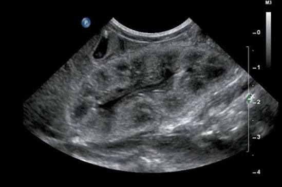
A kidney ultrasound — also called a renal ultrasound — is one of the most commonly ordered imaging tests in nephrology and urology. It uses high-frequency sound waves rather than X-rays to create real-time images of the kidneys, which means no ionizing radiation and no contrast dye injection. For patients with kidney disease, it is often the first imaging study ordered, and it provides a remarkable amount of information about kidney structure, size, and blood flow without any significant discomfort or risk.
How a Kidney Ultrasound Works
An ultrasound machine generates sound waves at frequencies between 2 and 5 MHz — far above the range of human hearing. A handheld device called a transducer is pressed against the skin after a thin layer of gel is applied; the gel ensures that sound waves travel efficiently between the transducer and the body. The sound waves penetrate the tissues, bounce off internal structures at different rates depending on tissue density, and return as echoes that the machine converts into a real-time image.
Different tissues appear in different shades of gray based on their echogenicity — how strongly they reflect sound waves. Dense, solid structures like kidney cortex appear brighter (hyperechoic). Fluid-filled structures like cysts appear black (anechoic). The test takes 20 to 30 minutes, involves no pain, no needles, and no radiation, and usually requires no fasting. A Doppler capability adds information about blood flow direction and velocity through the renal arteries and veins.
What a Normal Kidney Looks Like on Ultrasound
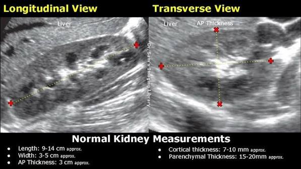
In a healthy adult, each kidney measures approximately 9 to 12 centimeters in length, 4 to 5 centimeters in width, and 3 to 4 centimeters in thickness. The left kidney is generally slightly larger than the right because the liver pushes the right kidney slightly downward. A size difference of more than 1.5 centimeters between the two kidneys is considered abnormal.
The kidney has a recognizable internal structure: the outer cortex has intermediate echogenicity similar to the adjacent liver; the inner medullary pyramids are darker (hypoechoic); and the central renal sinus — fat, vessels, and collecting system — appears bright white (hyperechoic). The sharp visual contrast between bright cortex and darker medulla is called corticomedullary differentiation. When CKD progresses and normal tissue is replaced by fibrosis, this contrast is lost — a finding called loss of corticomedullary differentiation.
Hydronephrosis: A Dilated Collecting System
Hydronephrosis is one of the most important and commonly detected findings on kidney ultrasound. It refers to dilation of the renal collecting system — the pelvis and calyces — from obstruction or backflow of urine. On ultrasound, it appears as a central anechoic (black) fluid-filled space replacing what is normally the echogenic renal sinus.
- Mild (Grade 1–2): Renal pelvis slightly dilated; parenchyma preserved
- Moderate (Grade 3): Pelvis and calyces clearly dilated; early cortical thinning
- Severe (Grade 4): Entire collecting system markedly dilated; significant cortical thinning; risk of permanent parenchymal loss
Common causes include ureteral stones, benign prostatic hyperplasia (bilateral hydronephrosis in men), pregnancy (particularly right-sided from uterine pressure), and tumors compressing the ureter. Bilateral unexplained hydronephrosis in a patient with elevated creatinine is an urgent finding that may represent post-renal AKI reversible with catheter or ureteral stenting.
Kidney Stones on Ultrasound
Kidney stones appear as hyperechoic (bright white) foci with a characteristic posterior acoustic shadow — a dark band trailing behind the stone where sound waves cannot penetrate. This combination of brightness plus shadowing allows radiologists to distinguish stones from other bright structures.
Ultrasound has a sensitivity of approximately 70% for detecting stones within the kidney itself. It is less accurate for small stones (under 5 mm) and ureteral stones — where overlying bowel gas interferes. For suspected ureteral colic, a non-contrast CT scan (CT KUB) remains the gold standard with near-perfect sensitivity. Ultrasound is preferred in pregnancy, children, and situations where radiation should be minimized. Even when the stone itself cannot be seen, detecting hydronephrosis on the side of the pain strongly suggests an obstructing stone.
Kidney Cysts: What They Mean
Kidney cysts are among the most common incidental findings on renal ultrasound, detected in roughly half of adults over age 50. The vast majority are benign simple cysts.
A simple renal cyst on ultrasound is perfectly round, completely anechoic (black), has a thin smooth wall, and shows posterior acoustic enhancement (the tissue behind it appears brighter). The Bosniak classification grades cysts by complexity and cancer risk:
- Bosniak I: Simple benign cyst — no follow-up needed
- Bosniak II: Mildly complex (thin septation, small calcification) — follow-up in 12 months
- Bosniak IIF: More complex features — follow-up imaging every 6–12 months
- Bosniak III: Indeterminate; malignancy rate approximately 50% — surgical consideration
- Bosniak IV: Solid enhancing components — treat as malignant; surgery recommended
Autosomal dominant polycystic kidney disease (ADPKD) presents as bilateral kidneys replaced by innumerable cysts of varying sizes, often with markedly enlarged kidneys. Diagnosis in adults with a positive family history requires only three or more unilateral cysts or two or more bilateral cysts before age 40.
Kidney Size and Echogenicity in CKD
Small kidneys (less than 9 cm) strongly suggest long-standing CKD. The scarring and fibrosis of chronic kidney disease causes the kidney to shrink as functioning nephron mass is progressively replaced. Bilateral small echogenic kidneys in a patient with elevated creatinine and proteinuria are practically diagnostic of advanced CKD.
Increased cortical echogenicity — when the kidney cortex becomes brighter than the adjacent liver — is a nonspecific but important finding correlating with parenchymal disease. It is seen in CKD from any cause, acute glomerulonephritis, nephrotic syndrome, and acute tubular necrosis. Higher grade echogenicity correlates with more advanced renal parenchymal disease.
Asymmetric kidney size (difference greater than 1.5 cm) suggests unilateral disease. The most important cause in a hypertensive patient is renal artery stenosis on the smaller side, causing ischemic nephropathy. For more on kidney function evaluation, see our guide to kidney function tests.
Renal Masses Detected on Ultrasound
A solid renal mass — one that has internal echoes and does not show features of a simple cyst — requires further characterization with CT or MRI.
Renal cell carcinoma (RCC) may appear as a solid, isoechoic or hyperechoic mass distorting the normal kidney contour. Small RCCs (less than 2 cm) may be nearly invisible against normal kidney tissue — which is why CT with contrast is preferred for definitive diagnosis and staging.
Angiomyolipoma (AML) is a benign tumor composed of fat, smooth muscle, and abnormal blood vessels. Because fat is highly reflective, AMLs appear as strikingly hyperechoic (very bright white) masses on ultrasound. Fat density below −20 Hounsfield units on CT is virtually diagnostic of AML. AMLs larger than 4 cm carry increased bleeding risk and are associated with tuberous sclerosis complex.
Doppler Ultrasound of the Kidneys
Renal artery stenosis is evaluated by measuring the peak systolic velocity (PSV) within the renal artery. A PSV above 180–200 cm/s combined with a renal-to-aortic ratio above 3.5 suggests hemodynamically significant stenosis. CT angiography or MR angiography provides more definitive assessment when Doppler is inconclusive.
The resistive index (RI) — calculated from the waveform of blood flow — measures resistance within the kidney’s small vessels. A normal RI is 0.60 to 0.70. An RI above 0.80 suggests significant intrinsic renal parenchymal disease with fibrosis and small vessel damage, consistent with advanced CKD or acute tubular injury.
Renal vein thrombosis — most common in nephrotic syndrome, particularly membranous nephropathy — appears as absent venous Doppler signal within the renal vein, combined with an enlarged, edematous kidney. This requires urgent anticoagulation. For more on conditions detected through kidney imaging and blood tests, see our guide to blood in urine test results.
What Kidney Ultrasound Cannot Show
- Small kidney stones and ureteral stones: CT without contrast is far more sensitive and is preferred when stone disease is strongly suspected
- Transitional cell carcinoma of the renal pelvis or ureter: CT urogram is the gold standard for urothelial tumors
- Early or small renal cell carcinoma: CT or MRI with contrast required for accurate detection and staging
- Kidney function: Ultrasound shows structure only — blood tests (creatinine, eGFR) and urine tests (ACR) remain essential; see our guide to kidney function monitoring
- Adrenal masses: CT is more sensitive for adrenal pathology
Frequently Asked Questions About Kidney Ultrasound
Is a kidney ultrasound the same as an abdominal ultrasound?
Not exactly. An abdominal ultrasound typically images the liver, gallbladder, spleen, pancreas, and kidneys together. A dedicated renal ultrasound focuses specifically on the kidneys and sometimes the bladder, often with more detailed images and Doppler assessment.
Can a kidney ultrasound detect cancer?
A kidney ultrasound can identify suspicious solid masses that require further evaluation, but CT or MRI with contrast is needed to characterize any suspected tumor, assess its size, and determine whether it has spread.
Is a kidney ultrasound safe during pregnancy?
Yes — ultrasound uses sound waves, not radiation, and has an excellent safety record in pregnancy. It is the preferred imaging method for evaluating kidney problems during pregnancy, including hydronephrosis, kidney stones, and pyelonephritis.
How accurate is ultrasound for kidney stones?
Ultrasound detects approximately 70% of kidney stones located within the kidney. It is less accurate for small stones (under 5 mm) and ureteral stones. A non-contrast CT scan is the gold standard with near-perfect sensitivity.
What does “echogenic” mean on an ultrasound report?
“Echogenic” means bright on the ultrasound image — the tissue reflects more sound waves than normal. When a kidney is described as echogenic, it usually means the cortex is brighter than expected, which is a nonspecific sign of kidney disease. See our guide to protein in urine for more on what kidney damage markers indicate.
This article is for educational purposes only. Always consult a qualified healthcare provider for diagnosis and treatment of any medical condition.
Kidney Ultrasound in Specific Clinical Situations
While the general principles of renal ultrasound interpretation apply broadly, certain clinical contexts warrant a more tailored understanding of what the test can and cannot contribute.
Acute flank pain: When a patient presents with sudden-onset severe flank pain radiating to the groin — the classic presentation of renal colic from an obstructing stone — the clinical question is whether a stone is present and whether it is causing obstruction. Kidney ultrasound can confirm the presence of hydronephrosis (the indirect sign of obstruction) even when the stone itself is not directly visible. Because ultrasound can be performed immediately at the bedside without radiation, it is often the first imaging study in emergency settings, especially for patients who are pregnant. However, a negative ultrasound does not exclude a stone; a non-obstructing stone in the kidney or a mid-ureteral stone may not be visible on ultrasound even when it is causing significant pain. In patients with classic renal colic and a negative ultrasound, a CT without contrast (CT KUB) is the definitive next step.
Recurrent urinary tract infections: In patients with recurrent UTIs — particularly children and women with frequent infections — a kidney ultrasound helps identify structural abnormalities that predispose to infection. Hydronephrosis suggests an obstruction that may be trapping bacteria in the collecting system. A duplicated collecting system (duplex kidney) is associated with vesicoureteral reflux, which allows bacteria to travel retrograde from the bladder to the kidney. Kidney stones can serve as a nidus for persistent infection, particularly struvite stones (formed from urease-producing bacteria). Identifying any of these structural factors changes the approach to prevention — it is not enough to treat each UTI in isolation if the underlying anatomy is predisposing the patient to recurrence.
Monitoring chronic kidney disease: For patients with established CKD, kidney ultrasound is not typically needed at every monitoring visit — it is not a test of kidney function, and a repeat ultrasound in a patient with known CKD and stable creatinine will usually show the same small echogenic kidneys as the prior scan. However, ultrasound is indicated in CKD when there is an unexplained acute deterioration in kidney function (to exclude new obstruction), when hematuria develops (to look for a new structural lesion), or when the diagnosis of CKD is being established for the first time and the clinician wants to confirm bilateral small kidneys consistent with chronic disease rather than bilateral acute obstruction that could be reversed.
Evaluating a transplanted kidney: Ultrasound is the primary imaging tool for post-transplant kidney monitoring. The transplanted kidney is placed in the pelvis — a superficial location that makes it particularly easy to image. The standard post-transplant ultrasound assesses the kidney’s size, parenchymal echogenicity, the presence of hydronephrosis (which may indicate ureteral obstruction from a stricture or blood clot), perinephric fluid collections (hematoma, urinoma, lymphocele), and the Doppler resistive index. In the early post-transplant period, a rising creatinine prompts urgent ultrasound to distinguish between vascular thrombosis (absent Doppler signal in the renal artery or vein — a surgical emergency), acute rejection (elevated RI, hypoechoic cortex — needs biopsy), urological obstruction (hydronephrosis — needs stenting), and perinephric collections (urinoma or hematoma — may need drainage). The ultrasound result from a post-transplant patient often directly determines the next clinical action.
Guiding kidney biopsy: Real-time ultrasound guidance is used for virtually all percutaneous kidney biopsies performed today. The sonographer or nephrologist images the kidney in real time, identifies the lower pole of the kidney (the preferred biopsy site because it is furthest from major vessels), and guides the biopsy needle to the cortex at the optimal angle and depth. This reduces the risk of hitting the renal vein, artery, or collecting system, and allows the operator to avoid visible cysts or masses. Post-biopsy ultrasound — typically performed 4 to 6 hours after the procedure — checks for perirenal hematoma and confirms the absence of arteriovenous fistula formation (detected as turbulent high-velocity Doppler signal at the biopsy site).
Understanding Your Kidney Ultrasound Report
Receiving a kidney ultrasound report can be confusing, particularly when it contains radiological terms that were not explained during the appointment. Here is a practical guide to the most common terms and findings patients encounter.
“No hydronephrosis”: This is a reassuring finding. It means the collecting system is not dilated, which makes an obstructing stone or tumor causing urinary tract obstruction unlikely on the day of the scan.
“Simple renal cyst, right kidney, 2.3 cm”: A simple cyst is benign. The report will typically classify it as Bosniak I or II. No follow-up is needed for a Bosniak I cyst. A Bosniak II cyst with a septation or small calcification typically gets a repeat ultrasound in 12 months to confirm stability. The size alone — even a large 6 or 7 cm simple cyst — does not determine whether it is benign or worrying; the internal features (thin wall, no solid component, no enhancement) are what matter.
“Echogenic kidneys, bilateral”: This means both kidneys are brighter than normal on the ultrasound image — the cortex reflects more sound waves than expected. This is a nonspecific finding that accompanies many forms of chronic kidney disease. It does not indicate the cause of the disease, only that the kidney parenchyma is abnormal. When this finding appears alongside abnormal kidney function tests, it supports the diagnosis of CKD but does not replace blood and urine testing for staging and management.
“Incidental hyperechoic lesion, right kidney, 1.1 cm — recommend CT for further characterization”: This means the radiologist found a small bright spot that could represent a benign angiomyolipoma (fatty tumor) or potentially something more worrisome. A CT scan with and without contrast can determine whether the lesion contains fat (making AML virtually certain) or shows enhancement characteristics that raise concern for a small renal cell carcinoma. Most small incidental hyperechoic lesions turn out to be benign AMLs, but CT confirmation is the appropriate next step.
“Mild left hydronephrosis — correlate clinically”: This means the left collecting system is slightly more dilated than normal, but the degree is mild and the finding may or may not be significant. Mild hydronephrosis can be entirely normal in a well-hydrated patient, in pregnancy (right more than left), or can represent an early obstruction that needs further workup. The phrase “correlate clinically” is the radiologist’s way of saying: combine this finding with the patient’s symptoms, kidney function tests, and history to determine whether further evaluation (CT urogram or cystoscopy) is warranted.
“Resistive index elevated bilaterally (right 0.78, left 0.80)”: A resistive index at the upper end of or above normal (0.80) on both sides suggests intrinsic small vessel disease within the kidney parenchyma, consistent with CKD, diabetic nephropathy, or long-standing hypertensive nephrosclerosis. An RI this high does not indicate obstruction — obstructed kidneys can also have elevated RI, but the pattern of elevation and the presence or absence of hydronephrosis helps distinguish the two. This Doppler finding, in a patient with already-known CKD, adds further evidence of significant parenchymal disease.
When any kidney ultrasound report raises questions, ask your provider to walk through each finding and what it means for your specific situation. Ultrasound findings are never interpreted in isolation — they are always viewed alongside the clinical history, blood tests, urine tests, and the findings from any previous imaging.
When Follow-Up Imaging Is Needed After a Kidney Ultrasound
Not every kidney ultrasound finding requires immediate action. Understanding the typical follow-up pathway for the most common findings helps patients know what to expect after the test.
Simple renal cyst (Bosniak I): No follow-up imaging is recommended. Simple cysts do not become cancerous and do not require any surveillance. Patients who receive this finding can be reassured and asked to mention it to future providers so it is not re-evaluated unnecessarily.
Mildly complex cyst (Bosniak IIF): A repeat ultrasound in 6 to 12 months is recommended to confirm that the cyst’s features have not changed in a way that would upgrade it to Bosniak III or IV. If two consecutive stable follow-up scans show no progression over 24 months, the surveillance interval may be extended or stopped based on the patient’s age and overall health.
Solid or indeterminate renal mass: A CT scan of the abdomen and pelvis with and without contrast — or an MRI with gadolinium — is the appropriate next step. This is not a reason to panic; the majority of incidentally found small solid renal lesions in adults are benign (angiomyolipoma, oncocytoma) or, if malignant, are detected at an early stage when surgery is curative.
Hydronephrosis without an identified cause: If the degree of hydronephrosis is moderate or severe, or if the patient has an elevated creatinine, a CT urogram is typically ordered next to identify the site and cause of obstruction. Mild hydronephrosis in a patient with normal kidney function and no symptoms can often be followed with a repeat ultrasound in 3 to 6 months to confirm resolution.
Asymmetric kidneys or Doppler findings suggesting renal artery stenosis: A CT angiogram or MR angiogram of the renal arteries provides definitive assessment of the degree of stenosis and whether intervention (renal artery stenting or surgical revascularization) is appropriate. Not all renal artery stenosis requires treatment — the decision depends on whether the stenosis is causing resistant hypertension or progressive ischemic nephropathy on the affected side.
In every case, the follow-up plan recommended by the radiologist and your provider is calibrated to the specific finding, the patient’s overall health, and the likelihood of finding something clinically significant on further testing. The goal is neither to over-investigate every incidental finding nor to dismiss findings that carry meaningful risk — and the kidney ultrasound, by providing a clear initial structural picture, sets the foundation for that decision-making process.
Sources: American College of Radiology Appropriateness Criteria; RadiologyInfo.org — Ultrasound of the Kidneys; NIDDK — Kidney Disease; AUA Microhematuria Guidelines.
I never fully understood kidney ultrasound: what it shows until I read this. The section on managing this condition day-to-day was especially useful for planning. Thank you for making complex medical information accessible without dumbing it down.
I never fully understood kidney ultrasound: what it shows until I read this. The article answered questions I didn’t even know I had until I started reading. Looking forward to reading more articles from this website.
Bookmarked this article on kidney ultrasound: what it shows immediately — going to reference it regularly. The connection between lifestyle choices and long-term outcomes is explained clearly here. I wish I had found this article earlier — would have saved a lot of confusion.