
Liver function tests — often called LFTs — are a panel of blood tests that measure specific enzymes and proteins produced by the liver, providing clinicians with a window into the liver’s health that no physical examination alone can match. These tests are ordered routinely in preventive health checks, systematically in patients with known liver disease, and urgently when symptoms suggest acute liver injury. Understanding what each marker measures, what it means when it is elevated or low, and how to interpret the panel as a whole is essential both for clinicians communicating results to patients and for patients trying to make sense of what their blood results mean for their health.
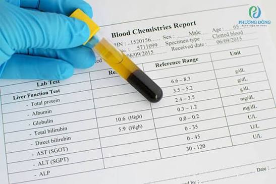
A standard liver function panel typically includes alanine aminotransferase (ALT), aspartate aminotransferase (AST), alkaline phosphatase (ALP), gamma-glutamyl transferase (GGT), total and direct bilirubin, albumin, and total protein. Each of these markers reflects a different aspect of hepatic physiology — ALT and AST indicate hepatocellular injury, ALP and GGT signal cholestatic or biliary pathology, bilirubin reflects the liver’s ability to process and excrete waste products, and albumin and total protein measure the liver’s synthetic function. Interpreting liver function tests requires understanding not just individual values but their relationships to each other — a pattern of isolated ALT/AST elevation points to a different set of conditions than a pattern of ALP elevation with normal aminotransferases.
ALT and AST — Markers of Liver Cell Injury
Alanine aminotransferase (ALT) and aspartate aminotransferase (AST) are enzymes found inside liver cells that leak into the bloodstream when hepatocytes are injured or destroyed. ALT is more specific to the liver than AST — AST is also found in muscle, heart, kidneys, and red blood cells, so AST elevation can arise from non-hepatic sources including vigorous exercise, muscle injury, and cardiac disease. For this reason, ALT is the better single marker of hepatocellular injury when evaluating the liver specifically. Normal ALT is generally defined as below 40–56 IU/L in most laboratories (some now advocate lower upper limits of normal, particularly for women), and normal AST as below 40 IU/L, though reference ranges vary slightly between institutions.
The degree of elevation provides clinical information about the likely underlying cause. Mild ALT elevation — one to three times the upper limit of normal — is found in a broad range of conditions including non-alcoholic fatty liver disease (MASLD), chronic viral hepatitis B and C, medication side effects, thyroid disease, and celiac disease, and is the most common incidental finding on routine blood panels. Moderate elevation — three to ten times normal — is associated with active viral hepatitis, autoimmune hepatitis, alcoholic hepatitis, and drug-induced liver injury. Marked elevation — more than ten times the upper limit of normal, sometimes exceeding 1000–2000 IU/L — occurs in acute viral hepatitis A, B, or E, ischemic hepatitis (shock liver from cardiovascular collapse), acute drug toxicity (particularly acetaminophen overdose), and Budd-Chiari syndrome. The ratio of AST to ALT also provides diagnostic information: an AST:ALT ratio greater than 2:1 is characteristic of alcoholic liver disease, whereas a ratio below 1 is typical of non-alcoholic fatty liver disease and chronic viral hepatitis. For more context on how liver cell injury presents clinically, see the article on fatigue as an early sign of liver disease.

ALP and GGT — Cholestatic and Biliary Markers
Alkaline phosphatase (ALP) is an enzyme found predominantly in liver bile duct cells, bone, intestine, and placenta. Elevated ALP in the context of liver disease indicates cholestatic pathology — impaired bile flow from intrahepatic or extrahepatic biliary obstruction. Conditions producing elevated ALP include primary biliary cholangitis (PBC), primary sclerosing cholangitis (PSC), bile duct stones, cholangiocarcinoma, pancreatic head cancer obstructing the bile duct, drug-induced cholestasis, and intrahepatic cholestasis of pregnancy. Because ALP is also elevated in bone disease (Paget’s disease, bone metastases, healing fractures, hyperparathyroidism), confirmation that elevated ALP is hepatic in origin is important. This is done by measuring GGT simultaneously: if GGT is also elevated, the ALP elevation is hepatic; if GGT is normal, bone disease or a non-hepatic source is more likely.
Gamma-glutamyl transferase (GGT) is a sensitive but non-specific marker of liver disease. It is elevated in virtually all forms of hepatic and biliary disease including MASLD, alcoholic liver disease, cholestasis, viral hepatitis, and drug reactions. GGT is also elevated by alcohol consumption even in the absence of liver disease — it is one of the most sensitive laboratory indicators of regular heavy alcohol use, normalizing within two to six weeks of abstinence. GGT elevation in the context of normal ALT and normal ALP, without other liver disease markers, is often related to alcohol, obesity, or medications (particularly anticonvulsants, barbiturates, and rifampicin), and requires clinical correlation rather than automatic concern for structural liver disease. The pattern of isolated GGT elevation with normal ALT, AST, and ALP, in a patient who is otherwise well and whose other blood tests are normal, is usually followed by lifestyle advice (alcohol reduction, weight loss) and repeat testing rather than immediate liver imaging or biopsy.
Bilirubin, Albumin, and Prothrombin Time — Function Markers
Bilirubin is a waste product of haemoglobin breakdown that the liver conjugates and excretes into bile. Total bilirubin above 17–20 μmol/L (or approximately 1–1.2 mg/dL) is elevated; visible jaundice typically appears when total bilirubin exceeds 35–50 μmol/L (approximately 2–3 mg/dL). The direct (conjugated) bilirubin fraction is elevated in cholestatic liver disease and biliary obstruction, while elevated total bilirubin with predominantly indirect (unconjugated) bilirubin points to pre-hepatic causes including haemolysis and, commonly, Gilbert’s syndrome — a benign genetic variant causing mild unconjugated hyperbilirubinaemia in otherwise healthy individuals. Understanding bilirubin fractions is key to interpreting the clinical significance of a raised bilirubin. The article on yellow skin and eyes in liver disease covers the clinical presentations of jaundice in detail.
Albumin — the major protein synthesized by the liver — reflects the liver’s synthetic capacity. Low albumin (hypoalbuminaemia) in the setting of liver disease indicates impaired synthetic function, typically occurring in chronic liver disease when hepatic reserve is substantially reduced. Normal albumin is 35–50 g/L; levels below 30 g/L in a patient with liver disease indicate significant synthetic failure, are associated with ascites (low albumin reduces plasma oncotic pressure, allowing fluid to leak into the peritoneum), and carry prognostic significance. Albumin is also reduced by malnutrition, nephrotic syndrome (protein loss in urine), protein-losing enteropathy, and systemic inflammation — so a low albumin is not specific to liver disease and requires clinical context to interpret correctly. Prothrombin time (PT) and its standardized form, the INR, measure the liver’s ability to synthesize the clotting factors that depend on adequate hepatic function — factors II, VII, IX, and X. A prolonged PT/INR in a patient with liver disease signals impaired synthetic function and is incorporated into prognostic scoring systems including the Child-Pugh score and MELD score for cirrhosis severity, as covered in the article on easy bruising and liver function.
Interpreting Liver Function Test Patterns
Recognizing patterns in the liver function panel rather than interpreting each marker in isolation is the most clinically productive approach. A hepatocellular pattern — predominantly elevated ALT and AST with relatively preserved ALP, GGT, and bilirubin — points to direct liver cell injury from viral hepatitis, drug toxicity, alcoholic hepatitis, autoimmune hepatitis, or ischaemia. A cholestatic pattern — predominantly elevated ALP and GGT, with variable ALT/AST elevation and raised bilirubin in more severe cases — points to biliary obstruction or cholestatic liver disease such as PBC or PSC. A mixed pattern, with both hepatocellular and cholestatic markers elevated, is seen in conditions such as drug-induced liver injury, alcoholic hepatitis, and some systemic diseases including sarcoidosis and heart failure.
The magnitude of elevation matters alongside the pattern. Mild transaminase elevation (below three times normal) in an asymptomatic patient with metabolic risk factors (obesity, diabetes, hypertriglyceridaemia) is most likely MASLD and warrants liver ultrasound, lifestyle advice, and repeat testing rather than immediate specialist referral. Moderate elevation (three to ten times normal) without an obvious cause requires investigation for viral hepatitis, autoimmune hepatitis, and drug causes. Severe elevation (more than ten times normal) requires urgent assessment to exclude acute liver failure — particularly when accompanied by jaundice, coagulopathy, or encephalopathy — because the management pathway for acute liver failure is time-critical and may include emergency liver transplant evaluation. The pattern of dark urine and pale stools alongside a cholestatic LFT pattern should prompt urgent biliary imaging to exclude bile duct obstruction from stones or malignancy.
Frequently Asked Questions About Liver Function Tests
Can a normal liver function test mean my liver is completely healthy?
Not necessarily. Liver function tests are useful markers of hepatic injury and function, but some significant liver conditions can be present with entirely normal LFTs for years. Non-alcoholic fatty liver disease (MASLD) is present in twenty-five percent of the global adult population and produces normal LFTs in many patients — the only reliable way to detect it is liver ultrasound showing increased hepatic echogenicity (fatty infiltration). Early hepatic fibrosis and even early cirrhosis can be present with normal ALT and AST. Hepatitis C can be present with normal or minimally elevated ALT during periods of low viral activity. A normal LFT panel reduces the probability of significant acute liver disease but does not exclude chronic liver disease or early fibrosis — which is why liver disease risk assessment for patients with metabolic syndrome, obesity, or significant alcohol use should include liver ultrasound in addition to blood tests.
Do I need to fast before a liver function test?
Fasting is not required for liver function tests — ALT, AST, ALP, GGT, bilirubin, albumin, and total protein are not significantly affected by recent food intake. However, if liver function tests are being drawn as part of a broader metabolic panel that includes fasting glucose or fasting lipids, the venepuncture appointment may be scheduled fasting for those components, and the LFTs are simply taken at the same time. Some laboratories recommend avoiding alcohol for at least twenty-four to forty-eight hours before testing to avoid artificially elevating GGT from recent alcohol consumption, which could confound interpretation. If you are taking any medications — including over-the-counter pain relievers such as acetaminophen (paracetamol), which even at therapeutic doses can mildly elevate ALT — inform your clinician, as many common drugs affect LFT results. Understanding your liver function test results in the context of the full clinical picture — including symptoms like when to seek medical attention for digestive symptoms — helps ensure the right follow-up happens promptly.
Sources: NIDDK — Liver Blood Tests · AASLD — Liver Disease Guidelines · Mayo Clinic — Liver Function Tests
MASLD and Metabolic Liver Disease — The Most Common Elevated LFT Cause
Metabolic dysfunction-associated steatotic liver disease (MASLD) — previously termed non-alcoholic fatty liver disease (NAFLD) — is now the most prevalent cause of mildly elevated liver function tests in adults in the United States, affecting an estimated eighty to one hundred million Americans. It encompasses a spectrum from simple hepatic steatosis (fat accumulation in more than five percent of hepatocytes without inflammation or fibrosis) through metabolic dysfunction-associated steatohepatitis (MASH, formerly NASH — steatosis plus inflammation and hepatocyte injury) to advanced fibrosis and cirrhosis. The majority of patients with MASLD have ALT between one and two times the upper limit of normal, with AST equal to or slightly lower than ALT. A substantial minority have entirely normal aminotransferases despite significant steatosis or early fibrosis — making liver ultrasound essential in high-risk patients with obesity, type 2 diabetes, dyslipidaemia, or hypertension, regardless of LFT results.
Monitoring liver function tests in established MASLD serves to track disease progression and detect complications. An AST:ALT ratio that rises above one in a patient with previously ratio-below-one MASLD suggests advancing fibrosis — the ratio rises as the aminotransferases decline in advanced cirrhosis due to loss of functional hepatocytes, making absolute values less meaningful at that stage. Platelet count decline alongside LFT changes indicates portal hypertension from cirrhosis. The development of hypoalbuminaemia, rising bilirubin, or prolonged PT/INR in a patient with MASLD marks decompensation — the transition from compensated cirrhosis (where liver synthetic function is preserved) to decompensated cirrhosis with ascites, encephalopathy, or variceal bleeding risk. Patients with MASLD-related cirrhosis require six-monthly liver ultrasound and AFP measurement for hepatocellular carcinoma surveillance, as covered in the article on swollen belly and signs of advanced liver disease.
Drug-Induced Liver Injury — When Medications Affect Your LFTs
Drug-induced liver injury (DILI) is an important and often underrecognized cause of abnormal liver function tests, responsible for over one thousand known hepatotoxic drugs, supplements, and herbal remedies. The pattern of DILI varies by agent: hepatocellular DILI (disproportionate ALT/AST elevation) is produced by isoniazid, nitrofurantoin, methyldopa, statins, and many antiretrovirals; cholestatic DILI (disproportionate ALP/GGT with variable aminotransferases) is produced by amoxicillin-clavulanate, flucloxacillin, erythromycin, and anabolic steroids; mixed DILI involves both patterns and is produced by carbamazepine, phenytoin, and some herbal products. Among the most clinically significant prescription medications affecting liver function tests are statins — commonly producing mild to moderate ALT elevation, usually asymptomatic and often self-resolving even if the statin is continued — and acetaminophen (paracetamol), where toxic doses (above 7.5–10 g acutely in adults) cause massive hepatocellular injury with ALT elevation often exceeding 3000–10,000 IU/L, representing a medical emergency requiring N-acetylcysteine treatment and potential transplant assessment.
Herbal and dietary supplements are an increasingly recognized cause of DILI that is frequently overlooked because patients do not consider them medications and may not disclose use unless specifically asked. Green tea extract, kava, black cohosh, comfrey, pyrrolizidine alkaloid-containing products (butterbur, coltsfoot), and numerous traditional herbal preparations have caused severe hepatotoxicity including acute liver failure. The Drug-Induced Liver Injury Network (DILIN) estimates that herbal and dietary supplements now account for approximately twenty percent of DILI cases in the United States. When evaluating unexplained liver function test elevation, a thorough medication history — explicitly including over-the-counter medications, vitamins, protein powders, weight-loss supplements, herbal teas, and traditional remedies — is essential. The Roussel Uclaf Causality Assessment Method (RUCAM) score helps estimate the probability that a specific agent caused DILI based on timing, course, risk factors, exclusion of other diagnoses, and prior literature. The article on itchy skin as a cholestatic liver symptom addresses clinical features that indicate biliary involvement in cholestatic DILI.
Acute Liver Failure — When LFT Elevation Becomes an Emergency
Acute liver failure (ALF) is a rare but life-threatening syndrome defined by the rapid development of severe hepatocellular injury, coagulopathy (INR ≥ 1.5), and hepatic encephalopathy in a patient without pre-existing liver disease. Recognition of acute liver failure is critical because its management requires urgent transfer to a specialist liver unit and potential emergency liver transplant assessment — and delay worsens outcomes. The laboratory hallmarks of acute liver failure are markedly elevated aminotransferases (typically thousands of IU/L), rising bilirubin, progressively prolonged PT/INR that does not correct with vitamin K administration, rising creatinine (indicating hepatorenal syndrome), and hypoglycaemia from failed hepatic gluconeogenesis. Importantly, a fall in aminotransferases in the context of worsening coagulopathy and rising bilirubin does not indicate improvement — it indicates hepatocyte exhaustion and progressing liver failure. This pattern of rising bilirubin, rising INR, and falling ALT is described as the “peak-fall-die” phenomenon in acetaminophen ALF and should prompt immediate specialist escalation.
The King’s College Criteria — developed for both acetaminophen and non-acetaminophen causes of ALF — identify patients at high risk of death without liver transplantation, incorporating arterial pH, INR, creatinine, and bilirubin into threshold-based scoring. In non-acetaminophen ALF, poor prognostic markers include INR greater than 6.5, age below ten or above forty years, jaundice-to-encephalopathy interval exceeding seven days, serum bilirubin above 300 μmol/L, and any non-A non-B hepatitis aetiology. These criteria inform urgent listing decisions, as the window for transplant surgery in acute liver failure is narrow and outcomes depend on timely escalation. For patients and families recognising that a relative may be deteriorating — developing jaundice, confusion, unusual tiredness, and bruising simultaneously — understanding the urgency of seeking emergency evaluation is essential. This overlaps with the article on easy bruising as a sign of impaired liver synthetic function.
Chronic Viral Hepatitis — Monitoring With Liver Function Tests
Chronic hepatitis B and chronic hepatitis C are among the most important causes of liver disease worldwide, and liver function tests play a central role in their monitoring and management. In chronic hepatitis B, the natural history involves distinct immunological phases — immune tolerance (high viral load, normal ALT, minimal liver damage), immune clearance (fluctuating or elevated ALT as the immune system attempts to control infection), immune control (HBeAg seroconversion, suppressed viral load, normal ALT), and reactivation (renewed viral replication and hepatitis activity). ALT levels during immune clearance phase can fluctuate widely, including episodes of marked elevation above ten times normal, and persistently elevated ALT above twice normal in the context of hepatitis B e antigen (HBeAg) positivity or elevated HBV DNA is a key criterion for initiating antiviral therapy. In chronic hepatitis C, ALT elevation is typically mild to moderate (one to three times normal) and may fluctuate or even normalize at intervals, leading to potential under-detection. All-oral direct-acting antiviral (DAA) regimens achieving sustained virological response (SVR) normalize ALT in the vast majority of treated patients — but patients with established cirrhosis prior to treatment remain at ongoing HCC risk and require continued six-monthly surveillance despite viral cure.
Alcoholic hepatitis — the acute hepatic inflammation superimposed on alcoholic liver disease from recent heavy alcohol consumption — produces a characteristic LFT pattern that differs from other hepatocellular causes. The AST:ALT ratio is typically greater than 2:1 (often 3:1 or higher), reflecting mitochondrial AST release preferentially over cytosolic ALT, combined with alcohol-induced GGT elevation and frequently raised bilirubin disproportionate to the degree of aminotransferase elevation. The Maddrey discriminant function — calculated from serum bilirubin and prothrombin time — and the MELD score identify patients with severe alcoholic hepatitis (Maddrey score ≥32 or MELD ≥20) who may benefit from corticosteroid therapy with prednisolone, though treatment decisions require exclusion of infection and careful risk-benefit assessment. Understanding how alcohol specifically alters LFT patterns helps differentiate alcoholic liver disease from other causes when taking a clinical history — and underscores why abstinence from alcohol is the single most effective intervention for reversing alcohol-related liver injury at all stages of the disease spectrum.
Liver function tests are a foundational tool in medical practice — but their greatest value comes from interpreting them systematically: pattern recognition over individual numbers, serial trending over single snapshots, and integration with clinical context and imaging rather than blood results in isolation. Whether you have received an abnormal LFT result that needs explanation, are managing a known liver condition, or are assessing risk in a patient with metabolic disease or significant alcohol use, understanding what each marker measures and what it means in combination is the foundation of informed decision-making. The articles in this series explore each individual marker in depth, from ALT and AST to bilirubin, albumin, and alkaline phosphatase, providing the detail behind each component of the panel that brief laboratory reports cannot convey.
This finally made sense of my lab results — my doctor mentioned my ALT was mildly elevated and I had no idea what that meant. The section on MASLD and why normal LFTs don’t rule out fatty liver was really eye-opening. I had no idea you could have significant fatty liver with completely normal blood tests.
Thank you for sharing that, Sandra — you’ve touched on one of the most important points in liver health. Fatty liver (MASLD) is genuinely very common and unfortunately a normal ALT does not mean the liver is fine, which is why liver ultrasound is recommended for anyone with metabolic risk factors like obesity or diabetes regardless of blood test results. If your doctor has identified mildly elevated ALT, asking about a liver ultrasound is a very reasonable next step.
The explanation of the AST:ALT ratio and what different ratios point toward was very useful. I’m a nurse and even I found the breakdown of hepatocellular vs cholestatic patterns here clearer than most textbooks put it. Bookmarking this for patient education.