
The sharp, stabbing heel pain that strikes the moment you take your first step out of bed in the morning — and then gradually eases as you walk around — is one of the most recognisable symptoms in musculoskeletal medicine. It is the hallmark of plantar fasciitis, the most common cause of heel pain in adults and one of the most frequent reasons adults visit their GP for foot-related problems. Affecting approximately one in ten adults at some point, plantar fasciitis is well understood and has a clear evidence-based treatment pathway — yet it remains frequently undertreated, leaving many patients managing avoidable years of heel pain.
What Is Plantar Fasciitis?
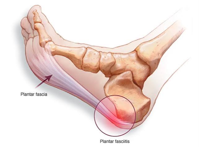
The plantar fascia is a thick, fibrous band — more precisely described as an aponeurosis — that runs along the sole of the foot. It originates from the medial calcaneal tuberosity (the underside of the heel bone) and fans out across the sole to attach to the base of each toe. Its primary mechanical function is to support the medial longitudinal arch of the foot during walking and running through what is known as the windlass mechanism: as the toes are forced upward during the push-off phase of gait, the plantar fascia tightens around the metatarsal heads, raising the arch and storing elastic energy for propulsion.
Plantar fasciitis is degeneration and pain at the proximal attachment of the plantar fascia to the heel bone — the point of maximum tensile load accumulation. Despite the “-itis” suffix, histological studies of plantar fascia tissue show degenerative collagen changes rather than acute inflammatory cell infiltrate, meaning the condition is more accurately described as plantar fasciopathy or plantar fasciosis — a pattern identical to the tendinopathy distinction discussed in our guide to tendinitis in adults. Anti-inflammatory treatments therefore have limited long-term effectiveness: the fundamental problem is degenerated collagen at the fascial origin, not ongoing acute inflammation.
A common misconception is that a heel spur causes plantar fasciitis pain. Heel spurs — bony protrusions at the calcaneal origin of the plantar fascia visible on X-ray — are present in approximately 50% of people with plantar fasciitis, but they are also found in around 20% of people without any heel pain. The spur is the result of chronic traction on the bone, not the cause of pain; treating the spur itself does not treat the pain.
Symptoms: The First-Step Pattern
The symptom that most reliably identifies plantar fasciitis is first-step pain — clinically described as post-static dyskinesia. After a period of rest (most dramatically after overnight sleep), the fascia shortens slightly and the degenerated tissue at the heel insertion becomes acutely painful when first loaded. Walking around for a few minutes loosens the fascia and redistributes load, and the pain eases — often significantly. This cycle of severe initial pain, gradual improvement with movement, and eventual return of discomfort with sustained weight-bearing across the day is the characteristic pattern.
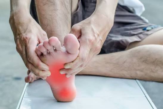
On examination, the tenderness is well-localised to the anteromedial aspect of the heel — the inner front corner of the heel pad, directly over the calcaneal origin of the plantar fascia. Pressing firmly on this spot with the thumb reproduces the pain accurately in plantar fasciitis; diffuse heel tenderness or central heel pad tenderness suggests fat pad atrophy rather than plantar fasciitis. The Windlass test — maximally dorsiflexing the toes while the ankle is in neutral, then palpating the fascia origin — increases fascial tension and may reproduce the pain.
- Pain is above the heel rather than on the undersurface — consider Achilles tendinopathy
- Pain is central under the heel pad rather than anteromedial — consider fat pad atrophy
- Pain does not ease with a few minutes of walking — consider calcaneal stress fracture
- There is burning, tingling, or numbness in the heel — consider tarsal tunnel syndrome
- Both heels are affected, especially in a younger adult with back stiffness — consider seronegative spondyloarthropathy (enthesitis)
What Causes Plantar Fasciitis?
Plantar fasciitis results from cumulative tensile overload at the fascial origin — when the plantar fascia is loaded more than it can repair between loading cycles. The risk factors reflect the mechanical and biological contributors to that load-repair imbalance:
Reduced ankle dorsiflexion from tight gastrocnemius and soleus muscles is the most consistently identified biomechanical risk factor. Tightness in the calf pulls the heel bone into plantarflexion, increasing the tensile load on the plantar fascia during walking and running.
BMI over 30 is the most important modifiable risk factor. Increased body weight directly multiplies the compressive and tensile forces transmitted through the plantar fascia with every step. Weight management is both a preventive and therapeutic measure.
Flat feet (pes planus) increase medial tensile load on the fascia. High-arched feet (pes cavus) reduce cushioning and increase impact loading. Both extremes of arch shape increase plantar fasciitis risk compared with a neutral foot posture.
Occupations requiring prolonged standing on hard surfaces — nursing, teaching, retail, factory work — sustain load on the fascia over many hours without adequate recovery periods. Plantar fasciitis is a well-recognised occupational hazard in these settings.
Rapid increases in running mileage, running on hard surfaces without adequate footwear, and running technique that increases heel strike force all raise the load on the plantar fascia beyond its repair capacity.
Flat, unsupportive footwear — flip-flops, worn-out trainers, and walking barefoot on hard floors — fails to cushion heel impact or support the arch. Both factors increase plantar fascia loading with each step.
Treatment: A Step-by-Step Approach
The large majority of plantar fasciitis cases — approximately 80–90% — resolve within 12 months with consistent conservative management. Treatment is most effective when the mechanical contributors (calf tightness, footwear, activity load) are addressed alongside symptomatic management.
Step 1: Stretching (Most Evidence-Based Conservative Treatment)
Stretching is the single best-evidenced conservative intervention for plantar fasciitis, outperforming heel cups, orthotics, and NSAIDs in controlled trials. The two most important stretching programmes are:
Plantar fascia-specific stretch (DiGiovanni 2003, Journal of Bone and Joint Surgery): sitting with the foot crossed over the knee, grasp the toes and pull them into maximal dorsiflexion (upward) before taking the first step in the morning, and repeat for 10 repetitions three times daily. This stretches the fascia at its most vulnerable moment — before the first load-bearing step — and has been shown in RCTs to produce significantly better outcomes than Achilles stretching alone at one year.
Gastrocnemius and soleus stretching: standing calf stretch with the affected leg behind, knee straight (gastrocnemius) and knee slightly bent (soleus), held for 30 seconds, three repetitions, twice daily. Restoring full ankle dorsiflexion removes the primary biomechanical driver of fascial overload in most patients.
These stretches should be performed consistently for at least 12 weeks. The most common reason for treatment failure is stopping the stretching programme when initial improvement occurs, before the fascial tissue has fully adapted.
Step 2: Footwear and Orthotic Modification
Replacing worn-out footwear is a simple, high-impact intervention. Running shoes should be replaced every 300–500 miles; after this point, the midsole cushioning is significantly degraded even if the shoe looks intact. Avoid walking barefoot on hard floors — keep supportive footwear on from the moment of first weight-bearing in the morning. Silicone heel cups (available without prescription) cushion heel impact and reduce the tensile load at the fascial origin. Custom orthotics are appropriate for patients with significant biomechanical issues (severe overpronation or high arch) but are not routinely required for straightforward plantar fasciitis.
Step 3: Night Splint
A night splint holds the ankle in a neutral or slightly dorsiflexed position during sleep, maintaining the plantar fascia at stretch throughout the night. This prevents the fascial shortening that occurs during sleep and is responsible for the severe first-step pain on waking. Randomised trials show night splinting reduces first-step pain significantly compared with stretching alone. The splint does require consistent use — most patients need several weeks of nightly wear to see sustained benefit. It can be uncomfortable initially, particularly if the patient is not used to maintaining dorsiflexion at rest.
Corticosteroid Injection
A single corticosteroid injection at the medial calcaneal origin of the plantar fascia is the most effective short-term pharmacological treatment, producing significant pain relief in most patients within one to two weeks. However, two specific risks warrant careful consideration:
- Plantar fascia rupture: repeated injections weaken the fascia; rupture is a recognised complication of multiple steroid injections. The general guidance is a maximum of two injections over the course of the condition.
- Fat pad atrophy: the heel fat pad is immediately deep to the injection site; repeated steroid delivery can cause atrophy of this cushioning structure, potentially replacing one painful problem (plantar fasciitis) with another (fat pad insufficiency).
Injection is appropriate as a bridge when pain is severe and preventing participation in rehabilitation, but stretching must continue alongside it. Ultrasound-guided injection improves accuracy and reduces the risk of fat pad delivery. See our guide to when joint pain needs medical evaluation for broader guidance on deciding between conservative and interventional management.
Shockwave Therapy (ESWT)
Extracorporeal shockwave therapy has Level 1 evidence for chronic plantar fasciitis (symptoms present for more than three months that have not responded to conservative measures). Multiple randomised controlled trials and systematic reviews confirm significant benefit compared with placebo, with success rates of 60–85% at three to six months. ESWT is now favoured over repeated corticosteroid injection for chronic plantar fasciitis by many specialist guidelines because it does not carry the fat pad atrophy or fascial rupture risks of injections and produces more durable results. Treatment is typically three to six weekly sessions.
Surgery
Surgery is very rarely needed — fewer than 5% of plantar fasciitis cases require it. When it is indicated, the most evidence-based procedure is gastrocnemius recession (for cases where tight calf is the primary driver and stretching has been insufficient to achieve adequate dorsiflexion), or endoscopic plantar fasciotomy (partial release of the fascial origin). Surgery is reserved for cases with at least 12 months of failed conservative management including ESWT. Partial fasciotomy carries a risk of longitudinal arch collapse if too much of the fascia is released, and outcomes are less predictable than for tendon release procedures elsewhere. Related patterns of foot and lower limb pain are covered in our article on injury pain versus chronic pain.
How Long Does Plantar Fasciitis Take to Heal?
Plantar fasciitis is a condition that requires patience. With consistent stretching, footwear modification, and load management, the majority of patients experience significant improvement within 3–6 months. The full course of recovery — to the point where the first-step pain is absent and sporting or occupational activities can be fully resumed — typically takes 12–18 months in established cases.
Factors that predict longer recovery include: obesity (particularly where weight reduction is not achieved), tight calf muscles that do not improve with stretching, occupations requiring prolonged standing that cannot be modified, bilateral involvement, and the presence of enthesopathy from an inflammatory arthritis condition. The 10–20% of patients who develop chronic plantar fasciitis (symptoms beyond 12 months despite conservative management) are the appropriate candidates for ESWT and, if that fails, surgical assessment.
Prevention of recurrence after recovery centres on maintaining the calf stretching programme indefinitely (not stopping when symptoms resolve), maintaining appropriate footwear, and avoiding sudden increases in activity that spike the load on the fascia. For related conditions at the Achilles tendon that can coexist with plantar fasciitis and share similar risk factors, see our guide to bursitis symptoms and causes, which covers retrocalcaneal bursitis at the heel.
Prevention: Keeping Plantar Fasciitis From Coming Back
Plantar fasciitis recurs in approximately 30% of patients who return to their pre-injury activity levels without making modifications. The most effective prevention strategies address the root causes rather than simply managing symptoms:
- Maintain calf stretching permanently: the gastrocnemius and soleus stretching programme should continue indefinitely after recovery — not stopped when symptoms resolve. Calf tightness is the single most consistent biomechanical predictor of plantar fasciitis recurrence.
- Replace footwear proactively: running shoes should be replaced every 300–500 miles; everyday shoes when midsole cushioning is visibly compressed. Worn-out footwear is one of the most common precipitants of a recurrence episode.
- Avoid sudden activity spikes: returning to running after a break, starting a new sport, or a dramatic increase in step count are common recurrence triggers. Increase activity by no more than 10% per week.
- Support foot posture at work: occupations requiring prolonged standing benefit from anti-fatigue mats, regular weight shifting, and footwear with adequate cushioning and arch support throughout the working day.
- Manage weight progressively: sustained weight management is the most impactful single modifiable factor for long-term prevention in overweight adults. Even modest sustained weight reduction maintains lower fascial load with every step.
Frequently Asked Questions
What is the fastest way to relieve plantar fasciitis pain?
The fastest immediate relief comes from the plantar fascia-specific stretch performed before the first steps of the morning — flexing the toes upward while seated, before weight-bearing, reduces the first-step pain within days for most patients. Adding a silicone heel cup and replacing worn footwear also reduces pain quickly. For more severe pain, a corticosteroid injection provides significant relief within one to two weeks, but should be accompanied by a stretching programme to address the underlying cause. Ice applied to the heel for 10–15 minutes after prolonged activity reduces post-activity discomfort.
Can I run with plantar fasciitis?
In most cases, yes — but with modifications. Complete rest from running is not routinely advised because it delays the return to full activity without improving the underlying fascial degeneration. The approach is to reduce mileage and intensity to a level that produces no more than mild discomfort during and for 24 hours after a run, then progressively increase as the fascia responds to treatment. Avoiding hard surfaces, ensuring adequate footwear cushioning, and shortening stride length to reduce heel strike force all reduce the fascial load per kilometre. Running through severe pain — or where pain increases progressively during a run — should prompt rest and reassessment.
Is plantar fasciitis the same as a heel spur?
No — they are related but distinct. A heel spur is a bony protrusion at the calcaneal origin of the plantar fascia, visible on X-ray, that forms as a result of chronic traction on the bone. Approximately 50% of people with plantar fasciitis have a heel spur, but 20% of people without heel pain also have one. The spur is not the source of pain — the degenerating fascial tissue at the calcaneal origin is. Treating the spur surgically, without addressing the fascial degeneration, does not reliably relieve plantar fasciitis pain. Heel spur removal is rarely performed because its removal alone is not beneficial.
Does weight loss help plantar fasciitis?
Yes — weight loss is one of the most effective interventions for plantar fasciitis in overweight and obese adults, and one of the most underused. Every step loads the plantar fascia at approximately 1.5 times body weight; during running, this multiplier increases. Reducing body weight directly and proportionally reduces the tensile load on the fascial origin with every step. Studies show that a 10% reduction in body weight produces a meaningful reduction in plantar fascia loading. In obese adults with plantar fasciitis, addressing body weight alongside stretching and footwear modification produces significantly better outcomes than stretching alone.
What is shockwave therapy and is it worth trying?
Extracorporeal shockwave therapy (ESWT) uses focused acoustic pressure waves to stimulate healing at the plantar fascia origin. It has Level 1 evidence for chronic plantar fasciitis — meaning the evidence is from multiple well-designed randomised controlled trials showing superiority to placebo. Success rates of 60–85% at three to six months are reported. It is most appropriate for chronic plantar fasciitis (symptoms for more than three months despite conservative management) and is increasingly preferred to repeated corticosteroid injection because it does not carry the fat pad atrophy or fascial rupture risks. ESWT is typically delivered as three to six weekly sessions and is available through physiotherapy and MSK specialist services.
Should I wear arch supports for plantar fasciitis?
Arch support can help, but the evidence is more nuanced than the orthotic industry often suggests. Off-the-shelf heel cups and cushioning insoles reduce heel impact and are a reasonable first-line adjunct for most patients. Custom orthotics are evidence-based for patients with significant biomechanical issues — severe overpronation, significant flat foot, or high arch — but are not required for everyone with plantar fasciitis. The most important footwear principle is adequate cushioning and arch support in everyday shoes, avoiding flat unsupportive footwear (flip-flops, worn trainers, bare feet on hard floors), and replacing running shoes before the midsole cushioning degrades.
When should I see a doctor about heel pain?
See a GP or physiotherapist if: heel pain has not improved after four weeks of consistent stretching and footwear modification; pain is severe enough to limit daily walking; the pattern does not fit plantar fasciitis (no improvement with a few minutes of walking, tingling or numbness, or central rather than anteromedial heel tenderness); or you have other symptoms suggesting an inflammatory condition (morning stiffness lasting more than one hour, back stiffness, eye inflammation, skin or bowel symptoms). Bilateral plantar fasciitis in a young adult should prompt assessment for seronegative spondyloarthropathy, which causes enthesitis at plantar fascia insertions as one of its features.
Summary
Plantar fasciitis is the most common cause of heel pain in adults — driven by cumulative tensile overload at the proximal attachment of the plantar fascia to the heel bone. The characteristic first-step pain that eases with walking is the reliable clinical hallmark. The most important modifiable risk factors are tight calf muscles, overweight, unsupportive footwear, and occupational prolonged standing — all of which are addressable. Treatment is most effective when centred on a consistent plantar fascia-specific stretching programme, combined with calf stretching, footwear modification, and progressive load management. Shockwave therapy is the intervention with the strongest evidence for chronic cases. Heel spurs visible on X-ray are incidental findings and not the cause of pain. For the majority of patients, full recovery is achievable within 12–18 months without surgery.
Medical disclaimer: This article is for general educational purposes and does not constitute medical advice. Consult a qualified healthcare professional for personalised assessment and management of plantar fasciitis or any foot condition.
References:
NHS. Plantar fasciitis. NHS. 2023.
NICE Clinical Knowledge Summary. Plantar fasciitis. NICE CKS. 2022.
DiGiovanni BF, Nawoczenski DA, Lintal ME, et al. Tissue-specific plantar fascia-stretching exercise enhances outcomes in patients with chronic heel pain. J Bone Joint Surg Am. 2003;85(7):1270–1277.
Goff JD, Crawford R. Diagnosis and treatment of plantar fasciitis. Am Fam Physician. 2011;84(6):676–682.
Versus Arthritis. Plantar fasciitis. versusarthritis.org. 2023.
NICE. Musculoskeletal conditions guidance. nice.org.uk.
I had plantar fasciitis for over a year and tried two corticosteroid injections that each gave me about six weeks of relief before the pain came back. My GP eventually referred me for shockwave therapy and the difference was significant — by the third session the first-step pain had reduced by about 70 percent and by the end of six sessions I was walking without limping. What I found helpful about this article is the explanation of why the injection wasn’t a lasting solution: the underlying fascial degeneration wasn’t being addressed, just the reactive inflammation around it. The stretching programme I should have been doing all along — the toe-flexing stretch before first steps — was never mentioned to me until a physiotherapist showed me after my second injection.
Sandra, your experience reflects a very common pattern: two injections providing temporary relief without addressing the underlying load fault, followed by referral for shockwave therapy that produced more durable improvement. The corticosteroid injection reduces the reactive tenosynovitis around the degenerated fascial tissue — which is why it relieves pain — but it does not reverse the collagen disorganisation at the calcaneal origin that is the structural basis of the condition. ESWT, in contrast, directly stimulates neovascularisation and collagen synthesis in the degenerated tissue, addressing the structural problem rather than the inflammatory response around it. The plantar fascia-specific stretch is the conservative intervention with the strongest evidence base — the DiGiovanni 2003 JBJS trial showing significantly better outcomes versus Achilles stretching alone — and the fact that it was not communicated to you earlier in your treatment course is a gap that better patient education could address. Daniel, your recovery trajectory — reducing mileage, softer surface, stretching protocol, footwear change, 14-week return to full training — is a model account of appropriate load management for running-related plantar fasciitis. The 10% weekly mileage increase rule you violated is precisely the load spike most consistently implicated in reactive plantar fasciitis in runners. The toe-flexion stretch before first steps works mechanistically by pre-tensioning the plantar fascia before the sudden eccentric load of first weight-bearing — mimicking the Windlass effect without the compressive impact — which reduces the load spike at the calcaneal origin that generates the acute post-static pain.
I’m a runner and had classic plantar fasciitis after increasing my weekly mileage from 25 to 45 miles over a single month. The first-step pain was exactly as described. I didn’t stop running but reduced to 15 miles per week on softer surfaces, started the calf stretching and plantar fascia-specific stretch twice daily, and switched to a shoe with more heel cushioning. It took about 14 weeks to get back to full training pain-free. The key message from my sports physio — which this article captures well — is that the stretch before the first steps of the day is the single most impactful intervention, particularly for runners, because it loads the fascia before the sudden tensile impact of first weight-bearing.