
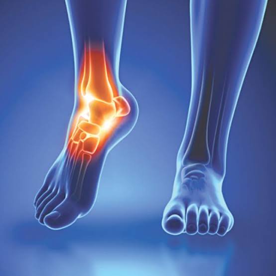
Foot and ankle pain what it may mean depends heavily on where the pain is located, how it started, and who is affected. The foot and ankle are the most mechanically stressed structures in the body — each step transmits forces of 1.5–2 times body weight through the foot, rising to 3–5 times during running. Despite this, foot pain is frequently dismissed or self-managed long after it should have been assessed. Some causes, like plantar fasciitis, resolve on their own with conservative management in most cases. Others — like a navicular stress fracture in a young athlete or a Charcot arthropathy developing silently in a person with diabetic neuropathy — require urgent recognition to prevent serious long-term harm. Understanding the difference starts with knowing what structures in the foot and ankle can hurt, and why.
Foot and Ankle Anatomy: Where the Pain Points
The foot contains 26 bones, 33 joints, and over 100 muscles, tendons, and ligaments — more per unit area than virtually any other region of the body. The most clinically important structures from a pain perspective are:
- Plantar fascia: a thick fibrous band running from the medial calcaneal tuberosity (base of the heel bone) to the proximal phalanges, supporting the medial arch. Its most vulnerable point — and the most common site of heel pain — is its calcaneal origin
- Achilles tendon: the largest tendon in the body, inserting into the posterior calcaneum; its watershed zone of poorest blood supply sits 2–6 cm above the insertion — the most common site of both tendinopathy and rupture
- Lateral ankle ligaments: the anterior talofibular ligament (ATFL) is the first and most commonly torn in lateral ankle sprains; the calcaneofibular (CFL) and posterior talofibular (PTFL) ligaments are torn in higher-grade injuries
- 1st metatarsophalangeal (MTP) joint: the big toe joint; site of gout (podagra), hallux rigidus (OA), and hallux valgus (bunion) — three very different conditions sharing one location
Pain location is the primary diagnostic guide: heel pain = plantar fasciitis or Achilles; lateral ankle = ligament sprain or peroneal tendons; forefoot = metatarsal stress fracture, Morton’s neuroma, or MTP joint pathology; medial arch = tarsal tunnel, tibialis posterior, or navicular; systemic burning/tingling in both feet = peripheral neuropathy.
Foot and Ankle Pain What It May Mean: Common Causes by Location
Foot and ankle pain is one of the most common reasons adults present to general practice. The distribution of causes varies significantly by age: plantar fasciitis dominates in adults aged 40–60, particularly those with obesity or a recent increase in activity. Ankle sprains are the most common cause of acute foot and ankle pain at any age, particularly in active individuals. Gout is most common in males over 40, with a strong association with dietary factors and diuretic use. Peripheral neuropathy becomes increasingly prevalent with longer duration of diabetes or other metabolic risk factors.
- Heel, worst first thing in the morning: plantar fasciitis (calcaneal origin); insertional Achilles tendinopathy
- Posterior heel and lower leg, with thickening: mid-portion Achilles tendinopathy
- Lateral ankle after twisting injury: ankle sprain; possible 5th metatarsal avulsion fracture
- Forefoot, “stone in shoe” feeling: Morton’s neuroma (3rd web space); metatarsal stress fracture
- Big toe joint, acute severe pain: gout (podagra); hallux rigidus (OA) if chronic and with restricted movement
- Both feet, burning/tingling/numbness: peripheral neuropathy; check for diabetes, B12 deficiency
Plantar Fasciitis
Plantar fasciitis is the most common cause of heel pain in adults, accounting for approximately 10% of all foot complaints in primary care. It is caused by repetitive tensile overload at the calcaneal origin of the plantar fascia — the point where the fascia attaches to the heel bone — leading to microtears and a chronic degenerative and inflammatory process. Despite the name, histopathological studies show that the tissue changes are more degenerative (fasciosis) than inflammatory in chronic cases; this does not change the management approach but explains why anti-inflammatory treatments alone are often insufficient.
The defining symptom pattern is pain in the heel that is worst with the first steps in the morning after getting out of bed, or after a period of rest (post-static dyskinesia). This is caused by the fascia contracting and shortening during sleep, then being stressed suddenly by the first weight-bearing steps. Pain typically improves after a few minutes of walking as the fascia warms up and lengthens, but may worsen again after prolonged standing. Tenderness is localized to the medial calcaneal tuberosity — pinpoint tenderness at this site is the key clinical finding. The Windlass test (passive dorsiflexion of the great toe while the patient stands) stresses the plantar fascia and reproduces pain at the calcaneal origin.
Risk factors include obesity (BMI ≥30 is the strongest risk factor), prolonged standing on hard surfaces, a sudden increase in physical activity (particularly running), flat feet or high arches (both can alter fascia loading mechanics), restricted ankle dorsiflexion from calf tightness, and middle age (peak incidence 40–60). Heel spurs — bony prominences visible on X-ray at the calcaneal origin — are a common finding in both symptomatic plantar fasciitis and asymptomatic heels; their presence does not determine the diagnosis or the prognosis.
Management follows a stepwise evidence-based approach. First-line: calf and plantar fascia stretching (the most consistent evidence; the towel/belt stretch — pulling the foot into dorsiflexion — and the stair-step calf stretch are both effective), supportive footwear with cushioned heel, and heel insoles. Night splints (maintaining the foot in dorsiflexion during sleep to prevent fascia shortening) are effective for nocturnal pain. Corticosteroid injection into the fascia origin provides short-term pain relief but carries a small risk of plantar fascia rupture with repeated injections. Extracorporeal shockwave therapy (ESWT) is supported by several randomized controlled trials for cases lasting over 6 months that have not responded to conservative measures. Surgery (plantar fasciotomy) is rarely needed — the natural history of plantar fasciitis is favorable, with approximately 80–90% of patients improving within 12 months with conservative management.
Achilles Tendinopathy and Rupture
Achilles Tendinopathy
Achilles tendinopathy describes pain, swelling, and functional limitation arising from degeneration of the Achilles tendon. The most common form — mid-portion tendinopathy — affects the watershed zone 2–6 cm above the calcaneal insertion, where blood supply is most limited and the tendon cannot heal as readily from overload injuries. Insertional tendinopathy, affecting the attachment itself, is a distinct entity with a different mechanical driver and a different management approach.
Symptoms of mid-portion Achilles tendinopathy include morning stiffness and posterior ankle pain that warms up during activity but worsens after exercise and the following morning. A fusiform (spindle-shaped) swelling of the tendon at the 2–6 cm zone is characteristically palpable. Risk factors include running (particularly with sudden increases in mileage), male sex, older age, and — importantly — fluoroquinolone antibiotics (ciprofloxacin and levofloxacin carry a black box warning for tendinopathy and tendon rupture, which can occur weeks after completing the antibiotic course). Any patient developing Achilles symptoms who has recently taken a fluoroquinolone should have this mentioned to their treating clinician.
The Alfredson heavy eccentric heel-drop protocol is the most extensively studied and evidence-supported physiotherapy intervention for mid-portion Achilles tendinopathy: 3 sets of 15 eccentric heel-drop repetitions (lowering the heel below the step with the affected leg, using the unaffected leg to return to the starting position), performed twice daily, seven days a week, for 12 weeks. The protocol produces good to excellent outcomes in 60–80% of patients. Extracorporeal shockwave therapy (ESWT) has supporting evidence for refractory mid-portion tendinopathy.
Achilles Tendon Rupture
Achilles tendon rupture typically presents with sudden severe pain in the posterior heel during a push-off or landing movement — often described as feeling like being kicked or struck from behind. An audible pop may be heard. The patient is usually a male aged 30–50 engaged in recreational sport (the “weekend warrior” profile). The Thompson test is the clinical diagnostic gold standard: with the patient prone and the knees flexed, squeezing the calf produces plantarflexion of the foot in an intact tendon; absence of this plantarflexion indicates a complete rupture.
Management is non-operative (functional weight-bearing in a progressive rehabilitation boot) or surgical repair. Multiple randomized controlled trials have shown equivalent re-rupture rates and functional outcomes between operative and non-operative management in appropriately selected patients. The decision depends on patient age and activity goals, surgeon experience, and patient preference. What is most important is avoiding prolonged non-weight-bearing immobilization — early functional rehabilitation (weight-bearing in equinus position from 2 weeks) produces better outcomes than traditional plaster casting for 8 weeks.
Ankle Sprain and Avulsion Fractures
Ankle sprains are the most common musculoskeletal injury in the world — approximately 23,000 ankle sprains occur per day in the United States alone. The vast majority (approximately 85%) are lateral ankle sprains caused by an inversion and plantarflexion mechanism, most commonly injuring the anterior talofibular ligament (ATFL). The assessment of any acute ankle injury must address the question: is this a ligament sprain or a fracture?
The Ottawa Ankle Rules are a validated, widely adopted clinical decision tool that specifies when X-ray is needed after an ankle injury. The rules require X-ray if there is: bone tenderness at the posterior edge or tip of either malleolus; bone tenderness at the base of the 5th metatarsal; bone tenderness at the navicular; or inability to weight bear both immediately after the injury and at the time of clinical assessment. These rules have near-100% sensitivity for clinically important fractures and can safely reduce unnecessary X-ray use. Any patient who cannot weight bear after an ankle injury should have X-rays.
An important missed injury in ankle inversion: the avulsion fracture of the 5th metatarsal base, where the peroneus brevis tendon pulls off a fragment of bone. This injury shares the exact same mechanism as a lateral ankle sprain. Without Ottawa-guided X-ray, it is easily dismissed as a sprain. Separately, the Jones fracture — at the junction of the proximal diaphysis and metaphysis of the 5th metatarsal (about 1.5 cm distal to the base) — is prone to nonunion and often requires surgical fixation, distinguishing it from the more proximal avulsion fracture which typically heals with conservative management.
For established ankle sprains, management has shifted away from prolonged immobilization: early weight-bearing (with a compression bandage and supportive boot), combined with a structured physiotherapy programme including proprioceptive exercises, produces better outcomes than immobilization. High-grade sprains (grade III, complete ATFL rupture) may develop chronic ankle instability in 20–30% of cases without adequate rehabilitation. Surgical ligament reconstruction (Broström procedure) is indicated for chronic instability that fails physiotherapy.
Foot Stress Fractures
Stress fractures of the foot occur when repetitive mechanical loading exceeds the bone’s ability to remodel — the result of either too much activity (overuse in a military recruit or distance runner) or insufficient bone strength (osteoporosis, vitamin D deficiency, the female athlete triad of low energy availability, menstrual dysfunction, and low bone density). The second metatarsal is the most common site — the so-called march fracture — followed by the third metatarsal. These present with progressive activity-related forefoot pain and point tenderness directly over the affected metatarsal shaft.
The navicular stress fracture is less common but more serious. The navicular (a tarsal bone on the medial foot) has poorer blood supply than the metatarsals and is prone to delayed healing and nonunion if not adequately managed. Navicular stress fractures present with vague medial midfoot pain during activity — they are frequently misdiagnosed as ankle sprains or generalized foot overuse. Plain X-ray is commonly normal for the first 2–3 weeks; MRI is the most sensitive investigation. The standard treatment for navicular stress fractures is strict non-weight-bearing cast immobilization for 6–8 weeks; elite athletes or displaced fractures are treated with surgical fixation.
Gout at the First MTP Joint (Podagra)
Gout at the first metatarsophalangeal joint — podagra — is one of the most dramatic and recognizable presentations in medicine. The classic description is exquisitely painful, red, hot, and swollen big toe joint that comes on suddenly, often overnight, reaching maximum intensity within hours. The pain is so severe that the weight of a bedsheet is reportedly unbearable — not merely an expression, but a clinical observation that reflects the extreme inflammatory state of an acute gout attack.
The first MTP joint is affected in approximately 50% of initial gout attacks. The ankle and midfoot are the next most commonly involved sites. Gout results from hyperuricaemia (elevated serum uric acid) leading to monosodium urate crystal deposition in joint spaces, with periodic acute inflammatory responses triggered by crystal phagocytosis by synovial neutrophils. Precipitating factors include dietary triggers (red meat, shellfish, alcohol — particularly beer), dehydration, sudden changes in serum uric acid level (from starting or stopping urate-lowering therapy), diuretic use, and renal impairment.
An important diagnostic point: serum uric acid is often paradoxically normal during an acute gout attack — the acute inflammation drives uric acid into the joint. The diagnosis is confirmed by joint aspiration and crystal analysis (needle-shaped, negatively birefringent monosodium urate crystals under polarized microscopy) or made clinically in a patient with the typical presentation and known hyperuricaemia. Acute attacks are managed with NSAIDs (most effective, first-line), colchicine (also first-line, particularly effective if started within 24 hours), or corticosteroids. Urate-lowering therapy (allopurinol, first-line) is started between attacks — not during an attack, as rapid changes in serum uric acid can precipitate further attacks.
Hallux Rigidus and Other Foot Osteoarthritis
Hallux rigidus is osteoarthritis of the first metatarsophalangeal joint — the most common site of symptomatic OA in the foot. The primary functional problem is loss of dorsiflexion at the 1st MTP joint. Normal gait requires approximately 65–70 degrees of 1st MTP dorsiflexion at toe-off; restriction forces the body to roll over the outer border of the foot, producing compensatory abnormal loading patterns and pain in the foot, ankle, and even the knee or hip.
Early hallux rigidus causes pain with extremes of 1st MTP movement, particularly dorsiflexion. Advanced disease causes pain throughout the range of movement and at rest. A dorsal osteophyte — a bony prominence on the dorsal (upper) aspect of the 1st MTP joint — is characteristic and may cause pain with shoe pressure. Midfoot OA (affecting the tarsometatarsal/Lisfranc joints) is another common presentation, often following a Lisfranc ligament injury or spontaneously in older adults.
Management of hallux rigidus: stiff-soled footwear (with a rocker-bottom modification) reduces the force through the 1st MTP joint during gait and is the most effective conservative measure. Corticosteroid injection for acute inflammatory flares. Cheilectomy — surgical removal of the dorsal osteophytes — restores some dorsiflexion in mild-to-moderate cases. First MTP fusion (arthrodesis) is the most reliable surgical option for end-stage hallux rigidus: it eliminates pain and is highly durable, though it permanently eliminates 1st MTP movement (which most patients tolerate well).
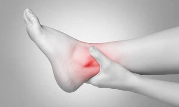
Other Foot and Ankle Pain Causes
Hallux Valgus (Bunion)
Hallux valgus is lateral deviation of the great toe at the first MTP joint with a corresponding medial prominence of the first metatarsal head — the “bunion.” It affects approximately 23% of adults aged 18–65 and up to 36% of adults over 65. The condition has a strong hereditary component; narrow, pointed footwear aggravates it but does not cause it. Symptoms include medial forefoot pain and tenderness over the bunion, difficulty fitting shoes, and secondary deformity of the second toe (hammer toe or overlapping toe). Management: wide-toed footwear, bunion pads, and orthotics can reduce symptoms but do not correct the deformity. Surgery — a metatarsal osteotomy combined with soft tissue balancing — is effective for symptomatic deformity that fails conservative management.
Morton’s Neuroma
Morton’s neuroma is a painful enlargement (perineural fibrosis, not a true neuroma) of the common plantar digital nerve, most commonly in the third web space between the third and fourth metatarsal heads. Symptoms are characteristic: a burning, tingling, or “electric shock” sensation in the forefoot and into the toes, a feeling like walking on a stone or a marble, and numbness between the affected toes. Tight, narrow footwear and high heels compress the metatarsal heads and aggravate symptoms. The Mulder’s click — a palpable or audible click reproduced by lateral compression of the metatarsal heads while compressing the neuroma from the plantar surface — is a useful clinical sign. Management: wide footwear, metatarsal pad (redistributes load away from the metatarsal heads), corticosteroid injection (50–70% success), surgical excision for refractory cases.
Peripheral Neuropathy and Diabetic Foot
Peripheral neuropathy — damage to the peripheral nerves, producing a length-dependent distal-to-proximal sensorimotor pattern — starts in the feet. The most common causes are diabetes (affecting ~50% of diabetic adults after 10 years of disease), excess alcohol, vitamin B12 deficiency, hypothyroidism, chronic kidney disease, and chemotherapy. Symptoms begin with tingling, burning, and numbness in the toes and balls of the feet, progressing proximally. Loss of protective sensation means that minor injuries — blisters, cuts, pressure sores — are not felt and progress to ulceration and infection.
Charcot arthropathy is a critical and frequently missed condition in patients with severe peripheral neuropathy, most commonly diabetic. It presents as an acutely warm, swollen foot or ankle — often without significant pain (because nociception is lost). The absence of pain in a swollen, hot foot is the warning sign: this should never be attributed to a simple sprain or cellulitis without excluding Charcot. Without urgent recognition and offloading (total contact casting or non-weight-bearing), the foot’s architecture collapses, producing the “rocker bottom” deformity that is extremely difficult to salvage and leads directly to chronic ulceration and amputation risk. Any warm, swollen foot in a patient with diabetic neuropathy is a Charcot arthropathy until proven otherwise.
Peroneal Tendinopathy
The peroneus longus and brevis tendons — the primary evertors of the foot — run posterior to the lateral malleolus, held in place by the superior peroneal retinaculum. Peroneal tendinopathy causes lateral ankle and retromalleolar pain, worsened by walking on uneven ground, running, and ankle dorsiflexion. It is commonly associated with chronic lateral ankle instability and is frequently underdiagnosed. Peroneus brevis split tears — a longitudinal split in the tendon at the level of the lateral malleolus — are a specific injury pattern seen in chronic ankle instability. Diagnosis is by ultrasound or MRI. Management: physiotherapy, support, corticosteroid injection; surgery for split tears that fail conservative management.
Foot and Ankle Pain Red Flags: When to Seek Urgent Care
- Warm, swollen foot or ankle in a diabetic patient with little or no pain — possible Charcot arthropathy; requires urgent assessment and offloading to prevent foot collapse and amputation
- Severe foot pain after ankle fracture with disproportionate swelling, tense shiny skin, and increasing pain with passive toe extension — possible compartment syndrome; surgical emergency
- Inability to weight bear after injury — X-ray required per Ottawa Rules to exclude fracture; do not assume it is a sprain
- Midfoot pain or lateral forefoot pain after a fall or twisting injury — possible navicular, 5th metatarsal avulsion, or Jones fracture; do not dismiss as sprain without X-ray
- Progressive numbness and tingling in both feet — peripheral neuropathy; check for diabetes (HbA1c), B12 deficiency
- Foot drop (inability to lift the front of the foot) — common peroneal nerve injury or L4-L5 radiculopathy; urgent neurological assessment
- Activity-related midfoot pain in a young athlete or military recruit — possible navicular stress fracture; requires MRI, not just rest
Self-Care Basics for Foot and Ankle Pain
Footwear is the single most important modifiable factor for most foot and ankle conditions. Rocker-bottom soles reduce forefoot loading in hallux rigidus and metatarsalgia. Wide-toed shoes reduce compression in Morton’s neuroma and hallux valgus. Cushioned heel insoles and arch supports reduce plantar fascia loading. A stiff sole with a slight heel raise is appropriate for Achilles tendinopathy. These changes cost relatively little and often provide sustained relief without any further intervention.
Stretching for plantar fasciitis and Achilles tendinopathy. The most evidence-supported home treatment for plantar fasciitis is the plantar fascia stretch (pulling the toes back to tension the fascia) and the calf stretch (stair-edge calf raises targeting both gastrocnemius and soleus). For mid-portion Achilles tendinopathy, the Alfredson eccentric heel-drop protocol performed consistently twice daily for 12 weeks is the most effective conservative intervention.
Weight management. Obesity (BMI ≥30) is the strongest independent risk factor for plantar fasciitis and is associated with worsened outcomes in ankle OA and foot stress fractures. Even modest weight reduction reduces heel loading during walking and is one of the most effective preventive measures for plantar fasciitis recurrence.
Related Articles on Horizon Health Guide
- Knee Pain: What Adults Should Know
- Hip Pain: Common Causes
- Joint Pain: Common Causes and When to Seek Care
- Bone Pain: What Adults Should Know
- Mobility and Healthy Aging: What Adults Should Know
Frequently Asked Questions
What are the most common foot and ankle pain causes?
Plantar fasciitis is the most common cause of heel pain in adults, affecting approximately 10% of people at some point in their life. Ankle sprains — particularly lateral ankle sprains (ATFL injury) — are the most common cause of acute foot and ankle pain at any age. In the forefoot, Morton’s neuroma, metatarsal stress fractures, and 1st MTP OA (hallux rigidus) are common in adults of working age. Gout (podagra) is the most common cause of sudden-onset 1st MTP joint pain in adults, particularly men over 40. In older adults, foot OA and peripheral neuropathy become increasingly prevalent.
What does plantar fasciitis feel like?
Plantar fasciitis produces a characteristic heel pain that is worst with the first steps in the morning or after sitting for a period — this “post-static dyskinesia” is the hallmark feature. The pain is typically located at the medial heel (the base of the heel bone, just inside the arch) and feels like a sharp or burning pain. It improves after a few minutes of walking but may worsen after prolonged standing. Tenderness on pressing the medial calcaneal tuberosity (the inside base of the heel) reproduces the pain. Around 80–90% of patients improve within 12 months with appropriate conservative management including stretching and footwear modification.
How can I tell if my ankle sprain is serious or might be a fracture?
The Ottawa Ankle Rules provide clinical guidance on when X-ray is needed. An ankle injury requires X-ray if there is bony tenderness at the tip or posterior edge of either malleolus, or at the base of the 5th metatarsal, or at the navicular bone — or if the patient cannot bear weight on the injured foot both immediately after the injury and at the time of assessment. If you cannot walk on the foot, do not assume it is “just a sprain” — get an X-ray. A common missed injury in lateral ankle sprains is an avulsion fracture at the base of the 5th metatarsal, which requires the same type of X-ray to detect.
Why is gout in the big toe so painful?
Gout in the first MTP joint — podagra — is one of the most painful conditions in medicine. The pain is caused by monosodium urate crystals depositing in the joint space and triggering a massive inflammatory response: neutrophils (white blood cells) engulf the crystals and release inflammatory mediators, causing rapid swelling, intense heat, redness, and exquisite tenderness. The 1st MTP joint is a high-pressure joint that moves with every step, and even minor stimuli — the weight of a sheet, vibration from footsteps — can set off severe pain during an acute attack. The attack typically resolves within a week even without treatment, but recurrence is likely without urate-lowering therapy (allopurinol).
What is Charcot arthropathy and why is it dangerous?
Charcot arthropathy (Charcot neuroarthropathy) is a destructive condition of the foot and ankle that develops in patients with severe peripheral neuropathy — most commonly diabetic neuropathy. The underlying mechanism involves loss of normal protective pain sensation: repetitive micro-trauma that would normally be painful is not perceived, and the joint and bone architecture is progressively destroyed from within. The danger is in its presentation: the foot is warm, swollen, and red — but relatively painless, because the patient cannot feel the damage occurring. This makes it easy to misdiagnose as cellulitis (a skin infection) or a simple sprain. If the patient continues weight-bearing without offloading, the foot collapses, producing the rocker-bottom deformity that is extremely difficult to manage and carries a high risk of ulceration, infection, and amputation. Any warm swollen foot in a diabetic patient with neuropathy should be evaluated immediately.
What is the Alfredson protocol for Achilles tendinopathy?
The Alfredson eccentric heel-drop protocol is the most evidence-supported physiotherapy treatment for mid-portion Achilles tendinopathy. The exercise involves standing with the ball of the foot on the edge of a step and the heel hanging off. The patient rises onto the ball of both feet (concentric phase), then slowly lowers the heel of the affected foot below the level of the step (eccentric phase), using the unaffected leg to return to the starting position. The programme is performed as 3 sets of 15 repetitions, twice daily, seven days per week, for a minimum of 12 weeks. The exercises are performed through pain — mild to moderate pain during the exercise is acceptable and expected. Results typically begin to improve after 6–8 weeks of consistent adherence. This protocol is effective for mid-portion tendinopathy; insertional tendinopathy requires a modified protocol without the drop below horizontal, as this compresses the tendon against the calcaneum and worsens symptoms.
Can foot pain be caused by a problem in the spine?
Yes. L4-L5 nerve root compression (most commonly from a lumbar disc herniation) can cause pain, tingling, and weakness in the dorsum of the foot — a pattern that can mimic peroneal tendinopathy or ankle sprain. L5 root involvement also produces foot drop (weakness of dorsiflexion and toe extension) — a sign that requires urgent investigation if new. S1 root compression refers pain to the lateral foot and heel and produces weakness of plantarflexion. Distinguishing features: lumbar movements (forward bending, extension) reproduce or worsen the foot symptoms; sensory and motor changes follow a dermatomal pattern rather than a peripheral nerve distribution; and passive foot and ankle examination is normal. For more on spinal causes, see our article on hip pain common causes, which also discusses lumbar referral patterns.
References
- Garrow AP, et al. The prevalence of foot pain and its associated factors in a United Kingdom population. Ann Rheum Dis. 2004;63(5):501-506.
- Alfredson H, et al. Heavy-load eccentric calf muscle training for the treatment of chronic Achilles tendinosis. Am J Sports Med. 1998;26(3):360-366.
- Stiell IG, et al. Decision rules for the use of radiography in acute ankle injuries. JAMA. 1993;269(9):1127-1132.
- Waterman BR, et al. The epidemiology of ankle sprains in the United States. J Bone Joint Surg Am. 2010;92(13):2279-2284.
- Rogers LC, et al. The Charcot foot in diabetes. Diabetes Care. 2011;34(9):2123-2129.
This article is for educational purposes only and does not constitute medical advice. Always consult a qualified healthcare professional for evaluation and treatment of foot and ankle pain.
I’ve had heel pain for six months and was convinced I needed a steroid injection or surgery. After reading the section on plantar fasciitis I realised I haven’t actually done the stretching consistently — just a few times here and there. Going to commit to the proper protocol for 12 weeks before I do anything else. Really helpful to know that most cases do resolve without surgery.
The warning about Charcot arthropathy is really important — my father has Type 2 diabetes and has mentioned his feet feeling ‘different’ recently. I had no idea a warm swollen foot without pain was actually an emergency. We’re booking an appointment tomorrow. Thank you for flagging this so clearly.
Tom, you’re absolutely right to book that appointment — it’s exactly the kind of thing that should not be left. The combination of diabetes, neuropathy, and new foot swelling warrants assessment same day or the next day, not in a few weeks. Ask the GP specifically to assess for Charcot arthropathy and request an X-ray if they haven’t already ordered one. Hope your father gets seen quickly and that it turns out to be something straightforward.