
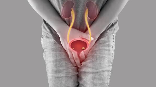
Incomplete Bladder Emptying: Causes, Consequences, and Management
Incomplete bladder emptying — the retention of a clinically significant volume of urine in the bladder after voiding — is one of the most common urological problems in adults and one of the least discussed. Unlike overactive bladder or stress incontinence, which produce symptoms that patients immediately connect to bladder dysfunction (urgency, leakage), incomplete emptying is often experienced indirectly: a persistent sensation that the bladder was not fully emptied after voiding, a need to return to the toilet within minutes of leaving it, recurrent urinary tract infections without an obvious explanation, or eventually the dull discomfort of chronic urinary retention. The medically measured correlate of this experience is the post-void residual (PVR) — the volume of urine remaining in the bladder immediately after voiding, measured by bladder ultrasound or catheterization. A PVR consistently above 100 mL is considered clinically significant; above 300 mL is associated with increased risk of upper urinary tract consequences including hydronephrosis and renal impairment; and acute retention — the complete inability to void — is a urological emergency requiring immediate catheterization. Understanding incomplete bladder emptying — its causes, its complications, the significance of different PVR levels, and its management options — is essential for any patient experiencing lower urinary tract symptoms (LUTS) and for clinicians evaluating voiding dysfunction in both men and women.
The bladder empties through a coordinated neuromuscular event: the detrusor (bladder wall muscle) contracts while the urethral sphincter and pelvic floor simultaneously relax, allowing urine to flow along a pressure gradient from the bladder through the urethra. Complete emptying requires adequate detrusor contractile force sustained throughout the voiding phase, unobstructed outflow through the urethra, and proper sphincter coordination that does not prematurely terminate the voiding reflex. When any of these elements fail — inadequate detrusor contraction, elevated urethral resistance from obstruction, or unsynchronized sphincter activity — the detrusor may fatigue or the voiding reflex may terminate before emptying is complete, leaving a residual volume. The mechanism that produces incomplete emptying determines both the appropriate evaluation and the most effective management, which is why accurate diagnosis — not just symptom management — is the foundation of effective care.

Common Causes of Incomplete Bladder Emptying
Bladder outlet obstruction (BOO) is the most common cause of incomplete emptying in men and refers to any condition that increases resistance to urine flow through the urethra, requiring the detrusor to generate higher voiding pressures to sustain flow. The most prevalent cause of BOO in men over 50 is benign prostatic hyperplasia (BPH), in which the enlarged prostate compresses the prostatic urethra and increases outlet resistance. With mild BPH, the detrusor compensates by generating higher pressures, and emptying may be reasonably complete; as obstruction worsens, the detrusor eventually fails to sustain the elevated pressure throughout the void, and a progressively larger residual remains. The relationship between BPH and the full spectrum of associated LUTS — including incomplete emptying, hesitancy, weak stream, and urgency — is detailed in the enlarged prostate and urination problems guide. Urethral stricture — narrowing of the urethral lumen by scar tissue from prior instrumentation, trauma, or infection — produces BOO through the same mechanism of elevated resistance; stricture is the most common cause of BOO in younger men without an enlarged prostate and should be specifically investigated in men under 45 with voiding dysfunction. Bladder neck obstruction — functional or anatomical narrowing at the bladder neck distinct from the prostate — causes incomplete emptying in young men with small prostates and normal urethral caliber, and is treated with alpha-blockade or transurethral incision of the bladder neck.
Detrusor underactivity (DU) — inadequate detrusor contractile force — is the other major mechanism of incomplete emptying, producing elevated PVR through insufficient voiding pressure rather than excessive outlet resistance. DU is underdiagnosed and often confused with obstruction because both produce similar symptoms (slow stream, hesitancy, incomplete emptying) and similar uroflowmetry results (low Qmax). The distinction matters clinically: surgical relief of obstruction improves emptying dramatically in BOO, but does not improve and may worsen emptying in DU. The causes of DU include diabetic cystopathy (autonomic neuropathy impairing the parasympathetic innervation of the detrusor), spinal cord injury or disease affecting the sacral voiding nucleus (S2-S4), aging-related detrusor myopathy (reduced detrusor contractility with age that is distinct from pathological DU), and medication effects from anticholinergics, opioids, and other agents that reduce detrusor contractility. Urodynamics — specifically pressure-flow studies measuring detrusor pressure at maximum flow rate — is the only way to definitively distinguish obstruction from DU when both are clinically plausible: obstruction produces high detrusor pressure with low flow, while DU produces low detrusor pressure with low flow. Neurogenic bladder dysfunction affects voiding through multiple mechanisms depending on the level and completeness of the neurological lesion. Upper motor neuron lesions (spinal cord injury above the sacral cord, MS plaques in the cervical or thoracic cord, spinal stenosis) characteristically produce detrusor-sphincter dyssynergia (DSD) — uncoordinated simultaneous contraction of the detrusor and sphincter that prevents sustained voiding and produces high post-void residuals and risk of high intravesical pressures. Lower motor neuron lesions (cauda equina injury, radical pelvic surgery damaging the pelvic nerves) produce a flaccid, poorly contracting detrusor with large PVRs and overflow incontinence. The NIDDK bladder control resource provides accessible evidence-based information on the full range of bladder dysfunction mechanisms.
Consequences of Chronically Elevated Post-Void Residual
A persistently elevated PVR is not merely a urodynamic finding — it carries concrete clinical consequences that progress if the underlying cause is not addressed. Recurrent urinary tract infections (UTIs) are the most common consequence of significant incomplete emptying. Stagnant urine remaining in the bladder after each void provides an ideal growth medium for bacteria, and the normal mechanical flushing effect of complete voiding — which clears bacteria from the bladder with each void — is absent. Women with elevated PVR from any cause frequently present with recurrent UTIs as their first symptom, sometimes before any voiding symptoms are noticed. In older men with BPH and incomplete emptying, recurrent UTIs are a well-recognized complication that often prompts the urological referral that leads to diagnosis. The management of recurrent UTIs in the setting of incomplete emptying requires addressing the underlying PVR rather than simply treating repeated infections with antibiotics, which provides only short-term bacterial clearance while the structural cause of the recurrence remains. Bladder stones develop from the crystallization of urinary solutes in stagnant urine, with calcium, oxalate, and urate being the most common components. Bladder stones cause dysuria, hematuria, and intermittency of stream (positional blockage of the bladder outlet), and require cystoscopic removal; they will recur if the incomplete emptying that created the stagnant urine environment is not corrected. Detrusor damage from chronic overdistension occurs when the bladder is chronically filled to above its normal capacity from retained residual urine. The detrusor muscle, stretched beyond its optimal working length by the combination of the incoming urine plus the large residual, loses its contractile efficiency — Laplace’s law: wall tension increases with the square of the radius — and over time the chronically overdistended detrusor develops fibrosis and irreversible loss of contractility. Recognizing and treating incomplete emptying before chronic overdistension has produced irreversible detrusor damage is critical for preserving the voiding function that treatment aims to restore. Upper urinary tract consequences — hydronephrosis and renal impairment — can develop when elevated bladder pressures from severe retention are transmitted retrograde to the ureters and kidney. High-pressure chronic retention (typically PVR above 500 mL with elevated resting bladder pressures) can cause bilateral hydronephrosis, ureteral dilatation, and ultimately obstructive nephropathy with renal function impairment. The AUA BPH guidelines classify upper tract compromise from retention as an absolute indication for surgical intervention in appropriate candidates, and the StatPearls BPH review provides the evidence base for management decisions in obstructive uropathy.
Evaluation: Measuring Post-Void Residual and Diagnosing the Cause
The clinical evaluation of incomplete bladder emptying begins with establishing the PVR — an objective measurement of the retained volume — and then identifying the mechanism responsible. Bladder ultrasound (bladder scan) is the standard non-invasive method for PVR measurement. A portable ultrasound device used immediately after voiding gives a PVR estimate that, while slightly less precise than catheterization, is adequate for clinical decision-making and requires no urethral catheterization. A single elevated PVR should be confirmed with a repeat measurement, since PVR varies with voided volume, fluid intake, and the completeness of voluntary effort at voiding; a consistently elevated PVR on two or more measurements is more clinically meaningful than a single high value. PVR classification guides management intensity: PVR under 100 mL is generally considered not clinically significant; 100–300 mL is significant and warrants evaluation for cause and monitoring; above 300 mL is high and requires active management; above 500 mL is severe and typically requires catheterization to relieve retention. Uroflowmetry measures the flow rate and pattern during a natural void. A reduced maximum flow rate (Qmax below 10 mL/second) combined with an elevated PVR is strong evidence of significant voiding dysfunction requiring further evaluation. The flow curve shape provides additional information: a plateau-shaped (obstructive) curve suggests BOO, while an intermittent staccato pattern may reflect straining, DSD, or detrusor underactivity with abdominal straining. Urodynamic studies (cystometry with pressure-flow studies and optional EMG of the sphincter) are indicated when the mechanism of incomplete emptying is unclear after initial evaluation, when neurogenic bladder is suspected, or when surgical intervention is planned. Pressure-flow studies provide the Bladder Outlet Obstruction Index (BOOI = PdetQmax − 2×Qmax), which quantifies the degree of obstruction on a validated scale: BOOI above 40 is obstructed, 20–40 is equivocal, below 20 is unobstructed. This distinction is essential before any surgical intervention, since deobstructive surgery in a patient with DU rather than BOO will not improve PVR and may worsen voiding function.
Management Options: From Behavioral Strategies to Surgical Intervention
The management of incomplete bladder emptying is matched to the mechanism and severity identified in the evaluation. Behavioral strategies can meaningfully improve emptying in patients with mild-to-moderate PVR and without severe obstruction or neurogenic dysfunction. Double voiding — remaining on the toilet for 3 to 5 minutes after the initial void and attempting a second void — allows residual urine that did not drain completely during the first void to shift to a gravitationally favorable position and drain on the second attempt, reducing PVR by 30–50% in many patients. Seated voiding for men with BPH reduces pelvic floor tension, optimizes the anatomical angle of the prostatic urethra, and consistently produces better emptying than standing voiding in studies of men with LUTS. Leaning slightly forward during voiding reduces the effective resistance of the bladder outlet by improving urethral alignment. Timed voiding — voiding on a regular schedule before the bladder becomes overdistended (every 2 to 3 hours) — prevents the detrusor from reaching the overdistended, poorly contracting state that produces the largest PVRs. For patients experiencing hesitancy alongside incomplete emptying, the difficulty starting urination guide covers the void initiation strategies that complement double voiding in patients with both hesitancy and incomplete emptying. Alpha-adrenergic blockers (tamsulosin, silodosin, doxazosin, terazosin, alfuzosin) are the first-line pharmacological treatment for BOO from BPH. By relaxing smooth muscle in the bladder neck and prostatic urethra, alpha-blockers reduce outlet resistance and improve both Qmax and PVR, typically within 2 to 4 weeks of initiation. Alpha-blockers are effective in the majority of men with symptomatic BPH, well-tolerated, and do not reduce prostate size; they are particularly effective for the storage symptoms and less effective for reducing PVR when the obstruction is severe or the detrusor has been significantly damaged by chronic retention. 5-alpha-reductase inhibitors (finasteride, dutasteride) reduce prostate volume by blocking dihydrotestosterone-mediated prostate growth, producing a 20–30% reduction in prostate volume and corresponding improvements in Qmax and PVR over 6 to 12 months of treatment. 5-ARIs are most effective in men with large prostates (above 40 mL by ultrasound), reduce the risk of acute urinary retention and the need for surgery, and have additive effects when combined with alpha-blockers in men with larger prostates and more severe symptoms. Clean intermittent self-catheterization (CISC) is the management cornerstone for patients with high PVR from detrusor underactivity, neurogenic bladder, or severe obstruction where immediate surgery is not appropriate. CISC — passing a small catheter into the bladder at regular intervals (typically 3 to 4 times daily) to empty the residual urine — reliably keeps PVR below clinically significant levels, prevents bladder overdistension and its consequences, dramatically reduces recurrent UTI risk, and preserves upper tract function in high-risk patients. Patients with neurogenic bladder and high PVR from DSD or flaccid bladder are trained in CISC as a lifelong bladder management technique rather than awaiting spontaneous improvement. Surgical management of BOO — transurethral resection of the prostate (TURP), laser enucleation (HoLEP), or open prostatectomy for very large glands — effectively reduces outlet resistance and dramatically improves PVR and Qmax in appropriately selected men with obstruction-dominant voiding dysfunction. The AUA guidelines specify the indications for surgical intervention in BPH: absolute indications include refractory urinary retention, recurrent UTIs from incomplete emptying, bladder stones or diverticulae from retention, and upper tract hydronephrosis; relative indications include significant LUTS not responding adequately to medical management. For women, surgical treatment of incomplete emptying addresses the specific cause identified: urethral dilation for stricture, surgical repair of pelvic organ prolapse if the prolapsed structure is compressing the urethra, or deobstruction of bladder neck obstruction. The prostate health and urinary symptoms guide covers the full prostate evaluation and management pathway for men with BPH-related voiding dysfunction including incomplete emptying.
Incomplete Emptying in Women and Older Adults
Incomplete bladder emptying is commonly associated with male BPH, but it is a clinically important problem in women and older adults that is frequently underdiagnosed. In women, the leading causes of elevated PVR include pelvic organ prolapse (cystocele, uterine prolapse, or rectocele) mechanically kinking the urethra and preventing complete drainage; detrusor underactivity from diabetic neuropathy, aging-related detrusor myopathy, or prior pelvic surgery damaging the parasympathetic innervation; urethral stricture or stenosis from prior catheterization or pelvic radiotherapy; and neurogenic bladder from multiple sclerosis, Parkinson’s disease, or spinal cord disease. Women with recurrent UTIs and no obvious anatomical reason for infection should have PVR measured as part of the evaluation — incomplete emptying as the driving mechanism is found in a significant proportion. In older adults of both sexes, detrusor underactivity becomes an increasingly prevalent cause of incomplete emptying alongside or instead of outlet obstruction; the combination of reduced detrusor contractility with aging and elevated outlet resistance from BPH in older men produces a particularly complex voiding dysfunction in which both mechanisms contribute to the elevated PVR. The weak urine stream guide addresses the flow rate component of voiding dysfunction that frequently accompanies incomplete emptying in both sexes, and the when bladder symptoms need evaluation guide provides the broader framework for deciding when urological referral is warranted for any lower urinary tract symptom including incomplete emptying. Any patient who consistently feels that their bladder has not fully emptied after voiding, who is experiencing recurrent UTIs without explanation, or who has been found to have an elevated PVR on any testing should discuss the finding and its implications with their clinician and understand that effective treatments exist for the full range of causes.
Sources: NIDDK — Bladder Control · AUA BPH Guidelines · StatPearls — BPH
Medications That Impair Bladder Emptying
Pharmacological impairment of bladder emptying is an underappreciated cause of elevated PVR that is reversible once identified. A thorough medication review is essential in any patient presenting with incomplete emptying, particularly when the onset is recent or temporally associated with starting a new drug. Anticholinergic medications are the most clinically important drug class for worsening incomplete emptying, because their mechanism — blocking muscarinic M3 receptors on the detrusor — directly reduces the contractile force that drives bladder emptying. Medications with anticholinergic activity include first-generation antihistamines (diphenhydramine, chlorpheniramine, available in OTC allergy and sleep preparations), tricyclic antidepressants (amitriptyline, imipramine, nortriptyline), antipsychotics (haloperidol, quetiapine, olanzapine, clozapine), antimuscarinics used for overactive bladder (oxybutynin, tolterodine, solifenacin — particularly relevant when prescribed to a patient who has both OAB and incomplete emptying), antiemetics (scopolamine, promethazine), and antispasmodics (dicyclomine, hyoscine). In patients with BPH or neurogenic bladder who already have marginal emptying, even modest anticholinergic exposure from an OTC sleep aid or first-generation antihistamine can significantly elevate PVR and precipitate acute urinary retention. Opioid analgesics impair bladder emptying through both central suppression of the pontine micturition center and peripheral opioid receptor-mediated reduction of detrusor contractility and bladder sensation. Opioid-induced urinary retention is particularly common with spinal and epidural opioids (used in perioperative analgesia) and with high-dose parenteral opioids, but is also clinically relevant with regular oral opioid therapy in patients with pre-existing voiding dysfunction. Dose reduction or opioid rotation may restore acceptable voiding function; CISC is used when PVR remains elevated despite opioid management adjustments. Alpha-adrenergic agonists — pseudoephedrine (in decongestant preparations), phenylephrine, and ephedrine — increase smooth muscle tone at the bladder neck and prostatic urethra, raising outlet resistance and elevating PVR in men with BPH or women with bladder neck dysfunction. Men with BPH should be specifically counseled to avoid pseudoephedrine-containing cold and sinus preparations, as these can precipitate acute urinary retention. Calcium channel blockers, used for hypertension and angina, may impair detrusor contractility through reduced smooth muscle calcium influx, contributing to PVR elevation in susceptible patients — a medication interaction that is worth considering when no other cause of new-onset incomplete emptying is identified. Recognizing medication-related incomplete emptying allows clinicians to recommend drug substitutions or discontinuation that resolve the voiding dysfunction entirely without urological intervention.
Monitoring and Long-Term Follow-Up
Incomplete bladder emptying requires ongoing monitoring rather than one-time evaluation, since PVR can change over time as the underlying cause progresses or responds to treatment, and since the consequences of inadequately managed retention accumulate gradually. For patients on watchful waiting or medical management for BPH-related incomplete emptying, annual PVR measurement alongside symptom reassessment (IPSS score) provides the longitudinal data needed to detect progression and guide the timing of escalated intervention. Men whose PVR is increasing despite alpha-blocker therapy — crossing from the 100–300 mL range toward 300 mL and above — warrant urology referral for consideration of adding a 5-ARI, switching to a phosphodiesterase-5 inhibitor, or proceeding to surgical evaluation. For patients on CISC, periodic measurement of the catheterized volumes at each self-catheterization and regular review of voiding diary data tracks whether spontaneous voiding is recovering (as sometimes occurs in incomplete retention from reversible causes) or whether CISC remains necessary. Monitoring renal function (serum creatinine and eGFR) annually is appropriate in patients with high PVR (above 300 mL) or recurrent hydronephrosis on imaging, since the upper tract consequences of chronic retention can be silent until significant renal impairment has developed. Patients who achieve successful reduction of PVR through treatment — whether behavioral, pharmacological, or surgical — should have the PVR confirmed at 3 and 12 months after treatment to establish the new baseline and confirm durability of the improvement. Regular monitoring converts incomplete emptying from a passively managed discomfort into an actively tracked condition with defined treatment endpoints and objective success criteria, which is the approach associated with the best long-term outcomes and prevention of the most serious consequences of chronic retention.
I’m a 67-year-old woman who has had three UTIs in the past eight months, which led to a bladder scan that showed a PVR of 185 mL consistently across two measurements. My GP had initially attributed the recurrent UTIs to postmenopausal changes and kept giving me antibiotics, which would clear the infection but they’d return within six to eight weeks. It was only when I pushed for further investigation that the incomplete emptying was identified. The article’s explanation of stagnant urine providing a growth medium for bacteria — and the point that you have to address the PVR rather than just keep treating infections — is exactly the situation I found myself in. I’ve since been referred to a urogynaecologist who identified a cystocele compressing my urethra on Valsalva. Understanding the mechanism has made the management path much clearer.
This is a well-structured guide that correctly emphasizes the critical distinction between obstruction and detrusor underactivity as the two major mechanisms — a distinction that is clinically essential before any surgical intervention. The Bladder Outlet Obstruction Index threshold of 40 for confirmed obstruction is correct per the International Continence Society definition. The section on medication-related incomplete emptying deserves emphasis: anticholinergic burden in elderly patients (who may be taking multiple agents with anticholinergic activity from different drug classes simultaneously) frequently contributes to PVR elevation in ways that completely reverse with appropriate deprescribing. Measuring the total anticholinergic burden across a patient’s complete medication list — not just looking for drugs prescribed ‘for the bladder’ — should be standard practice in the LUTS evaluation of any patient over 65. The chronic overdistension consequences section correctly highlights that the window for preserving detrusor contractility has limits, and that prolonged high-pressure retention produces fibrotic changes that are not fully reversible even after decompression.
Dr. Mehta, the total anticholinergic burden approach you describe is exactly the right framing — calculating the cumulative anticholinergic load across all medications (using validated scales like the Anticholinergic Cognitive Burden scale) rather than checking each drug in isolation frequently reveals that a patient is effectively on a high anticholinergic dose through the combination of multiple moderate-burden agents, none of which individually appears problematic. Patricia, your situation illustrates a very common and preventable diagnostic delay — recurrent UTI without an identified structural cause is an indication for bladder ultrasound and PVR measurement, and the mechanical cause (cystocele-related urethral compression in your case) being identified and treated will likely end the UTI recurrence cycle more effectively than any antibiotic regimen. A cystocele correction addressing the outlet compression should normalize your PVR and allow the normal flushing mechanism of complete voiding to protect against infection.