Liver cancer screening — more precisely called hepatocellular carcinoma surveillance — is one of the clearest examples in oncology of where screening genuinely saves lives. The reason it works is the same reason it is not recommended for the general public: liver cancer risk is not spread evenly across the population. It is highly concentrated in identifiable groups — people with cirrhosis, people with chronic hepatitis B, and a handful of specific hereditary conditions — and this concentration makes targeted surveillance far more efficient and cost-effective than any population-wide approach.
The distinction between detected-early and detected-late matters enormously for outcomes. Hepatocellular carcinoma detected through surveillance, before symptoms develop, is typically early-stage disease. Early-stage HCC is potentially curable — with liver transplantation, surgical resection, or ablation achieving five-year survival rates of 50 to 80 percent in selected patients. Liver cancer detected because of symptoms has almost always already reached a stage where those curative options are no longer available, and survival is measured in months rather than years. The technology that drives this difference is remarkably simple: an abdominal ultrasound and a blood test, performed twice a year.
Why Liver Cancer Screening Is Different From Other Cancer Screens
Most well-known cancer screening programs are applied broadly to defined age groups because risk is spread across many people. Liver cancer screening works differently because the risk is so much more concentrated. The foundation of HCC risk is liver damage: healthy livers almost never develop hepatocellular carcinoma. It is the cirrhotic liver — damaged by decades of viral infection, alcohol, metabolic disease, or hereditary conditions, and undergoing constant cycles of cell death and regenerative proliferation — that produces the environment where malignant hepatocytes emerge.
The 6-month surveillance interval is based on HCC tumor biology. The doubling time of HCC — the time required for a tumor to double in volume — averages approximately 3 to 6 months. Annual surveillance would miss a meaningful proportion of tumors before they reach a curable size. Every-6-month surveillance catches most tumors when they are still small enough to qualify for curative treatment. Quarterly intervals have not been shown to improve outcomes and increase the burden on patients and healthcare systems.
Patients enrolled in HCC surveillance programs are 2–3x more likely to receive curative treatment compared to patients who present with symptoms. Surveillance-detected HCC is associated with a 37% reduction in HCC mortality vs. no surveillance.
Who Qualifies for Liver Cancer Screening
Major hepatology guidelines from AASLD (US), EASL (Europe), and APASL (Asia-Pacific) agree on the core groups who should receive formal HCC surveillance. The threshold for recommending surveillance is an annual HCC incidence greater than 1.5 percent — above which surveillance is cost-effective.
All patients with cirrhosis qualify for surveillance, regardless of the cause. Whether cirrhosis developed from hepatitis C, hepatitis B, alcohol, NAFLD/MASH, autoimmune hepatitis, or any other etiology, the shared biology of cirrhosis produces sufficient HCC risk to warrant twice-yearly monitoring.
Hepatitis B carriers without cirrhosis qualify in specific subgroups:
- Asian or African men with HBV over age 40
- Asian or African women with HBV over age 50
- HBV-positive patients with a first-degree family member who had liver cancer
- HBV-positive patients with active hepatitis or liver fibrosis stage F2 or higher
- HBV-positive patients from sub-Saharan Africa over age 20
Hereditary hemochromatosis with established cirrhosis carries approximately 200 times the background HCC risk and warrants surveillance. Patients with primary biliary cholangitis (PBC) who have developed cirrhosis are similarly recommended for surveillance.
NAFLD/MASH without cirrhosis represents an evolving area. HCC can develop in fatty liver disease before cirrhosis is established, but surveillance is not yet universally recommended in non-cirrhotic NAFLD. Guidelines in this area are being revised, and patients with NAFLD and multiple metabolic risk factors should ask their hepatologist whether surveillance is appropriate. For a complete overview of who is at risk for liver cancer, see our guide to liver cancer.

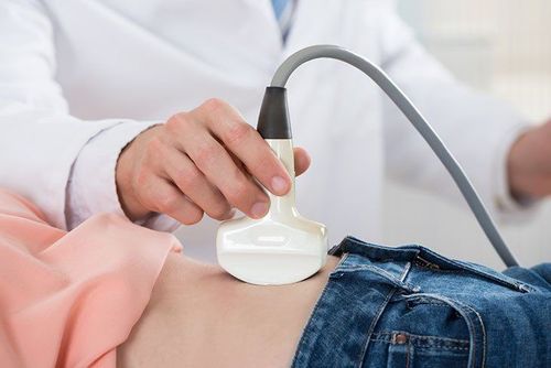
The Standard Screening Protocol — Ultrasound Every 6 Months
The backbone of liver cancer surveillance is abdominal ultrasound performed every 6 months. Ultrasound uses high-frequency sound waves to create real-time images of the liver and surrounding structures. It is non-invasive, involves no radiation, is widely available, and is far less expensive than CT or MRI. The standard surveillance ultrasound does not require intravenous contrast; it aims to identify new liver nodules or changes in nodule size since the previous scan.
The sensitivity of ultrasound for detecting early hepatocellular carcinoma is approximately 47 percent — meaning it detects fewer than half of early-stage HCC tumors when they are small. Overall sensitivity for HCC of any stage is approximately 84 percent. This gap is a known limitation: the background nodularity of cirrhosis makes new small lesions harder to distinguish. This is one reason AFP testing is added in most protocols.
AFP (Alpha-Fetoprotein) Blood Test — What It Adds
Alpha-fetoprotein is a protein elevated in approximately 60 to 70 percent of HCC cases. In adults without liver cancer, AFP levels are typically below 10 ng/mL. When AFP is combined with ultrasound in surveillance, the sensitivity for detecting early-stage HCC rises to approximately 63 percent, compared to 47 percent for ultrasound alone.
The AFP threshold most commonly used in surveillance is 20 ng/mL: values above this level in a cirrhotic patient, when not explained by acute hepatitis, prompt further evaluation with CT or MRI. AFP above 400 ng/mL in a patient with cirrhosis and a liver mass is considered strongly supportive of HCC diagnosis. AFP velocity — the rate of AFP rise over time — is clinically meaningful even at sub-diagnostic levels. A rise of more than 7 ng/mL per month, or more than 15 ng/mL over any 3-month surveillance period, is considered concerning.
AFP is one of several imperfect tumor markers used in oncology. Like the CA-125 test in ovarian cancer, AFP is most useful when tracked serially over time rather than interpreted as a single value, and serves as a surveillance adjunct rather than a standalone diagnostic test.
What Happens When Screening Finds Something
The algorithm for managing a liver nodule found on surveillance ultrasound depends primarily on the size of the lesion.
Nodule smaller than 1 cm: Very small lesions cannot be reliably characterized by any imaging modality. Current guidelines recommend short-interval follow-up ultrasound at 3 to 4 months. Most nodules under 1 cm that remain stable are not HCC.
Nodule 1 to 2 cm: Triggers multiphasic contrast-enhanced CT or MRI. Results are classified using the LI-RADS (Liver Imaging Reporting and Data System) scale — from LR-1 (definitely benign) to LR-5 (definitely HCC). A LR-5 result supports HCC diagnosis without biopsy. LR-3 or LR-4 findings require additional evaluation or biopsy.
Nodule larger than 2 cm: A lesion in a cirrhotic patient showing arterial enhancement and portal venous washout on CT or MRI is diagnosed as HCC based on imaging alone — no biopsy required. This is one important way liver cancer diagnosis differs from most cancers; compare this to pancreatic cancer diagnosis, where EUS biopsy is needed before treatment.
Rising AFP without a visible nodule: MRI is preferred over CT for its superior ability to detect very small HCC in cirrhotic livers, particularly with hepatobiliary contrast agents (gadoxetate disodium). If no lesion is found, the surveillance interval is shortened to 3 months and AFP is remeasured.
GALAD Score and Emerging Screening Tools
Standard AFP is an imperfect surveillance tool. A more sophisticated approach uses multiple biomarkers combined algorithmically.
The GALAD score combines five variables: Gender, Age, AFP-L3 (a liver cancer-specific AFP fraction), total AFP, and PIVKA-II (des-gamma-carboxyprothrombin, also called DCP). PIVKA-II is produced when vitamin K-dependent clotting factors are abnormally synthesized — a phenomenon that occurs in HCC. The GALAD model has been validated in multiple countries and outperforms AFP alone for early HCC detection. The FDA has cleared GALAD-based testing (GALADX) for use in the United States as a complement to imaging surveillance.
AFP-L3 — the lens culinaris agglutinin-reactive fraction of AFP — is more HCC-specific than total AFP and is elevated in some cases where total AFP is normal, making it useful for detecting AFP-negative tumors. Liquid biopsy approaches using circulating tumor DNA are under active investigation but remain research tools, not yet validated for clinical HCC surveillance.
Surveillance Barriers and How to Overcome Them
Despite robust evidence that surveillance improves HCC outcomes, real-world adherence is disappointingly low. Studies in the United States have found that only 10 to 25 percent of cirrhotic patients who qualify for HCC surveillance actually receive regular twice-yearly ultrasounds. This gap represents one of the most important unmet needs in liver cancer care.
Barriers include: lack of primary care awareness, patient education gaps, insurance and access issues, and hepatology waitlists. On the system side, automated patient recall programs and patient navigation interventions significantly improve adherence. What patients can do: ask your gastroenterologist or hepatologist directly — “Do I qualify for liver cancer screening? Am I enrolled in a surveillance program?” If you have NAFLD with multiple risk factors, ask about a fibrosis assessment (FibroScan or FIB-4 score).
The Impact of Early Detection on Survival
The case for liver cancer screening rests on hard evidence. A landmark meta-analysis found that surveillance was associated with a 37 percent reduction in HCC mortality compared to no surveillance. This improvement is driven by stage shift: patients enrolled in surveillance are far more likely to be diagnosed at BCLC 0 or A (early stage), where curative treatments achieve five-year survival rates of 50 to 80 percent.
At BCLC 0 (very early), five-year survival can approach 80 percent. At BCLC C (advanced, with portal vein involvement or distant spread), historical median overall survival was under 12 months. Patients in surveillance programs have a two to three times higher rate of receiving curative treatment compared to those who present symptomatically.
From a health economics perspective, HCC surveillance with ultrasound plus AFP every 6 months in cirrhotic patients is cost-effective by quality-adjusted life year (QALY) analysis in the US healthcare system. The National Cancer Institute’s liver cancer treatment summary and the AASLD HCC practice guidelines provide updated evidence summaries for clinicians and patients.
What to Do If You Are Not in a Surveillance Program
If you have cirrhosis, chronic hepatitis B, or another condition that may place you at elevated HCC risk, and you are not currently in a formal surveillance program, the most important next step is to raise this directly with your physician.
- Ask explicitly: “Do I have cirrhosis or significant liver fibrosis? Am I eligible for HCC surveillance?”
- Know your hepatitis status: If you have never been tested for hepatitis B or C, request testing. Knowing your HBsAg status and HCV antibody status is foundational to understanding HCC risk.
- Request a fibrosis assessment: FibroScan (transient elastography) or the FIB-4 blood test score can estimate whether you have significant fibrosis or cirrhosis without a liver biopsy.
- Enroll and stay enrolled: Once in a surveillance program, maintain the 6-month schedule. The protection of surveillance depends on its regularity — a missed scan is a window of undetected risk.
The American Liver Foundation provides patient resources on liver disease management, surveillance enrollment, and finding hepatology specialists.
Screening and Symptoms Together
Liver cancer screening and symptom recognition serve complementary but distinct roles. Screening — the twice-yearly ultrasound and AFP for high-risk patients — is the primary defense, catching disease before symptoms appear. Symptom awareness is the secondary safety net: for patients not in surveillance, or whose cancer emerges between surveillance intervals, recognizing liver cancer symptoms such as right upper quadrant pain, jaundice, unexplained weight loss, or new ascites can motivate the evaluation that leads to diagnosis. Together, regular surveillance and prompt symptom evaluation form the foundation of an effective liver cancer early detection strategy.
Sources: National Cancer Institute — Liver Cancer | AASLD HCC Practice Guidelines | American Liver Foundation
GALAD Score and Emerging Screening Tools
Standard AFP is an imperfect surveillance tool: it misses approximately 30 percent of HCC and generates false positives from liver inflammation. More sophisticated approaches use multiple biomarkers combined into an algorithm.
The GALAD score combines five variables: Gender, Age, AFP-L3 (a liver cancer-specific AFP fraction), total AFP, and PIVKA-II (des-gamma-carboxyprothrombin, also called DCP). PIVKA-II is produced when vitamin K-dependent clotting factors are abnormally synthesized — a phenomenon that occurs in hepatocellular carcinoma. The GALAD model has been validated in multiple countries and consistently outperforms AFP alone for both early HCC detection and reduction in false positives. The FDA has cleared GALAD-based testing (GALADX) for use in the United States as a complement to imaging surveillance in patients with cirrhosis.
AFP-L3 — the lens culinaris agglutinin-reactive fraction of total AFP — is more HCC-specific than total AFP and is elevated in some cases where total AFP remains normal, making it useful for detecting AFP-negative HCC tumors. AFP-L3 has been used in Japan for decades as part of multi-marker HCC screening and is increasingly incorporated into panel testing globally.
Liquid biopsy approaches — detecting circulating tumor DNA (ctDNA), circulating tumor cells, or tumor-derived exosomes in blood samples — are under active investigation for HCC surveillance. These technologies have the potential to detect HCC at the molecular level before it is visible on imaging, but they have not yet achieved the sensitivity and specificity needed for routine clinical use and remain research tools. The next decade of HCC surveillance innovation will likely center on blood-based multi-marker panels that outperform the current US + AFP standard without requiring imaging.
Surveillance Barriers and How to Overcome Them
Despite robust evidence that surveillance improves HCC outcomes, real-world adherence is disappointingly low. Studies in the United States have found that only 10 to 25 percent of cirrhotic patients who qualify for HCC surveillance actually receive regular twice-yearly ultrasounds. This gap between guideline recommendation and clinical practice represents one of the most important unmet needs in liver cancer care.
The barriers are multiple. Primary care physicians may not be aware of which patients qualify for HCC surveillance or may not have systems in place to order and track surveillance ultrasounds. Patients themselves often do not know they are at risk. Specialty hepatology care is not accessible to all patients, particularly those in rural areas, and gastroenterology or hepatology waitlists can delay timely enrollment. Insurance coverage gaps and cost concerns create additional obstacles for uninsured or underinsured patients.
On the system side, automated patient recall programs — where the electronic health record generates reminders at the 6-month mark and prompts both the patient and their provider — significantly improve adherence. Patient navigation programs that help patients with chronic liver disease access specialist care also improve surveillance rates. Hospitals and health systems that have implemented structured HCC surveillance programs report substantially higher adherence than those relying on spontaneous ordering.
What patients can do: If you have cirrhosis or chronic hepatitis B and are not currently in a surveillance program, ask your gastroenterologist, hepatologist, or primary care physician directly: “Do I qualify for liver cancer screening? Am I enrolled in a surveillance program?” If you have NAFLD and multiple metabolic risk factors — obesity, type 2 diabetes, hypertension — ask whether a fibrosis assessment (FibroScan or FIB-4 blood test score) is appropriate to determine whether you have significant fibrosis or cirrhosis.
The Impact of Early Detection on Survival
The case for liver cancer screening rests on hard evidence that it shifts the stage at diagnosis in ways that meaningfully improve survival.
A landmark meta-analysis evaluating HCC surveillance programs found that surveillance was associated with a 37 percent reduction in HCC mortality compared to no surveillance. This improvement is driven primarily by stage shift: patients enrolled in surveillance programs are far more likely to be diagnosed at BCLC 0 or A (early stage), where curative treatments — liver transplantation, surgical resection, and ablation — achieve five-year survival rates of 50 to 80 percent.
The numbers become vivid when compared across disease stages. At BCLC 0 (very early, single tumor under 2 cm), five-year survival after curative treatment can approach 80 percent. At BCLC A, five-year survival with appropriate treatment is 50 to 70 percent. At BCLC B (intermediate, multinodular), median survival with TACE is 16 to 26 months. At BCLC C (advanced, with portal vein involvement or distant spread), median overall survival was historically under 12 months — although newer immunotherapy combinations (atezolizumab plus bevacizumab) have improved this to approximately 19 months.
Patients in surveillance programs have a two to three times higher rate of receiving curative treatment compared to those who present symptomatically. Most symptomatic presentations are BCLC B or C at minimum — beyond the window for curative therapy.
From a health economics perspective, HCC surveillance with abdominal ultrasound plus AFP every 6 months in cirrhotic patients is cost-effective by quality-adjusted life year (QALY) analysis in the US healthcare system. The cost per QALY gained falls well within accepted thresholds, making the case for broader surveillance uptake not just clinically but economically compelling. The National Cancer Institute’s liver cancer resources and the AASLD HCC practice guidelines provide current evidence summaries for clinicians and patients.
What to Do If You Are Not in a Surveillance Program
If you have cirrhosis, chronic hepatitis B, or another condition that may place you at elevated HCC risk, and you are not currently in a formal surveillance program, the most important next step is to raise this directly with your physician. Specifically:
- Ask explicitly: “Do I have cirrhosis or significant liver fibrosis? Am I eligible for HCC surveillance?”
- Know your hepatitis status: If you have never been tested for hepatitis B or C, request testing. Knowing your HBsAg (hepatitis B surface antigen) status and HCV antibody status is foundational to understanding your HCC risk.
- Request a fibrosis assessment: FibroScan (transient elastography, which measures liver stiffness) or the FIB-4 score (calculated from age, AST, ALT, and platelet count) can estimate whether you have significant fibrosis or cirrhosis without an invasive liver biopsy.
- Enroll and stay enrolled: Once enrolled in a surveillance program, maintain the 6-month schedule. The protection of surveillance depends on its regularity — a missed scan is a window of undetected risk that cannot be recovered retroactively.
The American Liver Foundation provides patient resources on liver disease management, surveillance enrollment, and finding hepatology specialists.
Screening and Symptoms Together
Liver cancer screening and symptom recognition serve complementary but distinct roles. Screening — the twice-yearly ultrasound and AFP test for high-risk patients — is the primary defense: it catches disease before symptoms appear, when cure is most achievable. Symptom awareness is the secondary safety net: for patients who are not enrolled in surveillance, or whose cancer emerges between surveillance intervals, recognizing liver cancer symptoms such as right upper quadrant pain, new jaundice, unexplained weight loss, or worsening ascites can motivate the evaluation that leads to diagnosis. Together, regular surveillance and prompt symptom evaluation form the foundation of an effective liver cancer early detection strategy. If you are at high risk and not yet in a surveillance program, today is the right time to ask your doctor to enroll you.