The most important characteristic of skin cancer symptoms is that they are almost always visible. Unlike cancers that develop deep inside the body and produce vague internal symptoms, skin cancer develops on the body’s outermost surface where changes can be seen and felt directly. This visibility is the primary reason that skin cancer — even melanoma, the most dangerous type — has such high cure rates when caught early: the opportunity for early detection exists at every shower, every change of clothes, and every professional skin exam.
The challenge is knowing what to look for. Skin cancer symptoms vary significantly by type. Basal cell carcinoma, squamous cell carcinoma, and melanoma each have distinct visual characteristics. Some presentations are immediately alarming; others — particularly early melanoma or nodular melanoma — can be subtle enough to be overlooked for months. For a comprehensive overview of all three types including causes and treatment, see our guide to skin cancer types and treatment.
Basal Cell Carcinoma Symptoms
Basal cell carcinoma (BCC) most commonly develops on the face, head, neck, and other chronically sun-exposed areas. The five most common clinical presentations are:
1. Pearly or waxy bump: A small, shiny, flesh-colored or pale pink bump that appears pearly or translucent, often with tiny visible blood vessels (telangiectasias) on the surface. Most common on the face, particularly the nose, cheek, and around the eyes.
2. Flat, scar-like lesion: Flat, flesh-colored or slightly brownish lesions resembling scars without preceding injury. The surface appears slightly waxy, and the texture is firmer than surrounding skin. This morpheaform BCC subtype has indistinct margins and is the most difficult to delineate clinically.
3. Bleeding sore that heals and reopens: One of the most diagnostically significant BCC symptoms. Many patients report these sores being present for months, having attributed them to “a pimple that won’t go away.” This cyclical pattern of healing and reopening is a classic BCC red flag that warrants same-week evaluation.
4. Pink growth with raised edges: A pinkish, elevated growth with a rolled or pearlescent edge and a central crusted area. Common on the scalp and ears.
5. Ulceration (rodent ulcer): Advanced or neglected BCC develops central ulceration — an open sore with a crater-like center. This indicates deep invasion requiring more extensive treatment.
Squamous Cell Carcinoma Symptoms
SCC has a broader range of presentations than BCC, partly because it can arise in diverse skin environments — from chronically sun-damaged skin to chronic wounds to HPV-associated lesions.
Actinic keratoses (precancerous): The precursor to SCC on sun-exposed skin. AKs appear as rough, scaly, sandpaper-textured patches, usually 2–6mm, often more easily felt than seen. Multiple AKs indicate a “field of cancerization” with elevated SCC risk across the entire sun-damaged area.
Classic SCC presentations:
- A firm, flesh-colored or red nodule on sun-exposed skin
- A flat lesion with a scaly, crusted surface that may bleed when the scale is removed
- A new sore or raised area on an existing scar or area of chronic inflammation
- A rough, wart-like growth that may enlarge over weeks to months
- A red, sore area on the lower lip — specifically associated with heavy sun exposure
SCC in special locations: Oral cavity (persistent red or white patch inside mouth, especially in tobacco users); genital and perianal (HPV-related, wart-like or ulcerating); subungual (under nails — often mistaken for fungal nail infection).
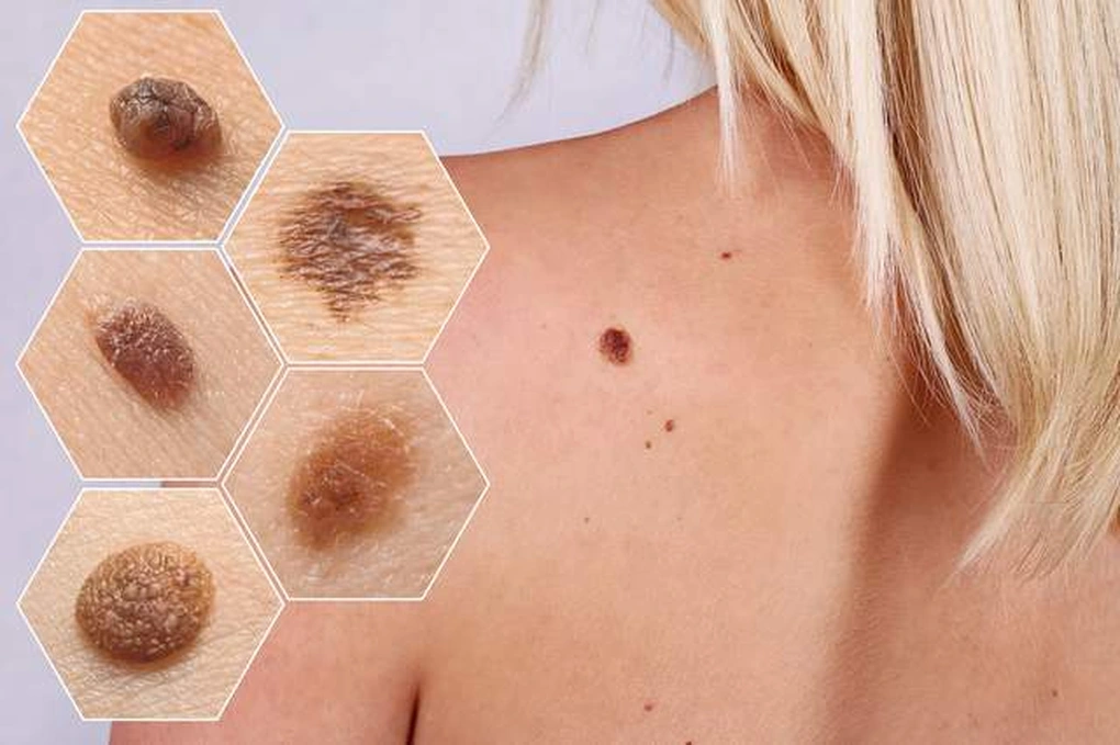
Melanoma Symptoms: The ABCDE Rule
The ABCDE rule is the internationally recognized framework for identifying potentially malignant pigmented lesions:
- A — Asymmetry: One half of the lesion does not match the other half in shape, pigment, or texture.
- B — Border: Ragged, notched, or blurred borders that seem to “melt” into surrounding skin rather than having a clear edge.
- C — Color: Multiple colors within the same lesion — shades of brown and black, areas of red, white, or blue-gray. The presence of black, white, or red within a predominantly brown lesion is particularly concerning.
- D — Diameter: Greater than 6mm (approximately the size of a pencil eraser), though small melanomas under 6mm are well-documented. Diameter alone should not dismiss concern.
- E — Evolution: Any change in a mole — size, shape, color, texture, or new symptoms (itching, bleeding, crusting) — warrants evaluation regardless of whether other criteria are met.
The E criterion captures what the others miss: a mole that has been stable for decades and then begins changing in middle age deserves prompt evaluation. A mole that changes but looks symmetric and small and single-colored can still be melanoma — the change is the signal.
The EFG Rule for Nodular Melanoma
Nodular melanoma — approximately 15% of all melanomas — does not follow the ABCDE pattern reliably. This is critically important: nodular melanoma is the most rapidly growing and most aggressive subtype, and it is specifically the type most likely to be missed by standard ABCDE-based detection.
The EFG rule was developed for nodular melanoma:
- E — Elevated: Presents as a raised bump, not a flat spreading lesion
- F — Firm: Feels firm and raised rather than flat and soft
- G — Growing: Grows quickly — 1mm or more per month — making growth history a key feature
Nodular melanomas can be amelanotic — lacking melanin pigment and appearing flesh-colored, pinkish, or reddish. This variant is among the most commonly missed skin cancers, often mistaken for an inflamed cyst, angioma, or pyogenic granuloma.
Symptoms by Body Location
Scalp: One of the most commonly missed skin cancer sites, particularly in men with thinning hair who have accumulated significant sun exposure. Any persistent scalp lesion, or a sore that bleeds when combing hair, warrants evaluation.
Lower lip: Specifically vulnerable to SCC due to direct downward sun exposure. Actinic cheilitis — rough, scaly, crusted condition of the lower lip — is the lip equivalent of AK. Any persistent roughness or non-healing sore on the lower lip deserves biopsy.
Palms, soles, and under nails: Acral lentiginous melanoma (ALM) develops at these sites independently of UV exposure. On the palm or sole, it appears as an irregularly pigmented patch. Under nails, it appears as a dark streak running the length of the nail (longitudinal melanonychia). Hutchinson’s sign — pigment extending onto the skin around the nail — is highly suspicious for subungual melanoma and warrants urgent dermatological evaluation.
Oral mucosa: Mucosal melanoma and oral SCC both occur on gums, palate, and inner cheeks. Any persistent pigmented or ulcerated lesion inside the mouth warrants dental or ENT evaluation, as oral cancers are frequently diagnosed late due to delayed recognition.
Non-Healing Wounds as Skin Cancer Symptoms
A sore that fails to heal over 4 weeks, or that repeatedly reopens after appearing to close, represents an abnormal wound healing process requiring evaluation. Normal skin wounds in healthy adults heal within 2–3 weeks. This pattern is most characteristic of BCC, where it reflects the tumor’s disruption of normal tissue architecture, but can occur with invasive SCC as well.
This symptom is particularly important in areas where patients might not expect to find skin cancer — the scalp under hair, the back, or areas covered by clothing. Any wound of more than 4 weeks’ duration without a clear resolving cause should be evaluated.
Symptoms of Advanced Skin Cancer
When skin cancer spreads beyond the primary site, symptoms reflect involved lymph nodes or distant organs:
- Regional lymph node enlargement: Enlarged, firm, non-tender lymph nodes near a primary skin cancer — axillary for upper extremity lesions, inguinal for lower extremity, cervical for head/neck — may represent regional metastasis.
- In-transit metastases (melanoma): Satellite clusters of pigmented or dark nodules appearing between the primary tumor and regional lymph nodes, tracking along the lymphatic drainage pathway.
- Distant metastases: Melanoma most commonly metastasizes to the brain (headache, neurological symptoms, seizures), bone (pain), liver (abdominal symptoms), and lungs (respiratory symptoms). Any neurological symptoms in a person with a history of melanoma warrant urgent imaging.
What Skin Cancer Does NOT Cause
- Pain is not a typical early symptom. Most early BCCs, SCCs, and melanomas are entirely painless. Waiting for a lesion to hurt is a common and significant delay.
- Fever, night sweats, and weight loss are not symptoms of localized skin cancer. These occur only with very advanced, metastatic disease.
- Most moles are benign. Having many moles does not mean you have skin cancer. Concern arises when a mole is new, changing, or displays ABCDE features.
- Sun spots and seborrheic keratoses are not cancers. These benign pigmented lesions are common in older adults and require no treatment, though new or changing spots always warrant evaluation to confirm their benign nature.
Dermoscopy: How Doctors See More
Dermoscopy uses a hand-held instrument combining 10x magnification with polarized light to visualize structures in the epidermis and superficial dermis invisible to the naked eye. Under dermoscopy, trained dermatologists identify features that substantially increase diagnostic accuracy: atypical pigment networks, blue-white veils, irregular streaks, and regression structures.
Meta-analyses show dermoscopy improves melanoma detection sensitivity by approximately 20–30% compared to naked-eye examination alone, while also improving specificity and reducing unnecessary biopsies of benign lesions. Dermoscopy is now standard practice for any suspicious pigmented lesion referred to a dermatologist.
- Any mole or skin spot that is changing in size, shape, or color
- A sore, scab, or wound that does not heal after 4 weeks
- A new growth that bleeds easily from minor contact
- A dark streak under a nail, especially if it involves the surrounding skin
- Any rapidly growing raised lesion (doubling in size over weeks)
- New lymph node enlargement near a previously treated skin cancer site
Frequently Asked Questions
Can skin cancer be painless?
Yes — and this is one of the most important facts about skin cancer screening. The majority of early BCCs, SCCs, and melanomas are entirely painless. Skin cancer often does not itch, burn, or hurt until it is relatively advanced. Waiting for pain as a signal to seek evaluation is a common and significant error.
What does a melanoma feel like to the touch?
Early superficial spreading melanoma feels flat or slightly elevated and may have a slightly rough texture at the edge. Nodular melanoma feels like a firm, raised bump. Melanoma does not feel distinctively different from many benign lesions — visual evaluation and biopsy are required for diagnosis.
How fast does skin cancer grow?
BCC is slow-growing — millimeters over years. SCC grows faster — weeks to months. Nodular melanoma can grow 1mm or more per month in diameter. Any change perceived over weeks to months in a pigmented lesion is a red flag requiring evaluation. The E (Evolution) criterion in ABCDE specifically captures this growth rate concern.
At what point do skin cancer symptoms become an emergency?
Urgent evaluation within days (not months) is needed for: any rapidly growing raised lesion doubling in size over weeks; visible satellite nodules near a known melanoma; new neurological symptoms (headache, weakness, vision changes) in a person with a history of melanoma; and any palpable new lymph node enlargement near a known skin cancer site.
Sources
- American Cancer Society. Skin Cancer: Signs and Symptoms.
- American Academy of Dermatology. What Does Skin Cancer Look Like?
- National Comprehensive Cancer Network. NCCN Guidelines: Melanoma v2.2024.
- American Academy of Dermatology. Moles: Overview.
- Vestergaard ME et al. Dermoscopy Compared with Naked Eye Examination for the Diagnosis of Primary Melanoma. Cochrane Database Syst Rev. 2008.
Recognizing Skin Cancer on Darker Skin Tones
A critical gap in public awareness about skin cancer symptoms is the assumption that they only affect people with fair skin. This misconception has real clinical consequences: skin cancer in people with darker skin tones is more often diagnosed at later stages, partly because the lesions present differently and partly because awareness of the risk is lower.
For people with darker skin tones, skin cancer symptoms to be aware of include:
- Palms, soles, and nails: Acral lentiginous melanoma (ALM) is proportionally the most common melanoma subtype in Black, Asian, and Hispanic individuals. It presents as an irregular, slowly expanding pigmented patch on the palm or sole, or a dark longitudinal streak under a fingernail or toenail. Because ALM is not UV-related, it is not prevented by sun avoidance, and checking these locations during self-examination is specifically important for people of all skin tones.
- Oral and mucosal melanoma: Mucosal melanoma can appear as a pigmented patch on the gums, palate, or inner cheeks. It is frequently recognized late because patients and clinicians may not look inside the mouth during skin cancer evaluations. Any persistent, new pigmented patch inside the mouth warrants evaluation.
- SCC on non-sun-exposed skin: In people with darker skin tones, SCC more commonly arises on areas of chronic inflammation, old scars (Marjolin’s ulcer), or burns rather than on sun-damaged skin. A non-healing sore or new growth on a chronic wound or old scar deserves biopsy.
The standard ABCDE criteria apply to all skin tones, but the background against which changes are recognized differs. A dermatologist familiar with skin cancer in patients with skin of color uses dermoscopy and a lower biopsy threshold when lesions appear in high-risk locations or display changes over time, regardless of pigmentation level.
How to Perform a Skin Self-Examination
Monthly skin self-examination is the foundation of skin cancer symptom detection between professional evaluations. A systematic approach ensures no body surface is overlooked.
Step 1: Examine your face, scalp, and neck in good lighting with a magnifying mirror. Part the hair in sections to inspect the scalp. Check the ears inside and out.
Step 2: Inspect your hands and forearms, including the spaces between the fingers, the backs of the hands, and the area under and around the fingernails. Look for dark streaks under nails (Hutchinson’s sign).
Step 3: Use a full-length mirror and a hand mirror to inspect the inner arms, armpits, and upper chest. Check the trunk front and back, including areas normally covered by swimwear.
Step 4: Inspect the back and buttocks using the hand mirror in front of the full-length mirror. The back and back of the legs are common sites for melanoma, particularly in men.
Step 5: Sit and examine the lower legs, the tops and soles of the feet, and between the toes. Check the toenails for dark streaks.
Step 6: Use a comb or blow-dryer to part the hair and systematically examine the scalp in all regions.
Any new, changed, or suspicious finding during self-examination warrants a dermatologist visit, not watchful waiting at home. The purpose of self-examination is detection, not diagnosis — diagnosis requires professional evaluation and biopsy when indicated. For guidance on how often professional evaluation should occur based on personal risk level, see our guide to skin cancer screening.
Documenting Changes Over Time
One of the most practical tools for early skin cancer symptom detection is photographic documentation. Photographing suspicious or borderline moles and skin changes at regular intervals allows direct comparison over weeks or months, making the E (Evolution) criterion far more reliable than relying on memory alone.
Smartphone cameras are adequate for this purpose. Use consistent lighting (natural daylight without direct flash), consistent distance, and include a ruler or coin for scale reference. Take photos from the same angle at each documentation interval.
If a photographically documented change is visible — a mole that looks even slightly larger, darker, or differently shaped compared to the photo taken 3 months prior — that change warrants dermatological evaluation. The threshold should be low: the cost of an unnecessary evaluation is minimal compared to the cost of a missed melanoma that progresses between observation periods.
Dermatology practices increasingly use digital dermoscopy systems that photograph and archive pigmented lesions at each visit, generating automated side-by-side comparisons at follow-up appointments. This “total body photography” approach has been shown to improve early melanoma detection rates in high-risk patients — those with many nevi, a personal or family history of melanoma, or a history of multiple skin cancers. Understanding your personal risk level, including risk factors explored in our guide to skin cancer causes and risk factors, helps determine whether you fall into the category where this more intensive surveillance approach is warranted.
Skin Cancer Symptoms vs. Benign Skin Conditions That Can Mimic Them
Many benign skin conditions can produce symptoms that overlap with skin cancer presentations. Knowing the most common look-alikes helps avoid unnecessary alarm while also clarifying when professional evaluation is genuinely needed.
Seborrheic keratoses vs. melanoma: Seborrheic keratoses — the waxy, “stuck-on,” often tan to dark brown growths common in adults over 40 — are among the most common conditions mistaken for melanoma. They can be darkly pigmented, irregular in shape, and multi-colored. Key distinguishing features: a rough, cerebriform (brain-like) warty surface; clearly defined edges; and a “stuck-on” appearance as if it could be lifted off. However, if in doubt, a dermatologist visit is always appropriate. Seborrheic keratoses that become inflamed, start bleeding, or change significantly also warrant evaluation.
Dermatofibromas vs. BCC: Dermatofibromas are firm, slightly raised, brownish papules most common on the legs. They dimple inward when pinched (dimple sign) — a feature not seen in BCC. They are benign but can be biopsied if there is clinical uncertainty.
Pyogenic granuloma vs. nodular melanoma: Pyogenic granulomas are rapidly growing, vascular, bright red nodules that bleed easily. They can grow within days to weeks and may resemble amelanotic (flesh-colored) nodular melanoma. Any rapidly growing skin nodule that bleeds easily should be biopsied to exclude melanoma. The “rapidly growing” feature is common to both — it is not reassuring in either direction.
When in doubt, the only definitive way to distinguish a skin cancer from its mimics is biopsy. Dermatologists are trained to identify the clinical situations where biopsy is essential versus where monitoring is appropriate — and a consultation is the appropriate first step whenever a lesion causes genuine concern.