Skin cancer is the most common cancer in the United States by a substantial margin. More than 9,500 people are diagnosed with some form of skin cancer every single day, and over 5 million cases are treated annually — more than all other cancers combined. Despite these staggering numbers, skin cancer carries a fundamentally different prognosis from most other malignancies: when caught early, the vast majority of cases are completely curable.
The reason for this combination of high incidence and high curability lies in biology. Most skin cancers grow slowly, remain localized for extended periods, and develop on the body’s most visible organ — which means they can be found at an early, treatable stage through vigilant self-examination and regular professional skin exams. Understanding the different types of skin cancer, their warning signs, and the risk factors that increase susceptibility is the foundation for preventing, detecting, and treating this disease effectively.
How Skin Cancer Develops
The skin has multiple layers and cell types, and the type of skin cancer that develops depends on which cells are affected. The epidermis — the outer layer — contains several important cell populations.
Keratinocytes are the most abundant cells. The basal layer (deepest) contains basal cells, which give rise to basal cell carcinoma (BCC). The squamous layer above contains squamous cells, the precursors to squamous cell carcinoma (SCC). Melanocytes are specialized cells scattered throughout the basal layer that produce melanin — the pigment responsible for skin color and UV-protective tanning. When melanocytes undergo malignant transformation, the result is melanoma.
The common mechanism across all three major types is DNA damage from ultraviolet (UV) radiation. UV light causes specific DNA mutations in skin cells that, if not repaired, lead to permanent genetic changes. Cumulative UV damage over a lifetime is the primary driver of BCC and SCC. Episodic intense UV exposure — particularly blistering sunburns in childhood and adolescence — is the strongest environmental risk factor for melanoma.
Basal Cell Carcinoma (BCC)
Basal cell carcinoma is the most common form of skin cancer — and the most common cancer of any type in humans. It accounts for approximately 80% of all non-melanoma skin cancers and is caused predominantly by cumulative UV radiation exposure. BCC is characteristically slow-growing and almost never metastasizes (risk below 0.1%). However, untreated BCC can erode deeply into underlying tissue, including cartilage and bone.
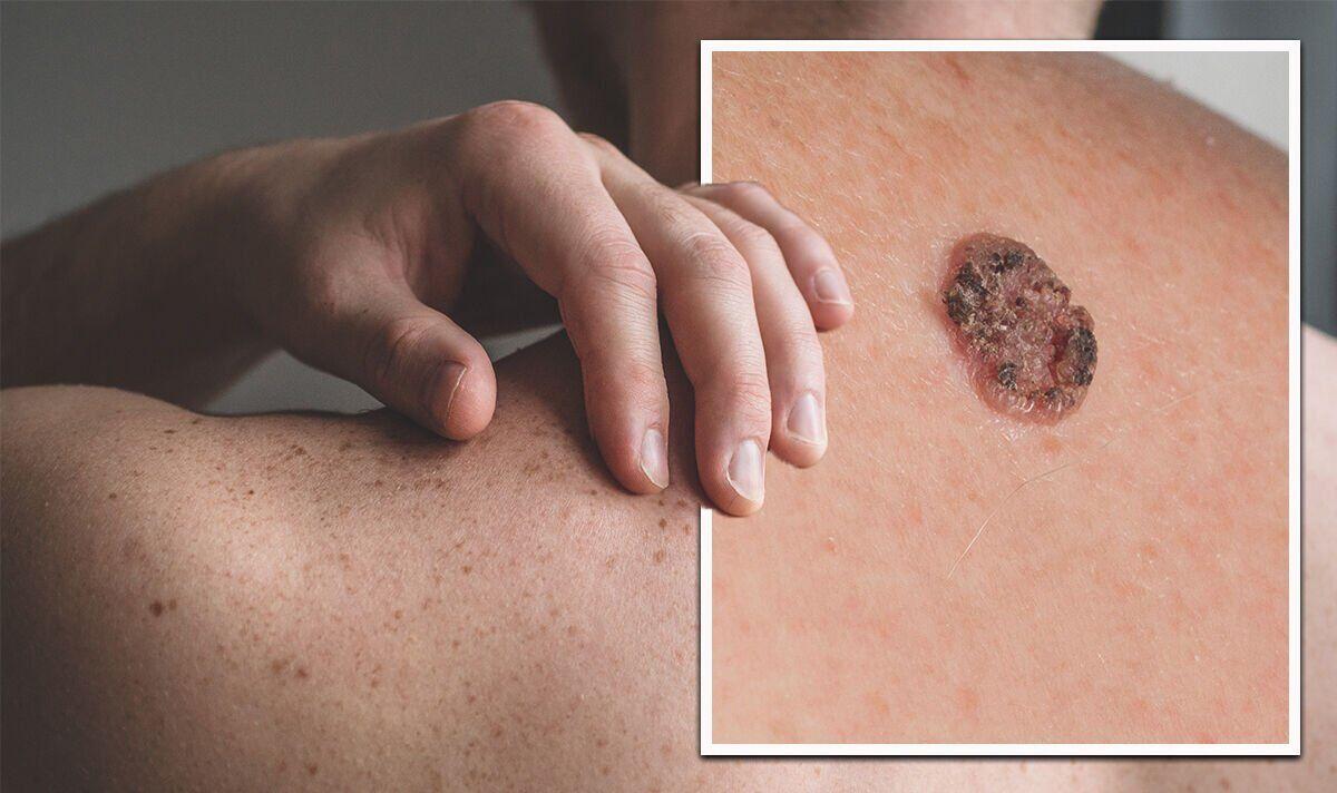
The typical presentations include a pearly or waxy bump with visible blood vessels, a flat, flesh-colored scar-like lesion, a bleeding or scabbing sore that heals and recurs, or a pinkish growth with raised edges. BCC most commonly occurs on the face, head, neck, and hands — areas of greatest lifetime sun exposure.
Treatment options include Mohs micrographic surgery (gold standard, >99% cure rate), standard excision, curettage and electrodesiccation, topical agents (imiquimod, 5-FU), and for rare advanced cases, hedgehog pathway inhibitors (vismodegib, sonidegib).
Squamous Cell Carcinoma (SCC)
Squamous cell carcinoma is the second most common skin cancer (~20% of cases). Unlike BCC, SCC carries a meaningful — though still relatively low — risk of metastasis (2–5% overall, higher for large or deeply invasive lesions and in immunocompromised patients).
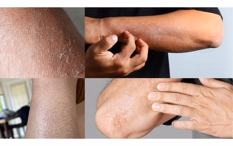
A key precancerous precursor is the actinic keratosis (AK) — a rough, scaly patch on sun-damaged skin representing early squamous dysplasia. Approximately 2–5% of individual AKs progress to SCC if untreated. AKs are treated with cryotherapy, topical 5-FU or imiquimod, or photodynamic therapy.
SCC presentation includes a firm red nodule, a flat lesion with scaly crusted surface, a new sore on old scar tissue, or a wart-like growth. Treatment is Mohs surgery or excision for most cases; cemiplimab (Libtayo) or pembrolizumab (Keytruda) — anti-PD-1 checkpoint inhibitors — are the standard of care for advanced or metastatic SCC.
Melanoma
Melanoma is the most dangerous skin cancer. Although it accounts for only about 1% of all skin cancer diagnoses, it is responsible for approximately 75% of skin cancer deaths. In 2024, the American Cancer Society estimated approximately 100,640 new melanoma diagnoses and 8,290 deaths in the United States.
Melanoma arises from melanocytes and can develop in existing moles or arise de novo. It can occur on any skin surface, including palms, soles, under nails, and on mucous membranes. Key subtypes:
- Superficial spreading melanoma: Most common (~70%); spreads horizontally before invading vertically
- Nodular melanoma: Most aggressive (~15%); grows rapidly and invades vertically from the start
- Lentigo maligna melanoma: Develops slowly in older, sun-damaged skin, often on the face
- Acral lentiginous melanoma: On palms, soles, or under nails; not UV-related; proportionally more common in people with darker skin tones
The ABCDE Rule for Melanoma Detection
The ABCDE rule is the standard framework for recognizing melanoma warning signs:
- A — Asymmetry: One half of the lesion does not match the other half
- B — Border: Ragged, notched, or irregular edges rather than smooth borders
- C — Color: Multiple shades of brown, black, red, white, or blue within a single lesion
- D — Diameter: Greater than 6 millimeters (roughly the size of a pencil eraser)
- E — Evolution: Any change in size, shape, color, or any new symptom (bleeding, itching, crusting)
The E criterion — evolution — is increasingly considered the most clinically important. A mole that is changing over weeks to months warrants dermatological evaluation regardless of whether it meets other ABCDE criteria. For a detailed guide to recognizing warning signs, see our companion article on skin cancer symptoms.
Risk Factors for Skin Cancer
Multiple factors increase skin cancer risk across all types:
- Ultraviolet radiation: The primary and most modifiable risk factor. Both cumulative sun exposure (more important for BCC/SCC) and episodic intense exposure with blistering sunburns (more important for melanoma) contribute to cancer risk.
- Tanning bed use: The International Agency for Research on Cancer (IARC) classifies tanning devices as Group 1 carcinogens — the same classification as tobacco smoke. Tanning bed use before age 35 increases melanoma risk by approximately 47–75%.
- Fair skin, light-colored eyes, red or blonde hair: Lower melanin levels reduce natural UV protection. People with very fair skin who burn easily and rarely tan are at highest risk.
- Personal or family history: A personal history of any skin cancer increases risk of additional skin cancers. First-degree family history of melanoma doubles lifetime melanoma risk.
- Large number of moles: Having more than 50 ordinary moles is a significant melanoma risk factor; atypical (dysplastic) moles confer additional risk.
- Immunosuppression: Organ transplant recipients and those on chronic immunosuppressive therapy have SCC rates 65–250 times higher than the general population.
- Chronic UV-related skin damage: Actinic keratoses and heavily sun-damaged skin indicate elevated SCC risk.
Diagnosis and Staging
When a skin lesion is suspicious for cancer, a skin biopsy is required for definitive diagnosis. This is performed as an in-office procedure under local anesthesia by a dermatologist.
For melanoma specifically, the most important prognostic factor from the initial biopsy is Breslow thickness — the depth of tumor invasion measured in millimeters. Breslow thickness is the primary determinant of clinical T stage, sentinel lymph node biopsy recommendations, and prognosis. After melanoma biopsy, staging involves sentinel lymph node biopsy (recommended for tumors >0.8mm) and imaging (CT, PET-CT) for suspected advanced disease.
5-year survival rates for melanoma by stage:
- Stage I (localized, thin): ~97–98%
- Stage II (localized, thick): ~65–93%
- Stage III (regional lymph nodes involved): ~40–78%
- Stage IV (distant metastasis): ~15–20% historically; now improving significantly with immunotherapy
Treatment Overview
Surgery is the primary treatment for most BCC, SCC, and early-stage melanoma. Mohs micrographic surgery achieves cure rates above 99% for appropriately selected BCC and high-risk SCC, preferred for tumors on cosmetically sensitive areas.
Immunotherapy has revolutionized the treatment of advanced melanoma and metastatic SCC. Anti-PD-1 checkpoint inhibitors (pembrolizumab, nivolumab) and anti-CTLA-4 agents (ipilimumab) have converted stage IV melanoma — historically uniformly fatal — into one with measurable long-term survival in a meaningful proportion of patients.
Targeted therapy with BRAF and MEK inhibitors (vemurafenib, dabrafenib, trametinib) is effective for the approximately 50% of melanomas harboring a BRAF V600E mutation. These agents produce rapid tumor responses but are prone to acquired resistance; they are often combined with immunotherapy in modern protocols.
Prevention: What the Evidence Actually Supports
Sunscreen: Broad-spectrum SPF 30 or higher, applied 15 minutes before sun exposure and reapplied every 2 hours. Randomized trial data (Nambour Skin Cancer Prevention Trial) show sunscreen reduces SCC risk by approximately 40% and melanoma risk by approximately 50%.
Protective clothing and shade: Wide-brimmed hats, UV-protective clothing (UPF 50+), and seeking shade during peak UV hours (10am–4pm) are the most effective physical barriers.
Tanning bed avoidance: The single most impactful modifiable melanoma risk factor for younger adults. There is no safe level of tanning bed use for cancer prevention purposes.
Vitamin D: A common concern is that sun avoidance causes vitamin D deficiency. Vitamin D requirements can be met entirely through dietary sources and supplements without any UV skin exposure. The American Academy of Dermatology does not recommend seeking sun exposure for vitamin D production.
For comprehensive information about recognizing warning signs, see our guide to skin cancer symptoms, and for information about who should be screened and how often, see our guide to skin cancer screening.
- Anyone with a personal or family history of melanoma or other skin cancer
- People with more than 50 moles or any atypical (dysplastic) moles
- Those with a history of frequent or blistering sunburns, especially in childhood
- Organ transplant recipients or anyone on chronic immunosuppressive therapy
- People who worked outdoors extensively for years
- Anyone who has used tanning beds regularly
Frequently Asked Questions
Is skin cancer curable?
Most skin cancers are highly curable when detected early. Basal cell carcinoma has a cure rate above 99% with appropriate treatment. Squamous cell carcinoma is cured in the vast majority of cases when treated before metastasis. Early-stage melanoma (Stage I) has a 5-year survival rate above 97%. Stage IV melanoma now has durable long-term responses in 20–40% of patients with modern immunotherapy regimens.
What does skin cancer feel like?
Most early skin cancers do not cause pain. Lesions may bleed after minor trauma, itch, or crust. The most reliable signs are visual: a new growth, a lesion that doesn’t heal, or a mole that is changing. Pain is more common in advanced, deeply invasive lesions.
Does a tan protect against skin cancer?
No. A tan represents DNA damage — the skin’s response to UV injury. Tanned skin provides no meaningful protection against skin cancer development. The “base tan” concept is not supported by evidence and is a myth that has contributed to tanning bed use among younger adults.
Who should see a dermatologist for a skin exam?
All adults benefit from periodic professional skin examination. High-priority individuals include those with personal or family history of skin cancer, those with many moles or atypical moles, those with a history of frequent sunburns, organ transplant recipients, and those who worked outdoors extensively. Our guide to skin cancer screening provides detailed guidance on screening frequency by risk level.
Sources
- American Cancer Society. Skin Cancer Facts & Statistics 2024.
- American Academy of Dermatology. Skin Cancer Information.
- National Comprehensive Cancer Network. NCCN Guidelines: Melanoma v2.2024.
- American Academy of Dermatology. Sunscreen FAQs.
- International Agency for Research on Cancer. IARC Monographs on UV Radiation — Vol 100D.
Precancerous Lesions: Actinic Keratoses
Actinic keratoses (AKs) — also called solar keratoses — are the most common precancerous skin lesions in the United States, affecting an estimated 58 million Americans. They represent the earliest visible stage of UV-induced squamous cell dysplasia and are considered obligate precursors to invasive SCC if left untreated over sufficient time.
AKs appear on chronically sun-damaged skin as rough, scaly, or crusty patches, typically 2–6mm in diameter. They often feel like sandpaper on the skin surface. Common locations include the scalp (particularly in bald or thinning-haired men), face, ears, lips, back of hands, and forearms — all areas that accumulate high lifetime UV exposure.
The risk of any individual AK progressing to SCC is approximately 2–5% over a lifetime, which sounds modest. But men and women with 20 or more AKs on their skin have a substantially elevated cumulative risk. Additionally, the same chronic UV damage that produces AKs simultaneously damages surrounding skin at a subclinical level, creating what dermatologists call a “field of cancerization” — a broad area of genetically altered skin at elevated SCC risk.
Treatment options for AKs include:
- Cryotherapy (liquid nitrogen): The most common office-based treatment for individual lesions; freezes and destroys abnormal cells
- Topical 5-fluorouracil (5-FU): Applied to the entire affected field 1–2 times daily for 2–6 weeks; treats both visible and subclinical AKs across a broad area
- Imiquimod cream: Immune modulator applied 2–3 times per week; effective for field treatment with a lower irritation profile than 5-FU
- Diclofenac gel: Less effective than 5-FU but better tolerated; used for mild to moderate AKs
- Photodynamic therapy (PDT): A photosensitizing agent applied to AK-affected skin is activated by a specific light wavelength, destroying abnormal cells; highly effective for field treatment of extensive AKs
Men over 50 with extensive sun-damaged skin and many AKs benefit from regular dermatological monitoring — typically every 6–12 months — with periodic field treatment to reduce the SCC burden.
Skin Cancer in People With Darker Skin Tones
A widespread misconception is that skin cancer is a disease that only or primarily affects people with fair skin. This misperception has real clinical consequences: skin cancer in people with darker skin tones is disproportionately diagnosed at later stages, partly because awareness of the risk is lower and partly because the lesions are harder to recognize against darker pigmented skin.
While the absolute incidence of BCC, SCC, and melanoma is lower in people with skin of color compared to fair-skinned individuals, the disease does occur — and when it does, the clinical features differ in important ways:
Acral lentiginous melanoma (ALM) — the subtype that occurs on palms, soles, and under fingernails — is proportionally the most common melanoma subtype in Black individuals and other people with darker skin tones. ALM is not caused by UV radiation, which means it is not prevented by sun protection behaviors. It often goes unrecognized because patients do not look for changes on their palms and soles during self-examination.
SCC in people with darker skin tones more commonly arises in the context of chronic wounds, old scars (Marjolin’s ulcer), lupus lesions, or areas of chronic inflammation rather than UV-exposed sun-damaged skin as in fair-skinned patients. This different etiology means UV protection is less protective for this population’s SCC risk.
Melanoma staging and survival disparities: Black patients with melanoma are more likely to be diagnosed at Stage III or IV compared to white patients, and have significantly lower 5-year survival rates for all stages. This disparity reflects both later stage at diagnosis and potential differences in access to care and immunotherapy eligibility.
Dermatological awareness campaigns and professional guidelines have increasingly emphasized that melanoma affects people of all skin tones, and that self-examination should include palms, soles, nail beds, and oral mucosa in all patients — particularly those of African, Asian, or Hispanic heritage.
Genetic Syndromes That Increase Skin Cancer Risk
Most skin cancers are sporadic — arising from environmental UV exposure in individuals without a hereditary predisposition. However, several genetic syndromes substantially elevate skin cancer risk and warrant heightened surveillance:
Familial atypical multiple mole melanoma (FAMMM) syndrome: Characterized by multiple atypical nevi (dysplastic moles), a family history of melanoma in two or more first-degree relatives, and an elevated personal lifetime melanoma risk of 70% or more. Surveillance involves professional full-body skin exams every 3–6 months and ophthalmological monitoring for uveal melanoma.
Xeroderma pigmentosum (XP): A rare autosomal recessive disorder affecting DNA repair enzymes. Individuals with XP cannot repair UV-induced DNA damage, making them exquisitely sensitive to sun exposure. They develop AKs and skin cancers — including aggressive SCCs and melanomas — often beginning in early childhood. Rigorous sun avoidance and intensive dermatological monitoring are lifelong requirements.
Gorlin syndrome (basal cell nevus syndrome): An autosomal dominant disorder caused by PTCH1 gene mutations that leads to the development of multiple BCCs beginning in adolescence or early adulthood, often numbering in the dozens to hundreds. Vismodegib is used to reduce BCC burden in Gorlin syndrome; ongoing dermatological surveillance is lifelong.
BRCA2 mutations: Beyond their well-known association with breast and ovarian cancer, BRCA2 mutations confer a modestly elevated melanoma risk. Individuals with BRCA2-associated hereditary breast and ovarian cancer syndrome should be aware of this and include skin monitoring in their surveillance program.
For those who want to understand how hereditary cancer syndromes interact with screening decisions more broadly, the principles discussed in our guide to cancer risk factors and genetics apply across multiple cancer types including melanoma.
Skin cancer, despite its high incidence, remains one of the most preventable and most curable cancers when approached with consistent sun protection habits, vigilant self-examination, and regular professional dermatological evaluation — a combination that reduces both the incidence of new cancers and the likelihood of late-stage diagnosis when cancers do develop.
The trajectory of skin cancer outcomes over the past two decades has been shaped above all by two developments: the widespread adoption of dermoscopy as a diagnostic tool that substantially improves early melanoma detection, and the introduction of immune checkpoint blockade that has fundamentally altered the prognosis for patients with metastatic melanoma. Neither breakthrough, however, changes the fundamental principle that prevention and early detection remain the most cost-effective and outcome-effective strategies for the individual patient.