Introduction
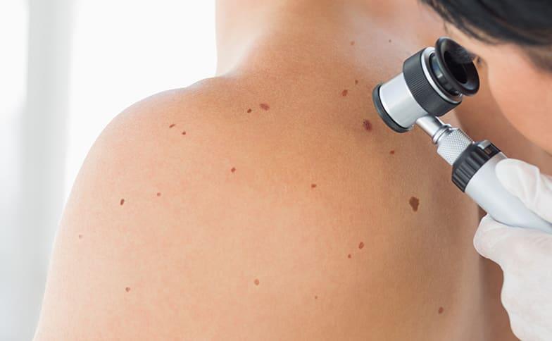
Skin cancer screening has a distinctive characteristic that sets it apart from most other cancer screening programs: it requires no specialized machinery, no laboratory testing, and no invasive procedures in its most basic form. The primary tool is direct visual inspection of the skin surface — an examination that every adult can perform on themselves monthly, and that a trained dermatologist can complete in 10–15 minutes using a hand-held dermoscope.
The rationale for skin cancer screening rests on two well-established facts: skin cancer is extremely common (affecting 1 in 5 Americans by age 70), and it is almost always curable when detected at an early stage. Early-stage melanoma (Stage I) has a 5-year survival rate above 97%. The same melanoma, if allowed to progress to Stage IV before diagnosis, has a historical 5-year survival rate of 15–20% — improved by immunotherapy, but still dramatically worse than early detection. The earlier a skin cancer is found, the more options exist for its removal and the better the outcome.
This guide covers who should be screened, how frequently, what screening involves at each level of risk, and what the current guidelines actually say — including why the USPSTF and AAD give different recommendations and what that means practically for patients.
Two Types of Skin Cancer Screening
Skin cancer screening operates at two distinct levels:
1. Skin self-examination (SSE): Monthly inspection of your own skin, performed at home, to identify any new, changed, or suspicious lesions. The American Academy of Dermatology recommends monthly SSE for all adults. SSE has the advantage of continuity — a person who examines their own skin monthly has far better baseline familiarity with their own lesions than a physician who sees them once a year.
2. Professional skin examination (PSE): A full-body examination performed by a dermatologist or trained physician. PSE includes direct visualization of the entire skin surface, use of a dermoscope for suspicious lesions, documentation of baseline moles and growths, and biopsy recommendation when indicated. PSE is recommended annually by the AAD for most adults and more frequently for high-risk individuals.
How to Perform a Skin Self-Examination
Monthly SSE should be systematic and cover the entire skin surface, including areas not easily seen without mirrors or assistance.
Materials needed: A bright lamp or natural light, a full-length mirror, a hand mirror, a comb or blow-dryer, and a chair for examining legs.
- Face, ears, scalp, and neck: Examine the face carefully with good lighting. Use the hand mirror for the ears (inner and outer). Use a blow-dryer on a low-heat setting or a fine-tooth comb to part the hair in sections and systematically inspect the scalp. Ask a partner to examine the posterior scalp.
- Hands and forearms: Inspect the backs of the hands, between the fingers, the fingertips, and the skin around and under the nails. Look for dark streaks running the length of any nail — longitudinal melanonychia — which warrants dermatological evaluation.
- Upper body: Raise arms and inspect the inner arms and armpits. Examine the chest and upper abdomen. Check the skin around the nipples and beneath the breasts.
- Back and buttocks: Use the hand mirror in front of the full-length mirror to inspect the upper back, lower back, and buttocks. The back is one of the most common melanoma sites in men, making this step particularly important.
- Lower legs and feet: Sit and examine the front and back of both legs. Inspect the soles, between the toes, the toenails, and the skin of the heel and ankle.
- Genitalia: Inspect the genital and perianal region. This area is less commonly affected but is a site for both SCC (particularly HPV-associated) and rare melanomas.
Any new lesion, changing lesion, or lesion that bleeds spontaneously found during SSE warrants a physician visit within days to weeks, not months. For a guide to the specific features that make a mole or skin growth concerning, see our overview of skin cancer symptoms.
What a Professional Skin Exam Involves
A professional skin examination by a dermatologist involves:
Full undressing: Patients are examined wearing a gown that allows systematic inspection of the entire skin surface. Some patients limit the exam out of modesty; a complete exam includes the scalp, ears, between the toes, genitalia, and perianal region — all sites where skin cancers can develop and be missed.
Dermoscopy: A hand-held dermoscope providing 10x magnification with polarized light is used for any lesion that appears suspicious. Dermoscopy allows visualization of subepidermal pigment patterns, vascular structures, and other features that substantially improve diagnostic accuracy beyond naked-eye examination.
Documentation: New lesions are documented in the patient’s chart. High-risk patients may have total body photography performed — standardized photographs of the entire skin surface used for comparison at subsequent visits.
Biopsy when indicated: If a lesion is suspicious for skin cancer, a biopsy is performed during the same visit or at an expedited follow-up appointment. The type of biopsy depends on the lesion characteristics and the level of suspicion for melanoma.
Who Should Be Screened and How Often
Average Risk (Every 1–3 Years)
Adults with no personal history of skin cancer, no first-degree family history of melanoma, fair to medium skin tone, and no significant additional risk factors can begin with:
- Monthly self-examination
- Professional skin examination every 1–3 years starting in early adulthood; annually after age 40 or 50
Moderate Risk (Annual)
Adults with one or more of the following should have annual professional skin examination and monthly SSE:
- Fair skin that burns easily and rarely tans
- History of multiple blistering sunburns, especially before age 20
- More than 50 ordinary moles
- A single atypical (dysplastic) nevus
- A first-degree relative with melanoma
- History of significant occupational sun exposure
High Risk (Every 3–6 Months)
Adults in high-risk groups require the most frequent surveillance:
- Personal history of melanoma: Every 3–6 months for the first 2 years, then every 6 months for years 3–5, then annually for life
- Multiple atypical nevi (dysplastic nevus syndrome): Every 3–6 months with total body photography
- FAMMM syndrome: Every 3–6 months beginning in adolescence; ophthalmology referral for uveal melanoma risk
- Organ transplant recipients: Quarterly in many transplant dermatology protocols; SCC risk is 65–250 times that of the general population
- Xeroderma pigmentosum or Gorlin syndrome: Monthly or more frequent evaluation beginning in early childhood
- Immunosuppressed individuals (HIV with low CD4, biologic therapy, long-term corticosteroids): Annual to biannual
USPSTF vs. AAD: Why the Guidelines Differ
The United States Preventive Services Task Force (USPSTF) issued a statement in 2023 assigning an “I” (Insufficient Evidence) grade to skin cancer screening for average-risk adults without signs or symptoms of skin cancer. This does not mean the USPSTF recommends against screening — it means the Task Force concluded that available evidence was insufficient to determine the net benefit or harm of a population-wide screening recommendation with certainty.
The American Academy of Dermatology (AAD), however, recommends annual professional skin examination for all adults. The AAD’s position reflects the clinical judgment that the low risk of skin examination (no radiation, no pain, no meaningful false-positive harms in the manner of a breast biopsy after a false-positive mammogram) makes the potential benefit of early detection worth pursuing even without RCT-level evidence for mortality reduction in average-risk adults.
The German SCREEN (Skin CancEr Research to provide Evidence for Effectiveness of screening) trial — a nationwide skin cancer screening program evaluated in a 2012 analysis — found a 48% reduction in melanoma mortality in the screened region after 5 years of screening compared to unscreened regions. This single-region study has methodological limitations but represents the largest real-world data on population-level skin cancer screening outcomes.
For individual patients, the practical implication is that dermatological societies worldwide recommend annual skin examination regardless of the USPSTF “I” grade. The USPSTF grade primarily affects insurance coverage and public health policy recommendations, not clinical practice guidelines for individual patient care.
Screening After a Melanoma Diagnosis
After successful treatment of melanoma, ongoing surveillance is essential because melanoma has a meaningful recurrence risk, patients with one melanoma have a 10-fold higher risk of developing a second primary melanoma, and satellite and in-transit recurrences are most commonly detected clinically during surveillance visits.
NCCN recommended follow-up intervals after melanoma treatment per the NCCN Melanoma Guidelines:
- Stage I: Clinical examination every 3–12 months for 5 years, then annually
- Stage II: Every 3–6 months for 2 years, then every 3–12 months for 3 years, then annually
- Stage III: Every 3–6 months for 2 years, then every 3–6 months for 3 years, then annually; imaging (CT chest/abdomen/pelvis or PET-CT) every 3–6 months for 2 years
- Stage IV (NED after immunotherapy): Clinical examination and imaging every 3–6 months
All stages require annual skin examination for life to detect new primary melanomas. Patients who have undergone screening and treatment for other cancers may find the follow-up structure familiar — for comparison, see how PSA test surveillance works after prostate cancer treatment, and the risk factors that guide prostate cancer screening intervals.
Total Body Photography and Technology-Enhanced Screening
Total body photography (TBP) is a technology-enhanced approach to melanoma surveillance in which standardized photographs of the entire skin surface are taken at baseline and compared at subsequent visits to identify any new or changing lesions.
TBP has been shown to improve early melanoma detection in high-risk patients while simultaneously reducing unnecessary biopsies of stable, monitored lesions — because the comparison provides objective evidence of change rather than relying on physician memory or patient report. When combined with serial dermoscopy (photographing individual lesions under dermoscopy at each visit), the sensitivity for early melanoma detection in high-risk populations is substantially higher than standard examination alone.
AI-assisted dermoscopy systems are increasingly available and FDA-cleared. These platforms analyze dermoscopic images using deep learning algorithms and provide diagnostic support scores. While AI tools do not replace physician judgment, studies have shown they approach or match dermatologist-level sensitivity for melanoma classification in high-quality dermoscopic images.
Biopsy: When and Which Type
When a lesion identified during screening is suspicious for skin cancer, a biopsy is performed for definitive diagnosis. The choice of biopsy technique matters significantly for melanoma:
Excisional biopsy (removal of the entire lesion with a 1–3mm margin) is the preferred approach for any lesion suspected of being melanoma. This ensures the pathologist receives the complete lesion for accurate Breslow thickness measurement and histological subtype classification — both essential for staging and treatment planning.
Punch biopsy (a circular 2–6mm full-thickness sample) is appropriate for evaluation of flat or minimally raised lesions when excisional biopsy is technically challenging due to lesion size or location.
Shave biopsy (superficial tangential removal) is appropriate for raised lesions suspected of being BCC or SCC but is not appropriate for lesions suspected of being melanoma — shave biopsy may transect the tumor and prevent accurate Breslow thickness determination.
Incisional biopsy (partial sampling of a large lesion) is acceptable for large lesions where complete excision would be cosmetically or anatomically impractical before confirming the diagnosis.
After biopsy, pathology results are typically available within 5–7 business days. Management — including wider surgical margins, sentinel lymph node biopsy, and imaging — is determined by the pathological diagnosis and stage. To understand the full spectrum of skin cancer types and how they differ in behavior and treatment, see our comprehensive skin cancer overview.
Chemoprevention for High-Risk Patients
For patients who develop multiple skin cancers despite regular screening and sun protection, chemoprevention offers an additional risk-reduction strategy:
- Nicotinamide (vitamin B3) 500mg twice daily: A randomized controlled trial (Chen AC et al., NEJM 2015) demonstrated that daily nicotinamide supplementation reduced new BCCs by 23% and SCCs by 30% over 12 months in patients with a history of at least two skin cancers in the prior 5 years. Nicotinamide is inexpensive, widely available, and well-tolerated. It does not reduce melanoma risk.
- Systemic retinoids (acitretin): Used in immunosuppressed organ transplant recipients with high SCC burden to reduce new SCC formation. Requires close monitoring due to teratogenicity and other side effects.
- Vismodegib (Erivedge): A hedgehog pathway inhibitor that reduces the formation of new BCCs and temporarily shrinks existing BCCs. Used primarily in Gorlin syndrome patients and in selected patients with a very high burden of new BCCs.
- Capecitabine: An oral chemotherapy agent used off-label in some very high-risk transplant recipients with rapidly developing multiple SCCs.
Who should not wait for annual screening: See a dermatologist within days — not at your next annual appointment — if you notice: a new spot that is bleeding without injury; a mole that has visibly changed in color, size, or shape in recent weeks or months; a sore that does not heal after 4–6 weeks; a new dark streak under a fingernail or toenail; any lesion that itches, crusts, or oozes persistently. Annual screening is appropriate for baseline monitoring; symptomatic lesions require prompt evaluation.
Frequently Asked Questions
Does skin cancer screening save lives?
For melanoma specifically, detecting it at Stage I rather than Stage IV is associated with a survival difference from ~97% to ~15–20% at 5 years. While large randomized controlled trials of skin cancer screening have not been performed, the biological rationale and the stage-specific survival data strongly support early detection. The German SCREEN trial provided population-level evidence consistent with a mortality reduction from systematic professional screening.
What does skin cancer screening cost?
Professional skin examinations are typically covered by health insurance as preventive visits or dermatology evaluations. Skin self-examination costs nothing. Biopsy procedures and pathology testing are typically covered when medically indicated. Total body photography may be a separate cost and coverage varies by insurer.
Is it worth seeing a dermatologist if I have no suspicious spots?
Yes — particularly once you are over 40, have a history of significant sun exposure, or have any of the high-risk factors described above. A dermatologist establishes a baseline examination, can identify lesions that warrant monitoring before they become concerning, and identifies early lesions that patients may not have noticed. A normal skin exam is not without value — it establishes the baseline against which future changes are measured.
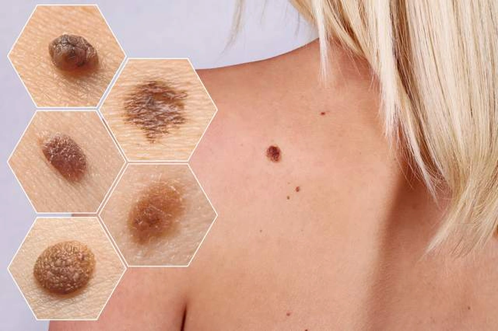
How do I know if a mole is normal?
Most moles are benign, uniform in color (one shade of brown or tan), symmetric, smooth-bordered, and smaller than 6mm. They remain stable for decades. The concern arises when a mole is new, changing, displays ABCDE features (asymmetry, irregular border, multiple colors, diameter >6mm, evolution), or produces any symptom such as bleeding or itching. For a comprehensive guide to recognizing concerning mole features, see our guide to skin cancer symptoms.
Sources
- U.S. Preventive Services Task Force. Skin Cancer: Screening (2023).
- American Academy of Dermatology. Skin Cancer Screening and Detection.
- National Comprehensive Cancer Network. NCCN Guidelines: Melanoma v2.2024 — Follow-up.
- Chen AC et al. A Phase 3 Randomized Trial of Nicotinamide for Skin-Cancer Chemoprevention. N Engl J Med. 2015;373(17):1618–1626.
- Katalinic A et al. Does Skin Cancer Screening Save Lives? An Observational Study Comparing Trends in Melanoma Mortality in Regions With and Without Screening. Cancer. 2012.
Skin Cancer Screening for People With Darker Skin Tones
A persistent and harmful misconception is that skin cancer screening is primarily relevant for people with fair skin. People with darker skin tones do develop skin cancer — and when they do, it is diagnosed at later stages on average, partly because screening is pursued less often and partly because the presentations differ from the classic sun-exposed-site patterns described in most patient education materials.
Clinicians experienced in skin cancer in patients with skin of color use a lower biopsy threshold for lesions in high-risk locations: the palms, soles, under and around the nails, and the oral mucosa. Acral lentiginous melanoma — the subtype most common among Black, Asian, and Hispanic patients with melanoma — is not caused by UV exposure and is not prevented by sun avoidance. Checking the palms, soles, and toenails as part of every skin self-examination is specifically important for people of all skin tones.
Similarly, squamous cell carcinoma in darker-skinned individuals more often arises on areas of chronic inflammation, old scars, or burns rather than on sun-damaged skin. Annual professional skin examination provides the opportunity for a trained clinician to identify these non-UV-related presentations before they progress. For more on how skin cancer presents differently across skin tones, see our guide to skin cancer symptoms.
When to Begin Skin Cancer Screening
For most average-risk adults, the AAD recommendation is to begin regular professional skin examinations in early adulthood, with annual visits after age 40–50. However, certain high-risk groups require earlier and more frequent surveillance:
- Children with xeroderma pigmentosum (XP): Skin cancer surveillance begins in infancy or early childhood. XP is a DNA repair disorder in which UV exposure causes accelerated mutation accumulation; affected children can develop multiple skin cancers by adolescence without rigorous photoprotection and surveillance.
- Gorlin syndrome (nevoid basal cell carcinoma syndrome): Children with Gorlin syndrome begin developing BCCs in adolescence and require regular examination and, in some cases, vismodegib therapy to reduce BCC burden.
- FAMMM syndrome: Surveillance for familial atypical multiple mole melanoma begins in adolescence, with ophthalmology evaluation for uveal melanoma risk.
- Organ transplant recipients: Surveillance begins shortly after transplantation and continues for life, with quarterly exams in many transplant dermatology protocols given the dramatically elevated SCC risk.
For average-risk individuals who have never had a baseline professional skin examination, starting one before age 40 provides a documented baseline against which future lesions can be compared. Earlier is better for establishing the normal topography of your own skin.
How to Talk to Your Doctor About Skin Cancer Screening
Many primary care physicians do not perform full-body skin examinations during annual physicals unless the patient specifically requests it. If skin cancer screening is not offered at your next preventive visit, you can request it directly: “Can we include a skin cancer check at today’s visit, or would you recommend a referral to dermatology?”
Patients who have specific concerns — a new or changing mole, a sore that is not healing, a family history of melanoma — should communicate these clearly at the start of the visit to ensure those lesions receive focused attention. Bringing a photograph of the lesion taken 3–6 months earlier to document change is particularly useful and may prompt a biopsy that a static clinical examination alone would not.
If you are in a high-risk group (personal history of melanoma, organ transplant, multiple atypical nevi), a referral to a dermatologist for ongoing surveillance is appropriate and worth requesting. Primary care-based screening, while valuable for average-risk patients, is not a substitute for dermatologist-level examination in patients who require more intensive surveillance. For comparison, understanding your risk profile for other cancers — including what makes someone high-risk for prostate cancer — follows a similar logic of matching screening intensity to individual risk level. See our overview of prostate cancer risk factors for context on how risk stratification works across different cancer types.