Horizon Health Institute | Cancer Education
Cancer Diagnosis Methods: How Doctors Detect and Confirm Cancer
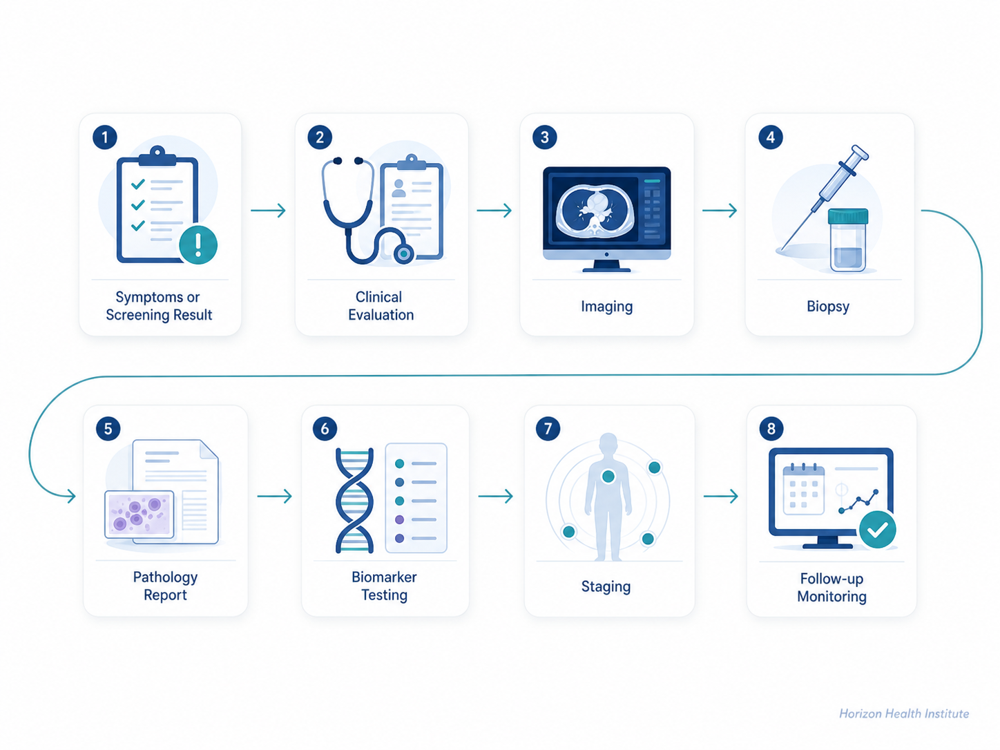
A cancer diagnosis usually does not come from one test alone. Doctors often combine a careful medical history, physical examination, imaging, laboratory testing, biopsy, pathology review, and sometimes advanced biomarker testing to understand what is happening in the body.
At Horizon Health Institute, we believe clear medical education helps patients and families feel more prepared when discussing cancer testing with a healthcare professional. This guide explains the major methods used to detect, confirm, classify, and monitor cancer in modern medicine.
Screening vs. Diagnosis: What Is the Difference?
Cancer screening and cancer diagnosis are related, but they are not the same. Screening means checking for cancer before symptoms appear. Diagnosis means evaluating a symptom, abnormal screening result, or suspicious finding to determine whether cancer is actually present.
Cancer Screening
Screening is used when a person may feel well but has an age, risk factor, or guideline-based reason to be checked. Examples include mammograms, Pap or HPV tests, colorectal cancer screening, and low-dose CT scans for certain people at high risk for lung cancer.
Cancer Diagnosis
Diagnosis is the process of confirming whether cancer is present, identifying the cancer type, and learning how far it may have spread. This often requires imaging, biopsy, pathology, and additional laboratory or molecular tests.
The CDC notes that regular screening can help find breast, cervical, colorectal, and lung cancers early, when treatment is more likely to work well. However, an abnormal screening result does not automatically mean cancer. It usually means more diagnostic testing is needed.
1. Medical History and Physical Examination
The diagnostic process often begins with a conversation. A clinician may ask about symptoms, how long they have been present, personal medical history, medications, tobacco or alcohol exposure, family history of cancer, infections, occupational exposures, and previous screening results.
A physical examination can help identify visible or palpable changes, such as a lump, enlarged lymph node, skin change, abdominal swelling, abnormal bleeding, weight loss pattern, or signs affecting breathing, digestion, urination, or neurological function.
Why this first step matters
A good clinical evaluation helps doctors choose the right next test. For example, persistent cough may lead to chest imaging, abnormal bleeding may lead to endoscopy or gynecologic testing, and a suspicious breast lump may lead to diagnostic mammography, ultrasound, and biopsy.
Sources: CDC cancer screening overview; National Cancer Institute information on cancer diagnosis and tumor markers.

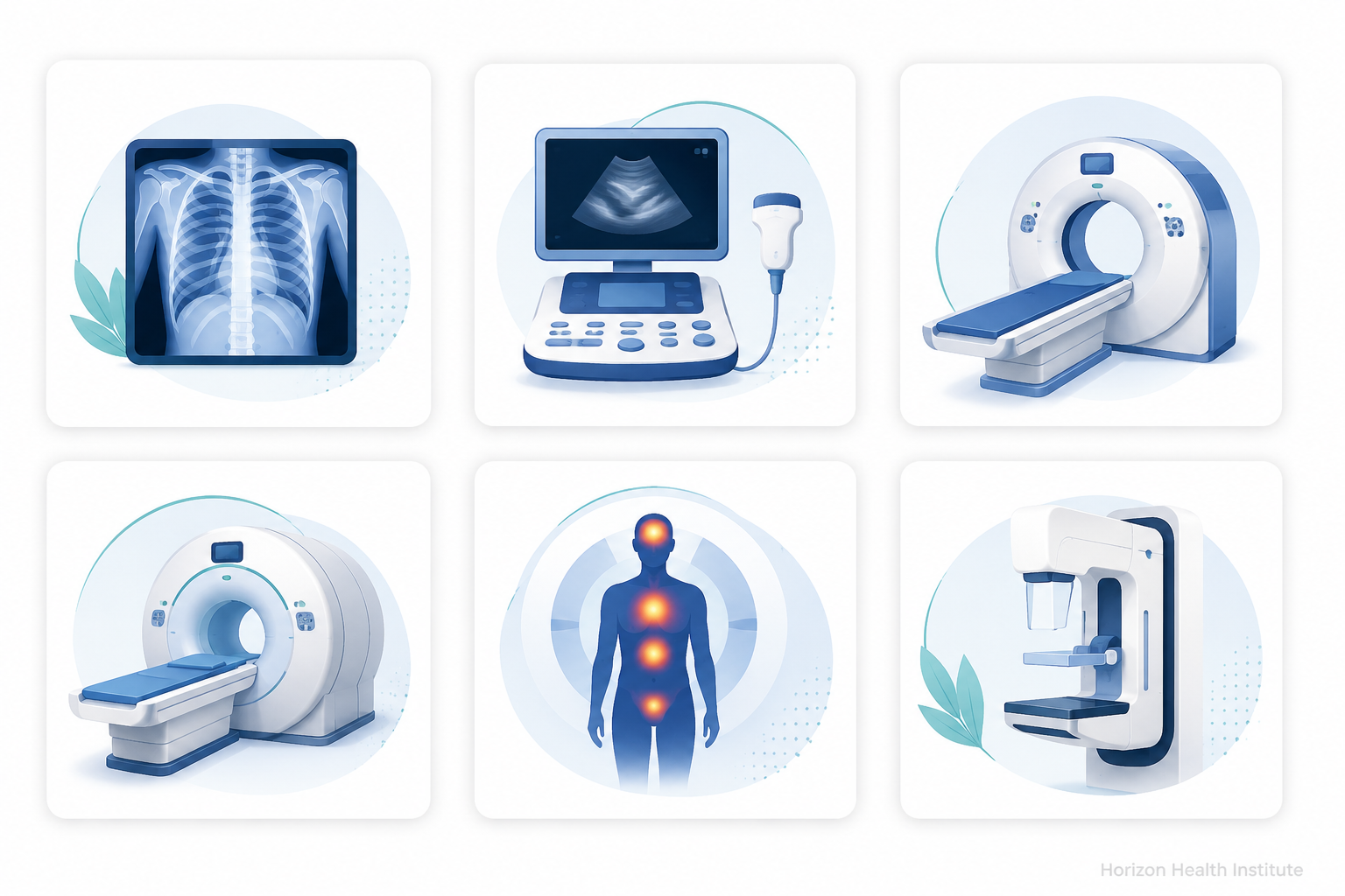
2. Imaging Tests Used in Cancer Diagnosis
Medical imaging helps doctors look inside the body without surgery. Imaging can show whether there is a mass, where it is located, whether nearby organs are affected, and whether cancer may have spread to lymph nodes, bones, lungs, liver, brain, or other areas.
X-ray
X-rays may be used to evaluate bones, lungs, or certain suspicious areas. A chest X-ray, for example, may reveal an abnormal lung shadow that requires further testing.
Ultrasound
Ultrasound uses sound waves and is commonly used to evaluate soft tissues, breast lumps, thyroid nodules, pelvic organs, liver lesions, and fluid-filled structures.
CT Scan
CT scans create detailed cross-sectional images and are often used to evaluate the chest, abdomen, pelvis, lymph nodes, and possible spread of cancer.
MRI
MRI provides detailed images of soft tissue and is often useful for the brain, spine, pelvis, breast, liver, prostate, and musculoskeletal areas.
PET Scan
PET scans can help show areas of increased metabolic activity. They are sometimes combined with CT or MRI to help with staging and treatment planning.
Mammography
Mammography is a specialized breast imaging test used for screening and diagnostic evaluation of breast changes, calcifications, or masses.
Imaging can suggest cancer, but it often cannot confirm cancer by itself
Imaging may show that a finding looks suspicious, but many noncancerous conditions can also appear abnormal on scans. In many cases, a biopsy and pathology review are needed to confirm the diagnosis.
Sources: National Cancer Institute; American Cancer Society; FDA information on imaging and in vitro companion diagnostics.
3. Biopsy: The Key Test for Confirming Many Cancers
A biopsy is a procedure that removes a small sample of tissue or cells so it can be examined under a microscope. For many cancers, biopsy and pathology provide the most important confirmation of diagnosis.
The type of biopsy depends on where the suspicious area is located, how large it is, whether it can be reached safely, and what information the medical team needs. Some biopsies are done with a needle, while others may require endoscopy, image guidance, or surgery.
Common types of biopsy
Needle Biopsy
A thin or core needle removes cells or tissue. Imaging such as ultrasound, CT, or mammography may help guide the needle.
Endoscopic Biopsy
A flexible scope is used to view areas such as the colon, stomach, bladder, lungs, or throat and collect tissue samples.
Skin Biopsy
A small sample of skin is removed to evaluate suspicious moles, lesions, or changes that may suggest skin cancer.
Surgical Biopsy
Surgery may remove part or all of a suspicious lump when a larger tissue sample is needed for diagnosis.
4. Pathology: What Happens After a Biopsy?
After a biopsy, the sample is sent to a pathology laboratory. A pathologist examines the cells and tissue structure under a microscope to determine whether cancer is present and, if so, what type of cancer it is.
Pathology reports may include the cancer type, grade, margin status, lymph node involvement, hormone receptor status, special stains, or molecular findings depending on the cancer. This information helps guide staging and treatment planning.
What a pathology report may help answer
- Is cancer present?
- What type of cancer is it?
- How abnormal do the cells look?
- Are cancer cells seen at the edge of the removed tissue?
- Are lymph nodes involved?
- Are there biomarkers that may affect treatment choices?
Sources: National Cancer Institute; American Cancer Society information on cancer diagnosis, biopsy, pathology, and biomarker testing.
5. Blood Tests and Laboratory Tests
Blood and laboratory tests can provide important clues about a person’s overall health and how the body is functioning. They may show anemia, infection, inflammation, abnormal liver or kidney function, blood cell changes, or other findings that help guide the next step.
However, routine blood tests usually cannot diagnose most cancers by themselves. Instead, they are used as part of a larger diagnostic picture that may include symptoms, imaging, biopsy, pathology, and specialist evaluation.
Common laboratory tests used during cancer evaluation
Complete Blood Count
A CBC measures red blood cells, white blood cells, and platelets. It may help detect blood cancers or show effects of bleeding, infection, or bone marrow problems.
Blood Chemistry Tests
These tests evaluate organs such as the liver and kidneys and may help doctors understand whether cancer or another condition is affecting body function.
Urine Tests
Urine testing may be used when symptoms involve the urinary tract, kidneys, bladder, or certain metabolic changes.
Genetic and Molecular Tests
Some tests look for inherited cancer risk, while others analyze cancer cells to help guide treatment decisions.
6. Tumor Marker Tests
Tumor markers are substances that may be found in blood, urine, body fluids, or tissue. Some are made by cancer cells, while others are made by normal cells in response to cancer or other conditions.
Tumor marker tests can be helpful, but they have limits. A high tumor marker level does not always mean cancer, and some people with cancer may not have elevated tumor markers. For this reason, tumor markers are usually interpreted alongside imaging, biopsy, pathology, and the patient’s clinical picture.
Important point
Tumor marker tests are often more useful for monitoring treatment response, checking for recurrence, or helping guide treatment than for diagnosing cancer alone.
7. Biomarker Testing and Precision Oncology
Biomarker testing looks for specific genes, proteins, or molecular changes that may help describe a cancer more precisely. In modern oncology, this information can help doctors understand the cancer’s behavior and, in some cases, choose targeted therapy, immunotherapy, or other treatment options.
Biomarker testing may be performed on tissue from a biopsy or surgery. In some situations, blood-based testing may be used to look for cancer-related DNA or other molecular signals, sometimes called a liquid biopsy. These tests do not replace a full clinical evaluation, but they can add important information after cancer is suspected or confirmed.
Sources: National Cancer Institute; American Cancer Society; FDA list of cleared or approved companion diagnostic devices.
8. Endoscopy and Direct Visualization
Endoscopy allows doctors to look directly inside certain parts of the body using a thin, flexible instrument with a camera. It can be used to evaluate symptoms, inspect suspicious areas, remove polyps, and collect biopsy samples.
Colonoscopy
Used to examine the colon and rectum. It can detect colorectal cancer and remove precancerous polyps during the same procedure.
Upper Endoscopy
Used to examine the esophagus, stomach, and upper small intestine when symptoms or imaging suggest a possible problem.
Bronchoscopy
Used to examine the airways and collect samples when lung cancer or another airway condition is suspected.
Cystoscopy
Used to examine the bladder and urinary tract when symptoms such as blood in the urine require evaluation.
9. Cancer Staging: Understanding How Far Cancer Has Spread
Once cancer is diagnosed, doctors often perform staging. Staging describes the size of the tumor, whether lymph nodes are involved, and whether cancer has spread to distant parts of the body. This information helps guide treatment planning and prognosis.
Staging may include imaging tests, pathology results, surgical findings, laboratory tests, and sometimes molecular information. A person’s stage can affect whether treatment includes surgery, radiation therapy, chemotherapy, immunotherapy, targeted therapy, hormone therapy, active surveillance, or a combination of approaches.
A simplified staging concept
- Local disease: cancer appears limited to the place where it started.
- Regional disease: cancer has reached nearby lymph nodes or tissues.
- Distant disease: cancer has spread to organs or tissues far from the original site.
10. Follow-Up Testing and Monitoring
Cancer diagnosis is not always a single moment. After the first diagnosis, follow-up testing may be needed to clarify the cancer type, confirm the stage, evaluate treatment response, or watch for recurrence.
Modern follow-up may include repeat imaging, blood tests, tumor marker trends, pathology review, biomarker testing, electronic health record tracking, and scheduled visits with the oncology team. The goal is to make decisions based on the most accurate and complete information available.
Sources: National Cancer Institute; American Cancer Society; CDC colorectal cancer screening information.
How Doctors Put the Pieces Together
Cancer diagnosis is like building a medical puzzle. Symptoms may raise concern. Imaging may locate a suspicious area. A biopsy may confirm whether cancer cells are present. Pathology may identify the cancer type. Biomarker testing may reveal molecular features that guide treatment. Follow-up testing may help monitor progress over time.
Key takeaways
- Cancer screening checks for cancer before symptoms appear.
- Cancer diagnosis confirms whether cancer is present and identifies the cancer type.
- Imaging can find suspicious areas but often cannot confirm cancer alone.
- Biopsy and pathology are central to confirming many cancer diagnoses.
- Tumor markers can be useful but are usually interpreted with other tests.
- Biomarker testing is increasingly important in modern cancer care.
- Follow-up testing helps doctors monitor treatment response and recurrence risk.
When to Discuss Cancer Testing With a Healthcare Professional
It is reasonable to discuss cancer testing if you have a new lump, unexplained bleeding, persistent pain, ongoing cough, difficulty swallowing, unexplained weight loss, unusual fatigue, changing skin lesion, abnormal screening result, or a strong family history of certain cancers.
The right test depends on the person, symptoms, age, risk factors, and clinical findings. A healthcare professional can help decide whether screening, diagnostic imaging, laboratory testing, biopsy, specialist referral, or follow-up monitoring is appropriate.
Trusted Medical Sources
- Centers for Disease Control and Prevention. Cancer Screening Tests.
- National Cancer Institute. Tumor Markers and Cancer Diagnosis Information.
- American Cancer Society. Biomarker and Tumor Marker Tests.
- U.S. Food and Drug Administration. Cleared or Approved Companion Diagnostic Devices.
- CDC. Colorectal Cancer Screening Information.
Horizon Health Institute
Horizon Health Institute provides clear, evidence-informed health education designed to help readers better understand prevention, early detection, diagnosis, and long-term health monitoring.