Breast cancer screening is among the most studied, most debated, and most clinically consequential topics in women’s health. The debate is not whether mammography reduces breast cancer mortality — it does, with 20–35% mortality reductions documented across multiple large randomized controlled trials. The debate is about when to start, how often to screen, and which women need more than a standard mammogram.
In 2024, those debates shifted significantly. The United States Preventive Services Task Force (USPSTF) updated its breast cancer screening recommendation, lowering the recommended starting age from 50 to 40 for average-risk women — one of the most important changes to population-level screening guidance in nearly a decade. This change aligned the USPSTF more closely with the American Cancer Society, the American College of Radiology, and clinical oncology guidelines that had long recommended beginning at 40.
This guide explains the breast cancer screening modalities available, what the major guidelines actually say and how they differ, what dense breasts mean for your screening plan, which women need high-risk protocols, and what to expect when a mammogram result requires follow-up.
Breast Cancer Screening Methods
The standard first-line tool for breast cancer screening is mammography. Several additional modalities play important supplemental roles, particularly for women with dense breasts or elevated risk.
Mammography
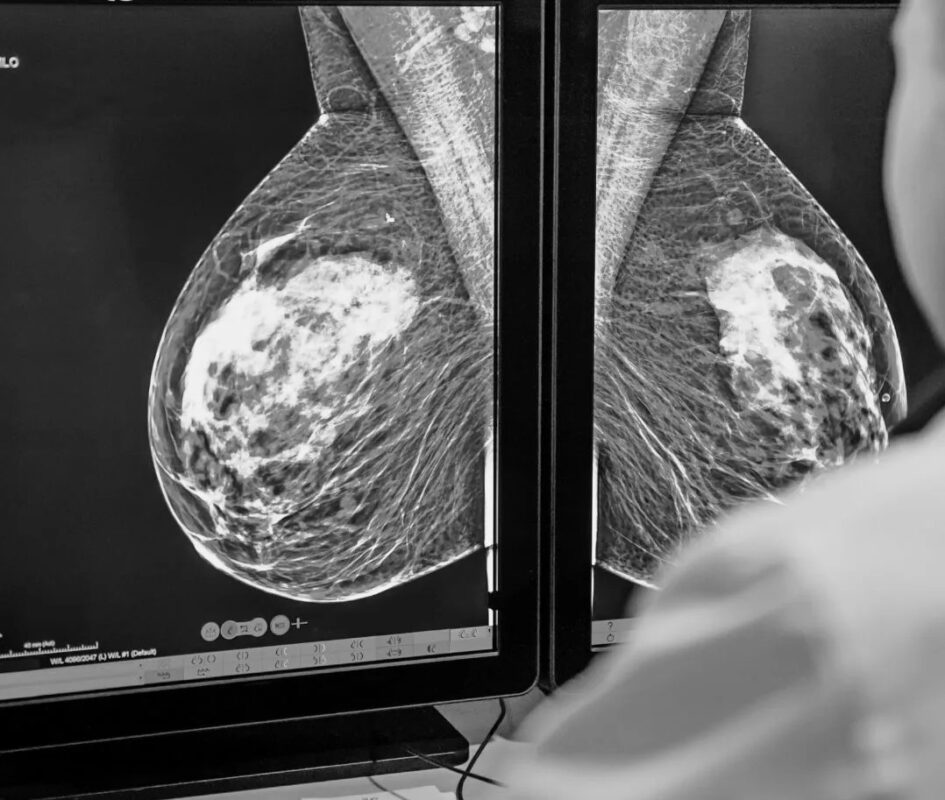
2D Digital Mammography (FFDM): Standard digital mammography remains the foundation of breast cancer screening and is available at virtually all mammography centers. It captures flat, two-dimensional X-ray images of the breast tissue.
3D Mammography (Digital Breast Tomosynthesis / DBT): DBT acquires multiple low-dose X-ray images from different angles and reconstructs them into a three-dimensional dataset that radiologists can scroll through layer by layer. In a study of nearly 460,000 screening examinations (Friedewald SM et al., JAMA 2014), DBT detected 27–41% more invasive cancers than 2D alone and reduced callback rates by approximately 15%. DBT has become the standard at many breast imaging centers and is recommended over 2D by the American College of Radiology when available.
Breast Ultrasound
Breast ultrasound uses sound waves rather than radiation. Its main roles in screening include: supplemental screening for women with dense breasts who cannot have MRI; evaluation of palpable abnormalities; and distinguishing cysts from solid masses. Whole-breast ultrasound as supplemental screening detects approximately 4 additional cancers per 1,000 women screened beyond mammography, but with a higher false-positive rate.
Breast MRI
Breast MRI with gadolinium contrast has sensitivity approaching 90–95% — significantly higher than mammography. It is the standard supplemental imaging for high-risk women (annual MRI + annual mammography). Abbreviated breast MRI (Fast MRI, 3–10 minute protocol) is emerging as a practical option for intermediate-risk women with dense breasts, detecting approximately 9–16 additional cancers per 1,000 screened beyond mammography.
Breast Cancer Screening Guidelines — What Each Organization Says
Breast cancer screening guidelines have historically differed on starting age (40 vs. 45 vs. 50) and frequency (annual vs. biennial). The 2024 USPSTF update significantly narrowed those differences. Here is what each major organization currently recommends:

Start: Age 40
Frequency: Every 2 years (biennial)
Stop: Age 74
Grade: B
40–44: Optional annual
45–54: Annual
55+: Biennial or annual
Stop: When life expectancy <10 yrs
Start: Age 40
Frequency: Annual
Modality: 3D preferred over 2D
Most aggressive guideline
Start: Age 40
Frequency: Annual
Risk-stratified: Earlier + MRI for high-risk women
Benefits and Harms of Mammography Screening
The ongoing debate over screening recommendations exists because mammography — like all medical screening — produces both benefits and harms. An informed decision requires understanding both.
Benefits
Mortality reduction: A meta-analysis of randomized controlled trials (Gøtzsche PC & Jørgensen KJ, Cochrane 2013) estimated a 20% relative reduction in breast cancer-specific mortality with mammography screening; other analyses have estimated 25–35%. NCI estimates that for every 10,000 women screened annually for 10 years, approximately 3–5 breast cancer deaths are prevented.
Stage shift: Screening detects cancers at earlier stages when they are more treatable. Screened populations have significantly higher proportions of Stage I cancers and lower proportions of Stage III–IV at diagnosis compared to unscreened populations. Earlier-stage diagnosis means more treatment options, including less aggressive surgery, and substantially better survival.
Harms
False positives: The most commonly discussed harm. In a study of approximately 170,000 women (Hubbard RA et al., Ann Intern Med 2011), 61% of women screened annually for 10 years had at least one false-positive mammogram; 7% had at least one false-positive biopsy recommendation. The great majority of false positives resolve with additional imaging rather than surgery.
Overdiagnosis: Detection of cancers that would never have caused symptoms or death in the patient’s lifetime. Estimates range from approximately 11–22% of screen-detected cancers. This is the most contested harm of mammography. Current pathology cannot reliably identify which screen-detected cancers would have remained indolent — so all detected cancers are treated.
Radiation: Approximately 0.4 mSv per mammogram — equivalent to roughly 7 weeks of background radiation. The lifetime risk from screening radiation is considered negligible compared to the mortality benefit.
False negatives: Mammography misses approximately 10–20% of breast cancers; miss rates are higher in dense breast tissue (see below). False negatives can delay diagnosis in women with a new symptom they attribute to their recent “normal” mammogram.
For most average-risk women beginning at 40, the mortality reduction benefit of regular mammography substantially outweighs the harms — particularly when women understand what a callback means before they experience one.
Dense Breasts and Supplemental Screening
What Breast Density Means
Breast density describes the proportion of fibroglandular tissue vs. fat in the breast, as seen on mammography. Radiologists categorize density using the ACR BI-RADS system:
Lowest density; highest mammogram sensitivity; lowest cancer risk from density
Some fibroglandular tissue scattered throughout predominantly fatty tissue
Moderately dense; may reduce mammogram sensitivity; discuss supplemental screening
Highest density; 4–6× higher cancer risk; mammogram sensitivity ~50%; supplemental screening strongly recommended
Approximately 40–50% of US screening-age women have dense breasts (Category C or D). Density is not something you can feel — it is a radiological finding. You should be notified if you have dense breasts (required by law in most US states; FDA mandated notification as of 2023).
Supplemental Screening Options for Dense Breasts
- Supplemental breast ultrasound: Detects approximately 4 additional cancers per 1,000 screened women beyond mammography; higher false-positive rate; no radiation; no contrast; widely available
- Abbreviated breast MRI (Fast MRI): Detects approximately 9–16 additional cancers per 1,000; significantly better than ultrasound for supplemental screening; requires IV gadolinium contrast; 3–10 minute protocol; emerging as a practical option as cost and availability improve
- Full diagnostic MRI: Most comprehensive; reserved for high-risk women (see below)
The decision about supplemental screening depends on your overall risk level, preferences, and insurance coverage. If you have Category C or D density, discuss with your provider whether supplemental screening is appropriate for you.
High-Risk Breast Cancer Screening
Who Qualifies as High-Risk?
The following individuals qualify for enhanced screening protocols:
- BRCA1 or BRCA2 pathogenic variant carrier (or untested first-degree relative of a known carrier)
- Lifetime breast cancer risk ≥20–25% by a validated risk assessment model (Tyrer-Cuzick, BRCAPRO, or similar)
- History of chest wall or breast radiation between ages 10–30 (e.g., mantle field for Hodgkin lymphoma)
- Li-Fraumeni syndrome (TP53 mutation), Cowden syndrome (PTEN), or Bannayan-Riley-Ruvalcaba syndrome
The High-Risk Screening Protocol
Per NCCN and ACS high-risk guidelines: annual mammography (3D preferred) AND annual breast MRI, typically beginning at age 25–30 or 10 years before the youngest affected relative’s age at diagnosis, whichever is earlier. Clinical breast examination every 6–12 months. Discussion of risk-reduction strategies: chemoprevention (tamoxifen, raloxifene, exemestane) and prophylactic risk-reducing mastectomy (reduces breast cancer risk by >90% in BRCA carriers).
What Happens After an Abnormal Mammogram
Receiving a callback after a screening mammogram is anxiety-producing — but it is critical to understand that the overwhelming majority of callbacks do not result in a cancer diagnosis.
The callback statistics: Approximately 10% of screened women are called back for additional evaluation. Of those: approximately 90% are cleared with additional imaging alone; approximately 10% are recommended for biopsy; of those biopsied, approximately 20–25% are found to have cancer. In other words: for every 100 women called back, approximately 1–2 are ultimately diagnosed with breast cancer.
Understanding BI-RADS Categories
| BI-RADS | Meaning | Next Step |
|---|---|---|
| 0 | Incomplete — additional imaging needed | Diagnostic mammogram, ultrasound, or MRI |
| 1 | Negative — no abnormality found | Routine next scheduled screening |
| 2 | Benign finding — clearly not cancer | Routine next scheduled screening |
| 3 | Probably benign — <2% chance of malignancy | 6-month short-interval follow-up mammogram |
| 4 | Suspicious — biopsy recommended | Core needle biopsy (4A/4B/4C = increasing suspicion) |
| 5 | Highly suspicious for malignancy | Biopsy required |
| 6 | Known biopsy-proven malignancy | Treatment planning |
If biopsy is recommended: core needle biopsy — guided by ultrasound or stereotactic (mammogram-guided) technique — is the standard outpatient procedure performed under local anesthesia. No surgical incision is required. Results typically take 2–5 business days. For a guide to what questions to ask at every step of the process, see the cancer questions for doctor guide.
For a complete guide to what breast cancer is, its types, stages, and treatment options, see the breast cancer overview article. For a guide to breast symptoms that may prompt earlier evaluation between screening appointments, see the breast cancer symptoms article.
Frequently Asked Questions
- Friedewald SM et al. — Breast cancer screening using tomosynthesis in combination with digital mammography; JAMA 2014
- Gøtzsche PC & Jørgensen KJ — Screening for breast cancer with mammography; Cochrane Database Syst Rev 2013
- Hubbard RA et al. — Cumulative probability of false-positive recall or biopsy recommendation after 10 years of screening mammography; Ann Intern Med 2011
- USPSTF 2024 — Breast cancer screening recommendation statement
- Oeffinger KC et al. (ACS) — Breast cancer screening for women at average risk: 2015 guideline update; JAMA 2015
- National Cancer Institute — cancer.gov screening fact sheet
- American Cancer Society — cancer.org
This article is for educational purposes only and does not constitute medical advice. Screening decisions should be made in consultation with a qualified healthcare provider based on individual risk factors, preferences, and clinical context.
Breast Cancer Treatment: Key Advances and Approaches
Breast cancer treatment has become increasingly personalized over the past two decades, driven by advances in tumor biology characterization that distinguish meaningfully different breast cancer subtypes with different biological behaviors and optimal treatment approaches. The four major molecular subtypes of breast cancer — Luminal A (hormone receptor-positive, HER2-negative, low-grade), Luminal B (hormone receptor-positive, HER2-negative, high-grade or HER2-positive), HER2-enriched (HER2-positive, hormone receptor-negative), and triple-negative (estrogen receptor-negative, progesterone receptor-negative, HER2-negative) — each have distinct prognoses, responses to systemic therapy, and optimal treatment sequencing.
Hormone receptor-positive (HR+) breast cancer: HR+ breast cancer, which accounts for approximately 70% of cases, is treated with endocrine therapy (hormone-blocking treatment) as the cornerstone of systemic therapy. For premenopausal women, tamoxifen (5–10 years) or ovarian suppression plus an aromatase inhibitor is standard. For postmenopausal women, aromatase inhibitors (anastrozole, letrozole, exemestane) have superseded tamoxifen as the preferred endocrine therapy due to superior efficacy. In metastatic HR+ breast cancer, the CDK4/6 inhibitors (palbociclib, ribociclib, abemaciclib) combined with an aromatase inhibitor or fulvestrant have transformed outcomes: the MONARCH, PALOMA, and MONALEESA trials established this combination as first-line standard of care, substantially improving progression-free and overall survival compared to endocrine therapy alone.
HER2-positive breast cancer: HER2-positive breast cancer, which accounts for approximately 15–20% of cases, was once associated with a poor prognosis but is now one of the most treatable breast cancer subtypes due to the development of HER2-targeted therapies. Trastuzumab (Herceptin) was the first anti-HER2 agent and remains a cornerstone of treatment. For early-stage HER2-positive breast cancer, neoadjuvant pertuzumab + trastuzumab + chemotherapy followed by adjuvant T-DM1 (if residual disease) is standard, based on the APHINITY and KATHERINE trials. In metastatic HER2-positive disease, trastuzumab deruxtecan (T-DXd / Enhertu) has demonstrated remarkable efficacy even in patients who have progressed through multiple prior lines of HER2-directed therapy, with response rates exceeding 60% in heavily pretreated patients (DESTINY-Breast01/02/03 trials).
Triple-negative breast cancer (TNBC): TNBC — which accounts for approximately 10–15% of breast cancers and is disproportionately common in younger women and Black women — was historically treated with cytotoxic chemotherapy alone. Several advances have improved outcomes: pembrolizumab (Keytruda) added to neoadjuvant chemotherapy for early-stage, high-risk TNBC improved event-free survival in the KEYNOTE-522 trial and is now standard for eligible patients. Olaparib (for BRCA1/2 germline mutation carriers) and sacituzumab govitecan (Trodelvy, an antibody-drug conjugate targeting Trop-2) have improved outcomes in metastatic TNBC.
For authoritative information on breast cancer, the American Cancer Society’s breast cancer resource provides patient-friendly comprehensive guides. The National Cancer Institute’s breast cancer PDQ offers evidence-based clinical summaries. The NCCN Breast Cancer Guidelines are the most widely used clinical practice standards among U.S. oncologists. For information about breast cancer symptoms that often lead to initial evaluation, see our guide to breast cancer symptoms. For information about recommended breast cancer screening approaches — including mammography and supplemental MRI for high-risk women — see our comprehensive guide to breast cancer screening. For information about what a breast lump means and how it is evaluated, see our article on breast lumps.
Breast cancer survivorship — the period after completion of primary treatment — brings its own set of considerations. Long-term follow-up with an oncology team, adherence to prescribed adjuvant endocrine therapy (which may continue for 5–10 years for hormone receptor-positive breast cancer), monitoring for late effects of treatment (including bone loss from aromatase inhibitors, cardiac effects from anthracyclines or trastuzumab, and lymphedema from axillary surgery), and attention to lifestyle factors associated with reduced recurrence risk (physical activity, healthy weight maintenance, limiting alcohol) are all important components of survivorship care. Women with a personal history of breast cancer should continue annual mammography and, if high-risk, annual breast MRI. Discussing a personalized survivorship care plan with your oncology team helps ensure that surveillance, screening, and ongoing health needs are clearly defined after active treatment is complete.
Pingback: Mammogram: What to Expect, Types, and How to Read Results - Horizon Health Guide
Pingback: Mammogram Screening: A Complete Guide by Age and Risk Level - Horizon Health Guide
Pingback: Breast Cancer Risk Factors: Genetic, Hormonal, and Lifestyle - Horizon Health Guide