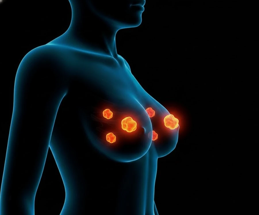
Each year, approximately 39 to 40 million mammograms are performed in the United States — more than any other cancer screening test. Yet approximately 36% of women over 45 who should be having regular mammograms are not current with their screening. That underscreening gap — not the absence of available mammography, but the gap between eligibility and participation — represents the most significant remaining opportunity to reduce breast cancer mortality in the U.S.
The reasons women skip mammograms are well-studied: uncertainty about when to start or how often to go, confusion about conflicting guidelines, anxiety about callbacks, concerns about cost, and the logistical friction of scheduling. This guide addresses those barriers directly — not the theoretical case for mammography screening, which has been settled — but the practical information that turns a vague intention to “get a mammogram” into a consistent, optimized screening habit.
This guide covers who should begin mammogram screening and when, how to decide between annual and biennial scheduling, what to discuss with your doctor to ensure you are in the right protocol for your risk level, what mammogram screening looks like at different life stages, when standard mammography is not enough, and how to build screening into a sustainable routine.
Who Should Get Mammogram Screening and When
The controversy about when to begin mammogram screening has largely resolved. All major U.S. guidelines now support starting at or before age 45 for average-risk women, and most recommend beginning at 40.
All major guidelines support starting at 40. First mammogram establishes baseline — critical for all future comparisons. Higher density common in younger women.
Peak benefit period. Higher proportion of aggressive subtypes — particularly in 40–49. Annual catches more interval cancers. Most clinicians recommend annual for this group.
Continue as long as overall health is good and life expectancy ⁉10 years. Slower-growing HR+ tumors predominate; biennial sensitivity is higher relative to tumor growth rates.
USPSTF: insufficient evidence; not recommending against. ACS: continue if life expectancy ⁉10 years. Decision depends on health status, comorbidities, and patient values.
The first mammogram at 40 establishes a critical baseline against which all future images are compared. Many findings that are straightforward to dismiss in later years — a stable benign-appearing density — require additional imaging in the first year simply because there is nothing to compare them to. Starting at 40 rather than 45 or 50 allows more years of comparative data to accumulate before cancer risk increases in the late 40s. For a detailed comparison of what each guideline says, see the breast cancer screening guidelines article.
Annual vs. Biennial Mammogram Screening — How to Decide
For women who have decided to screen, the annual vs. biennial question is the most consequential remaining decision. Both are evidence-based. The right choice depends on your risk profile and your values.
- Women ages 40–54: higher rates of aggressive subtypes; more interval cancers with biennial
- Women with dense breasts (Category C/D): lower mammogram sensitivity; value in frequency
- Women with elevated (not high) risk: family history, prior atypical biopsy
- Women who strongly value maximum early detection
- Catches ~15.6% more cancers over 10 years vs biennial (Myers JAMA 2015)
- Average-risk women 55+ with predominantly slow-growing HR+ tumors
- Women with fatty or scattered density: better sensitivity, less gained from frequency
- Women who have had multiple false-positive callbacks and find the anxiety disproportionate
- Women who prefer minimizing medical touchpoints while maintaining evidence-based care
- ~42% cumulative false-positive rate at 10 years vs 61% annual (Hubbard 2011)
What to Tell Your Doctor Before Mammogram Screening
The information you share with your provider before starting mammogram screening determines whether you receive a standard or enhanced protocol — a decision that can significantly affect your outcomes.
Family History Details That Matter
- First-degree relatives (mother, sister, daughter) with breast cancer — and their age at diagnosis (premenopausal diagnosis at 42 carries different implications than postmenopausal at 72)
- Second-degree relatives: maternal/paternal grandmothers, aunts
- Ovarian cancer in the family — particularly relevant for BRCA mutation probability
- Known genetic mutations: BRCA1, BRCA2, PALB2, ATM, CHEK2
- Ashkenazi Jewish ancestry — carries higher BRCA1/2 prevalence
Personal Breast History
- Prior breast biopsies: atypical ductal hyperplasia (ADH), atypical lobular hyperplasia (ALH), or LCIS significantly elevates lifetime risk
- Prior breast cancer (personal history, not family)
- History of chest wall or breast radiation between ages 10–30 — qualifies for high-risk protocol
- Dense breast history from prior mammograms
How Risk Models Work
When your provider collects this information, they may calculate your lifetime breast cancer risk using a validated model — most commonly the Tyrer-Cuzick model (preferred by NCCN; incorporates family history, hormonal factors, breast density, prior biopsies, and BMI) or BRCApro (focused on estimating BRCA1/2 mutation probability). A lifetime risk ≥20–25% qualifies you for a high-risk protocol: annual mammogram plus annual breast MRI beginning at age 25–30. Understanding which category you fall into is the most important outcome of your initial screening conversation with your provider.
Mammogram Screening by Life Stage
What a first mammogram at 42 means, feels like, and produces is different from a mammogram at 52 or 68. Understanding what is typical at each stage helps normalize the experience and manage expectations throughout your screening journey.
The first mammogram establishes your baseline. Because no prior images exist for comparison, your radiologist cannot evaluate whether findings are new or have been stable for years — a limitation that drives more BI-RADS 0 (incomplete) results in first-time mammograms. Breast tissue in younger women also tends to be denser, which reduces sensitivity and increases callback likelihood. If called back after a first mammogram, it is more likely to reflect the absence of a baseline than a suspicious finding. Bring any prior breast imaging from other contexts if available, and ask whether your images will be stored electronically for easy access at future facilities.
Breast density often decreases around menopause as fibroglandular tissue is replaced by fat — mammogram sensitivity typically improves in this transition. However, hormone replacement therapy (HRT), particularly combined estrogen-progesterone, can maintain or increase breast density. Inform your radiologist and scheduling facility if you are on HRT. Combined HRT also modestly increases breast cancer risk; discuss with your provider how this affects your individual screening plan.
Women in their 60s should be in a consistent screening rhythm. At age 65, a U.S. woman has an average life expectancy of approximately 20 additional years — ample time to benefit from early cancer detection. Medicare Part B covers annual screening mammograms at 100% with no cost-sharing when the provider accepts Medicare assignment, removing the financial barrier entirely for this group. Underscreening in this age group is driven more by inertia than by any clinical rationale.
Screening decisions for women 75 and older should be individualized. The relevant questions: How is your overall health? Do you have significant comorbidities that would affect your ability to tolerate breast cancer treatment if found? What are your values regarding medical intervention? Women in good health with a life expectancy of 10 or more years likely continue to benefit. Women with serious chronic illness or limited life expectancy have less to gain from early detection. This decision belongs with your primary care provider.
When Standard Mammogram Screening Isn’t Enough
Dense Breasts
Approximately 40–50% of U.S. screening-age women have heterogeneously dense or extremely dense breasts (BI-RADS Category C or D). Dense tissue reduces mammogram sensitivity from approximately 85–90% in fatty breasts to 50–65% in dense breasts. The FDA’s 2023 updated MQSA rule requires all mammography facilities to notify patients about their breast density in plain language. If you have dense breasts, discuss supplemental screening options with your provider:
- Supplemental breast ultrasound: adds approximately 4 additional cancer detections per 1,000 screened women; higher false-positive rate; no radiation or contrast required
- Abbreviated breast MRI: adds approximately 9–16 additional detections per 1,000; higher sensitivity than ultrasound; 3–10 minute protocol; requires IV gadolinium contrast
High-Risk Women
Women with BRCA1/2 mutations, lifetime breast cancer risk ≥20–25% by validated model, or history of thoracic radiation at ages 10–30 qualify for annual mammogram (3D preferred) plus annual breast MRI, typically beginning at age 25–30. For a complete breakdown of high-risk criteria and the evidence behind supplemental screening, see the breast cancer screening article.
Women with Breast Implants
Mammography with implants requires a modified technique: 4 standard views plus 4 Eklund displacement views (implant displaced posteriorly; breast tissue pulled forward) — 8 total images. Most trained mammography technologists perform this routinely; inform the scheduling facility when booking that you have implants.
Making Mammogram Screening a Sustainable Habit
Schedule your next appointment before leaving the current one. Link it to your birth month or annual well-woman exam. Add a calendar reminder the same date each year.
Request a copy of your images and report after each exam. Essential when switching facilities. Keep dates, BI-RADS scores, and follow-up notes in your health records.
All U.S. facilities must be MQSA-certified (searchable at FDA website). Look for ACR Breast Imaging Center of Excellence or NAPBC designation. Ask if 3D mammography is available.
Mobile mammography units travel to workplaces, community centers, and rural areas. If access or scheduling is a barrier, contact your local hospital or health department about mobile programs.
Overcoming common barriers:
- Cost: ACA-compliant plans cover screening mammograms without cost-sharing; Medicare Part B covers annual screening at 100%; NBCCEDP provides free/low-cost mammograms for uninsured low-income women
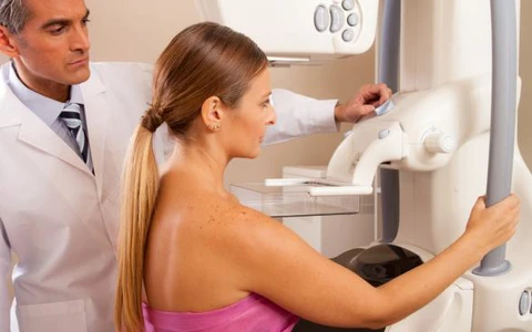
- Callback anxiety: Know the statistics in advance — approximately 10% of screenings lead to a callback, and 90% of callbacks are resolved by additional imaging alone. See the mammogram procedure guide for what to expect at every step.
- Pain concerns: Schedule the week after your period begins; inform the technologist if compression is too tight
- Lack of provider recommendation: Ask your primary care provider or gynecologist directly — “Should I be getting a mammogram, and how often?”
What Mammogram Screening Has Achieved
The Stage Shift
In the pre-screening era (prior to the 1980s), the majority of breast cancers were diagnosed at Stage II or later. Today, approximately 65% of newly diagnosed breast cancers in regularly screened U.S. women are caught at Stage I or II — before regional spread, when treatment is less aggressive and outcomes are dramatically better.
| Stage at Diagnosis | 5-Year Relative Survival | Treatment Context |
|---|---|---|
| Stage I (localized) | ~99% | Often lumpectomy ± radiation; excellent outcomes |
| Stage II (regional, small) | ~86% | Lumpectomy or mastectomy; adjuvant therapy likely |
| Stage III (regional, advanced) | ~57% | Often neoadjuvant chemotherapy + surgery + radiation |
| Stage IV (distant/metastatic) | ~28% | Systemic therapy; not curative in most cases |
Mortality Decline
U.S. breast cancer mortality declined by approximately 40% between 1989 and 2020 (NCI data). Researchers attribute approximately half of this decline to widespread mammography screening and half to improvements in systemic therapy. The combination of earlier detection and more effective treatment represents one of the most significant oncologic mortality reductions in modern medicine.
Approximately 36% of eligible U.S. women are not current with mammogram screening. Closing that gap — through better provider communication, reducing cost barriers, normalizing the process, and addressing anxiety about results — remains the primary frontier in breast cancer outcomes improvement.
For details about what happens during a mammogram appointment and how to interpret your results, see the mammogram procedure guide. For a comparison of all major screening guidelines, see the breast cancer screening guidelines article. For an overview of breast cancer types and treatment, see the breast cancer overview. If you notice any new breast cancer symptoms between screening appointments, see your provider promptly.
Frequently Asked Questions
- Hubbard RA et al. — Cumulative probability of false-positive recall or biopsy recommendation after 10 years of screening mammography; Ann Intern Med 2011;155(8):481–492
- Myers ER et al. — Benefits and harms of breast cancer screening: a systematic review; JAMA 2015;314(15):1615–1634
- Gøtzsche PC & Jørgensen KJ — Screening for breast cancer with mammography; Cochrane Database Syst Rev 2013
- NCI SEER — Breast cancer stage distribution and 5-year survival statistics
- CDC BRFSS 2020 — Breast cancer screening utilization rates
- USPSTF 2024 — Breast cancer screening recommendation update
- National Cancer Institute — cancer.gov/types/breast/screening-fact-sheet
- American Cancer Society — cancer.org mammogram recommendations
This article is for educational purposes only and does not constitute medical advice. All screening decisions should be made in consultation with a qualified healthcare provider based on individual risk factors, history, and clinical context.
Breast Cancer Treatment: Key Advances and Approaches
Breast cancer treatment has become increasingly personalized over the past two decades, driven by advances in tumor biology characterization that distinguish meaningfully different breast cancer subtypes with different biological behaviors and optimal treatment approaches. The four major molecular subtypes of breast cancer — Luminal A (hormone receptor-positive, HER2-negative, low-grade), Luminal B (hormone receptor-positive, HER2-negative, high-grade or HER2-positive), HER2-enriched (HER2-positive, hormone receptor-negative), and triple-negative (estrogen receptor-negative, progesterone receptor-negative, HER2-negative) — each have distinct prognoses, responses to systemic therapy, and optimal treatment sequencing.
Hormone receptor-positive (HR+) breast cancer: HR+ breast cancer, which accounts for approximately 70% of cases, is treated with endocrine therapy (hormone-blocking treatment) as the cornerstone of systemic therapy. For premenopausal women, tamoxifen (5–10 years) or ovarian suppression plus an aromatase inhibitor is standard. For postmenopausal women, aromatase inhibitors (anastrozole, letrozole, exemestane) have superseded tamoxifen as the preferred endocrine therapy due to superior efficacy. In metastatic HR+ breast cancer, the CDK4/6 inhibitors (palbociclib, ribociclib, abemaciclib) combined with an aromatase inhibitor or fulvestrant have transformed outcomes: the MONARCH, PALOMA, and MONALEESA trials established this combination as first-line standard of care, substantially improving progression-free and overall survival compared to endocrine therapy alone.
HER2-positive breast cancer: HER2-positive breast cancer, which accounts for approximately 15–20% of cases, was once associated with a poor prognosis but is now one of the most treatable breast cancer subtypes due to the development of HER2-targeted therapies. Trastuzumab (Herceptin) was the first anti-HER2 agent and remains a cornerstone of treatment. For early-stage HER2-positive breast cancer, neoadjuvant pertuzumab + trastuzumab + chemotherapy followed by adjuvant T-DM1 (if residual disease) is standard, based on the APHINITY and KATHERINE trials. In metastatic HER2-positive disease, trastuzumab deruxtecan (T-DXd / Enhertu) has demonstrated remarkable efficacy even in patients who have progressed through multiple prior lines of HER2-directed therapy, with response rates exceeding 60% in heavily pretreated patients (DESTINY-Breast01/02/03 trials).
Triple-negative breast cancer (TNBC): TNBC — which accounts for approximately 10–15% of breast cancers and is disproportionately common in younger women and Black women — was historically treated with cytotoxic chemotherapy alone. Several advances have improved outcomes: pembrolizumab (Keytruda) added to neoadjuvant chemotherapy for early-stage, high-risk TNBC improved event-free survival in the KEYNOTE-522 trial and is now standard for eligible patients. Olaparib (for BRCA1/2 germline mutation carriers) and sacituzumab govitecan (Trodelvy, an antibody-drug conjugate targeting Trop-2) have improved outcomes in metastatic TNBC.
For authoritative information on breast cancer, the American Cancer Society’s breast cancer resource provides patient-friendly comprehensive guides. The National Cancer Institute’s breast cancer PDQ offers evidence-based clinical summaries. The NCCN Breast Cancer Guidelines are the most widely used clinical practice standards among U.S. oncologists. For information about breast cancer symptoms that often lead to initial evaluation, see our guide to breast cancer symptoms. For information about recommended breast cancer screening approaches — including mammography and supplemental MRI for high-risk women — see our comprehensive guide to breast cancer screening. For information about what a breast lump means and how it is evaluated, see our article on breast lumps.
Pingback: Breast Cancer Risk Factors: Genetic, Hormonal, and Lifestyle - Horizon Health Guide