
Heart Valve Disease: What Adults Should Know
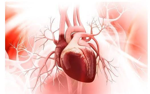
Heart valve disease encompasses any condition that impairs the function of one or more of the heart’s four valves — the mitral, tricuspid, aortic, and pulmonary valves that serve as one-way gates controlling blood flow through the cardiac chambers and into the great vessels. When a valve functions normally, it opens fully to allow unobstructed forward flow and closes completely to prevent backward leakage. Heart valve disease disrupts one or both of these functions: stenosis narrows the valve opening, obstructing forward flow and forcing the upstream chamber to generate higher pressure to push blood through; regurgitation (or insufficiency) prevents complete closure, allowing backward leakage that forces the heart to handle a larger volume with each beat. Both mechanisms impose abnormal loads on cardiac chambers that, over time, cause pathological remodeling and ultimately heart failure if the valve lesion is not corrected.
Heart valve disease is common and increasing in prevalence as the population ages. Approximately 2.5 percent of the U.S. population has moderate or severe valve disease, and prevalence increases dramatically with age — affecting approximately 13 percent of adults over 75. The most common clinically significant valve lesions in developed countries are aortic stenosis (degenerative calcification in older adults), mitral regurgitation (degenerative prolapse or coronary disease), and aortic regurgitation. Rheumatic heart disease — which historically dominated valve disease epidemiology globally — remains the primary cause of mitral stenosis and multivalvular disease in developing countries, where rheumatic fever following streptococcal pharyngitis is still prevalent. Advances in valve intervention over the past two decades — particularly the development of transcatheter heart valves that allow valve repair or replacement without open-heart surgery — have transformed the treatment landscape and extended intervention eligibility to patients who previously could not safely undergo cardiac surgery.
Aortic Stenosis — The Most Common Severe Valve Lesion in Adults
Aortic stenosis (AS) is the most common severe valve lesion in developed countries, affecting approximately 2 to 5 percent of adults over 65 and 8 to 10 percent of those over 80. Degenerative (calcific) aortic stenosis — caused by progressive calcium deposition on the aortic valve leaflets that gradually stiffens and narrows the valve opening — is the dominant etiology in older adults, sharing many risk factors with coronary artery disease (hypertension, dyslipidemia, diabetes, smoking, male sex). Bicuspid aortic valve, a congenital abnormality present in 1 to 2 percent of the population in which the normally three-leaflet aortic valve has only two leaflets, accelerates calcific degeneration, causing severe AS several decades earlier than tricuspid calcific disease (typically presenting in the 50s and 60s rather than 70s and 80s).
Aortic stenosis progresses through a long asymptomatic phase, during which the left ventricle compensates for the increased pressure load by developing concentric hypertrophy (thickened walls, preserved ejection fraction). This compensated phase can last decades, with patients remaining asymptomatic despite progressively worsening stenosis. The clinical transition point is the onset of symptoms — the classic triad of angina (chest pain with exertion), syncope (fainting, particularly with exertion), and heart failure (dyspnea, reduced exercise tolerance). Once symptoms develop, prognosis without intervention is poor: median survival with angina or syncope is 2 to 3 years; with heart failure symptoms, 1 to 2 years. The onset of symptoms is therefore a critical inflection point that triggers referral for valve replacement evaluation.
Severity grading in aortic stenosis uses echocardiographic Doppler parameters: peak velocity across the valve (normal below 2 m/s; mild AS 2 to 3 m/s; moderate AS 3 to 4 m/s; severe AS above 4 m/s), mean pressure gradient (severe AS generally above 40 mmHg), and calculated valve area (severe AS below 1.0 cm²). “Low-flow, low-gradient” AS — where a failing LV cannot generate high gradients despite severe valve narrowing — requires additional assessment with dobutamine stress echocardiography or CT calcium scoring to confirm anatomic severity and distinguish true severe AS from pseudo-severe AS in a patient with a primary cardiomyopathy.
Mitral Regurgitation — The Most Prevalent Valve Lesion Overall
Mitral regurgitation (MR) is the most common valve lesion by prevalence, affecting approximately 10 percent of the general adult population in mild degrees and 2 to 3 percent in moderate or severe degrees. The mitral valve is a complex structure consisting of two leaflets, the annulus (fibrous ring), chordae tendineae (tendon-like cords supporting the leaflets), and papillary muscles (myocardial projections to which the chordae attach) — any component of which can be affected by disease to produce MR.
Degenerative (primary) MR — caused by intrinsic abnormality of the valve leaflets and supporting structures — is the dominant etiology in developed countries. Mitral valve prolapse (MVP) is the most common cause: the leaflet tissue is myxomatous (thickened, abnormally elastic) and billows back into the left atrium during systole, disrupting coaptation and allowing regurgitation. MVP affects 2 to 3 percent of the general population, is more common in women, and in most cases produces only mild-to-moderate regurgitation that is well tolerated for decades. A subset of MVP patients develop severe MR requiring intervention, either from progressive leaflet disease or chordal rupture (which causes acute severe MR, a cardiovascular emergency presenting with acute pulmonary edema and hemodynamic instability).
Functional (secondary) MR results not from intrinsic valve disease but from left ventricular remodeling that displaces the papillary muscles and prevents normal leaflet coaptation despite anatomically normal leaflets. Functional MR complicates approximately 20 to 30 percent of patients with dilated cardiomyopathy or prior myocardial infarction involving the posterior LV wall; it worsens heart failure by increasing regurgitant volume and is associated with worse prognosis in HFrEF patients. The management of functional MR is more nuanced than primary MR — GDMT for HFrEF (which can reduce LV size and functional MR severity through reverse remodeling) is the foundation, and transcatheter edge-to-edge mitral repair (MitraClip) has been shown to improve survival and reduce hospitalizations in selected patients with persistent severe secondary MR despite optimal medical therapy.
When Does Valve Disease Require Intervention?
Valve intervention — whether surgical valve repair or replacement, or transcatheter valve repair or replacement — is indicated when the hemodynamic burden imposed by the valve lesion is about to cause or has already caused irreversible cardiac damage, or when symptoms significantly impair quality of life despite medical management. The general principles that guide intervention timing differ by valve lesion type and the relative risk of the intervention versus the risk of continued observation.
For aortic stenosis, intervention is indicated at the onset of symptoms (angina, syncope, or heart failure) regardless of anatomic severity indices, because symptomatic severe AS carries a high short-term mortality risk without intervention. Intervention is also indicated in asymptomatic patients with very severe AS (peak velocity above 5 m/s) or those undergoing other cardiac surgery (where concomitant aortic valve replacement adds modest operative risk), and is reasonable for those with rapid disease progression or results from exercise testing showing symptom onset or hemodynamic deterioration with exertion.
For severe primary mitral regurgitation, intervention is indicated in symptomatic patients (NYHA Class II or above) and in asymptomatic patients with evidence of early LV dysfunction (LVEF 60 percent or below, which in the context of the volume overload of severe MR represents already significantly impaired myocardial contractile reserve) or LV dilation (LVESD 40 mm or above). Early intervention before overt LV dysfunction and before severe LV dilation develops is associated with better long-term outcomes — waiting for symptoms alone in primary MR risks allowing irreversible LV impairment to develop before the trigger for intervention is met.
The decision between surgical and transcatheter approaches is increasingly complex and patient-specific. Surgical mitral valve repair — at experienced centers where repair rates for primary MR exceed 95 percent — produces durable, excellent outcomes and is preferred over replacement when feasible, since preserving the native valve maintains better LV geometry and function. Transcatheter mitral edge-to-edge repair (MitraClip, PASCAL) is used in patients with prohibitive surgical risk and anatomy suitable for the device. Surgical aortic valve replacement (SAVR) remains preferred in younger patients (particularly under 65 to 70) due to longer durability of surgical valves and concerns about the need for valve-in-valve TAVR procedures as transcatheter valves degenerate — a consideration that matters more in patients with decades of expected life remaining.
Monitoring Valve Disease — Living With a Known Valve Problem
Most adults with mild-to-moderate valve disease are managed conservatively — with periodic echocardiographic surveillance to detect progression, medical management of comorbidities that can accelerate valve disease (blood pressure control is particularly important in aortic stenosis, aortic regurgitation, and functional MR), and education on symptoms that should prompt earlier evaluation. The appropriate surveillance interval depends on the lesion type and severity: mild aortic stenosis is typically monitored echocardiographically every 3 to 5 years; moderate AS every 1 to 2 years; severe asymptomatic AS annually. Similarly, moderate MR warrants echocardiography every 1 to 2 years, while severe asymptomatic primary MR is monitored every 6 to 12 months.
Patients with prosthetic heart valves — either mechanical or bioprosthetic — require specific ongoing management. Mechanical valves require lifelong anticoagulation with warfarin (maintaining INR in the target range specific to valve position and patient risk factors) because their non-biological materials are thrombogenic without anticoagulation. Bioprosthetic valves (made from porcine or bovine pericardium) do not require long-term anticoagulation in most patients after the early post-implant period, but degenerate over time — typically requiring re-intervention after 10 to 20 years depending on patient age and valve type, with younger patients (whose higher metabolic activity accelerates bioprosthetic degeneration) experiencing shorter durability.
The American Heart Association’s heart valve disease resources provide comprehensive guidance for patients living with valve conditions. The CDC valvular heart disease overview covers risk factors, prevalence, and management basics. The NHLBI heart valve disease guide addresses all four valves, symptoms, testing, treatment options, and living with valve disease.
Related reading: Heart Failure | Cardiomyopathy Guide | Coronary Artery Disease | What Causes Heart Disease? | Major Risk Factors for Heart Disease
Sources
- Otto CM, et al. 2020 ACC/AHA Guideline for the Management of Patients With Valvular Heart Disease. J Am Coll Cardiol. 2021;77(4):e25-e197.
- Mack MJ, et al. Transcatheter Aortic-Valve Replacement with a Balloon-Expandable Valve in Low-Risk Patients (PARTNER 3). N Engl J Med. 2019;380(18):1695-1705.
- Stone GW, et al. Transcatheter Mitral-Valve Repair in Patients with Heart Failure (COAPT). N Engl J Med. 2018;379(24):2307-2318.
- Nkomo VT, et al. Burden of Valvular Heart Diseases: A Population-Based Study. Lancet. 2006;368(9540):1005-1011.
- Baumgartner H, et al. 2021 ESC/EACTS Guidelines for the Management of Valvular Heart Disease. Eur Heart J. 2022;43(7):561-632.
- Nishimura RA, et al. 2014 AHA/ACC Guideline for the Management of Patients With Valvular Heart Disease. J Am Coll Cardiol. 2014;63(22):e57-e185.
Aortic Regurgitation — When the Aortic Valve Leaks
Aortic regurgitation (AR) — also called aortic insufficiency — occurs when the aortic valve fails to close completely during diastole, allowing blood to regurgitate back from the aorta into the left ventricle. The LV must then handle an increased stroke volume with each beat (the normal forward stroke volume plus the regurgitant volume), imposing a chronic volume overload that causes progressive LV dilation (eccentric hypertrophy) and, eventually, systolic dysfunction and heart failure if the regurgitation is not corrected before irreversible myocardial damage occurs.
Common causes of aortic regurgitation include bicuspid aortic valve (which can cause pure AR or mixed stenosis and regurgitation), aortic root dilation (which widens the annulus and prevents leaflet coaptation — seen in hypertension, Marfan syndrome, bicuspid aortopathy, and inflammatory aortitis), infective endocarditis (which destroys leaflet tissue), and rheumatic fever (which causes leaflet scarring and retraction). Acute severe aortic regurgitation — caused by aortic dissection involving the aortic root, infective endocarditis with leaflet perforation, or trauma — is a cardiovascular emergency producing sudden catastrophic volume overload on an unprepared LV, causing acute pulmonary edema and hemodynamic collapse requiring emergency surgical intervention.
Chronic severe AR has a characteristically long asymptomatic phase — patients can remain symptom-free for years or decades because the LV gradually dilates to accommodate the volume overload. The LV dilation itself produces symptoms when it becomes extreme: exertional dyspnea, reduced exercise tolerance, and ultimately rest symptoms as LV systolic function declines. The clinical signs of severe chronic AR — wide pulse pressure (large difference between systolic and diastolic blood pressure), visible carotid pulsations, and the classic decrescendo diastolic murmur best heard at the aortic area and left sternal border — are among the most dramatic in cardiology, reflecting the large regurgitant volume crossing the aortic valve with each diastole. Intervention timing in chronic severe AR targets LV dimensions (surgical aortic valve replacement or repair when LVEDD exceeds 55 mm or LVESD exceeds 50 mm) and systolic function (when LVEF falls below 55 percent), before irreversible myocardial damage from chronic volume overload occurs.
Mitral Stenosis — An Obstructed Valve Causing Pulmonary Hypertension
Mitral stenosis (MS) obstructs blood flow from the left atrium to the left ventricle during diastole, producing elevated left atrial pressure that backs up into the pulmonary circulation, causing progressive pulmonary hypertension, dyspnea, and right heart failure. Rheumatic fever remains the primary cause globally — the post-streptococcal inflammatory response damages the mitral leaflets, causing thickening, calcification, fusion of leaflet tips, and subvalvular apparatus involvement that progressively narrows the valve orifice over years to decades following the initial rheumatic episode. In developed countries, mitral stenosis due to rheumatic disease has become substantially less common (reflecting streptococcal pharyngitis treatment with antibiotics preventing rheumatic fever), and calcific mitral annular stenosis in older adults is increasingly recognized as a distinct entity driven by heavy calcium deposition in the mitral annulus that stiffens and narrows the valve orifice.
Symptoms of mitral stenosis develop insidiously: exertional dyspnea (as elevated LA pressure transmits to the pulmonary circulation and limits exercise capacity), palpitations (from the atrial fibrillation that develops in 30 to 50 percent of patients with significant MS, as chronic LA pressure elevation dilates the atrium and promotes atrial arrhythmias), and — in severe cases — hemoptysis (from rupture of dilated pulmonary veins), signs of right heart failure (peripheral edema, ascites), and low cardiac output symptoms. The classic auscultatory finding of mitral stenosis is a low-pitched rumbling diastolic murmur best heard at the apex in the left lateral decubitus position, accompanied by an opening snap (an early diastolic sound produced by the sudden tensing of the fused but still mobile leaflets as they snap to maximal opening).
Percutaneous mitral balloon commissurotomy (PMBC) — a catheter-based procedure that dilates the fused mitral valve leaflets using a balloon inflated in the mitral valve orifice — is the preferred intervention for rheumatic mitral stenosis with favorable valve anatomy (pliable, non-calcified leaflets, limited subvalvular involvement). PMBC achieves immediate valve area increase with outcomes comparable to open surgical commissurotomy in appropriate anatomy, avoiding open-heart surgery. Surgical mitral valve repair or replacement is required when anatomy is unfavorable for PMBC, when PMBC has failed, or when significant mitral regurgitation coexists with the stenosis.
Infective Endocarditis — When Valves Become Infected
Infective endocarditis (IE) is a microbial infection of the endocardial surface of the heart — most commonly involving the cardiac valves — that produces friable vegetations (masses of fibrin, platelets, and microorganisms) that adhere to valve leaflets, cause progressive valve destruction, embolize to distant organs, and require prolonged antibiotic therapy. IE is a serious condition with 30-day in-hospital mortality of 15 to 30 percent even with optimal treatment, and is the third or fourth most common life-threatening cardiovascular infection after bacteremia, pneumonia, and urinary tract infection.
The most common organisms causing IE in different clinical contexts: Staphylococcus aureus (the most common overall, particularly in healthcare-associated IE, injection drug use, and intravascular device-related IE — producing aggressive, rapidly destructive disease); viridans group streptococci (classically associated with dental procedures in patients with predisposing valve abnormalities, though this association has been increasingly questioned as the dominant causative mechanism); Enterococcus species (associated with gastrointestinal and genitourinary procedures and healthcare exposure); and Staphylococcus epidermidis (prosthetic valve endocarditis, healthcare-associated). Fungal endocarditis (Candida, Aspergillus) is rare but carries exceptionally high mortality and almost uniformly requires surgical intervention.
The AHA 2007 and 2021 updated guidelines on IE prophylaxis represent an important conceptual shift from previous recommendations. Current guidelines restrict antibiotic prophylaxis before dental procedures to patients at the highest risk of adverse outcomes from IE — those with prosthetic cardiac valves or prosthetic material used for valve repair, prior IE, certain congenital heart disease categories, and cardiac transplant recipients with valvulopathy — rather than the broader population of all patients with any valve abnormality previously recommended. The restriction reflects evidence that the absolute risk of IE from dental procedures is very low even in patients with valve disease, that transient bacteremia from daily activities (chewing, flossing, brushing) likely contributes more to cumulative IE risk than dental procedures, and that the risks and costs of widespread prophylaxis outweigh benefits in lower-risk patients. Patients with prosthetic valves or other high-risk features should ensure their dental providers know their cardiac history so appropriate prophylaxis (amoxicillin 2g, or clindamycin/azithromycin if penicillin allergic, 30 to 60 minutes before the procedure) can be administered when indicated.
How Valve Disease Is Detected and Graded
Most significant valve disease is detected through its characteristic murmur on cardiac auscultation — either as an incidental finding on routine examination or during evaluation of symptoms. The murmur characteristics (systolic vs. diastolic timing, location, quality, radiation, response to maneuvers) provide important diagnostic clues: systolic murmurs at the right upper sternal border radiating to the neck suggest aortic stenosis; a holosystolic murmur at the apex radiating to the axilla suggests mitral regurgitation; a diastolic decrescendo murmur at the left sternal border suggests aortic regurgitation; and the low-pitched apical diastolic rumble of mitral stenosis is an auscultatory finding requiring careful examination technique in a quiet room to detect. Many valve lesions are now discovered incidentally during echocardiography performed for other indications.
Echocardiography is the definitive diagnostic and grading tool for all valvular heart disease. Doppler echocardiography measures blood flow velocities and pressure gradients across stenotic valves; regurgitation severity is graded using multiple complementary measures including regurgitant jet area, vena contracta width, flow reversal in downstream vessels, and quantitative parameters (regurgitant volume, fraction, and effective regurgitant orifice area). Grading of severity as mild, moderate, or severe determines surveillance intervals and intervention thresholds. Stress echocardiography (exercise or dobutamine) is useful for unmasking symptoms or hemodynamic deterioration at physiological heart rates in patients with apparently discordant symptom-severity relationships at rest.
Regular follow-up with a cardiologist familiar with valvular heart disease is essential for all patients with moderate or greater valve lesions — the combination of symptom monitoring, periodic echocardiographic surveillance, and proactive intervention timing (before irreversible cardiac damage or severe symptoms) defines the management approach that produces the best long-term outcomes in heart valve disease.
Bookmarked this article on heart valve disease: what adults should immediately — going to reference it regularly. What I liked most was that the article didn’t just say what to avoid — it also gave alternatives. This gave me real confidence going into my next specialist appointment.
I have been reading about heart valve disease: what adults should for weeks and this is the most thorough guide I have found. The section on managing this condition day-to-day was especially useful for meal planning. Forwarding this to others in my support group who are dealing with similar issues.
Thank you for covering heart valve disease: what adults should so thoroughly without being overly technical. I appreciated how the article addressed both the clinical side and the practical lifestyle adjustments. Thank you for making complex medical information accessible without dumbing it down.
My doctor recommended I look into heart valve disease: what adults should and this article covered it perfectly. The article answered questions I didn’t even know I had until I started reading. Forwarding this to others in my support group who are dealing with similar issues.
I never fully understood heart valve disease: what adults should until I read this. I especially valued the explanation of why these recommendations exist, not just what they are. Appreciate the effort that went into researching and writing this — it shows.
I never fully understood heart valve disease: what adults should until I read this. I appreciate that the article is careful about distinguishing between what is known and what is still being researched. Keep up this kind of thorough health journalism — it genuinely helps patients like me.