
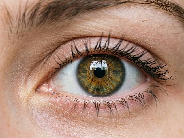
Blurry Vision and High Blood Sugar: What It Means
The connection between blurry vision and high blood sugar operates through two distinct mechanisms that differ fundamentally in reversibility, urgency, and clinical significance. The first — osmotic fluid shifts in the lens of the eye — produces blurry vision that fluctuates with blood glucose levels, improves when glucose is controlled, and is fully reversible in early-stage blood sugar elevation. The second — diabetic retinopathy, the damage to blood vessels in the retina caused by years of elevated glucose — produces vision changes that are progressive, cumulative, and ultimately irreversible without treatment, representing one of the most significant long-term complications of diabetes and the leading cause of new blindness in working-age adults in the United States. Understanding which mechanism is responsible for a given visual change — and what the difference means for urgency and management — is among the most practically important pieces of information for anyone with elevated blood sugar or a diabetes diagnosis.
The Reversible Mechanism: Osmotic Lens Changes
The lens of the eye is a transparent, biconvex structure that focuses light onto the retina by changing its shape through a process called accommodation. Its ability to focus precisely depends on maintaining a specific shape and water content, regulated by the movement of water and solutes between the lens and the surrounding aqueous humor.
When blood sugar rises significantly above normal, the elevated glucose diffuses into the lens (the lens does not require insulin for glucose uptake), where it is converted by the enzyme aldose reductase into sorbitol — a sugar alcohol that does not easily cross the lens cell membrane and therefore accumulates within lens fibers. Sorbitol accumulation raises the osmotic pressure inside the lens, drawing water in from the surrounding aqueous humor. This water influx swells the lens, changing its shape and refractive index — and therefore its focal length. The result is a change in the prescription of the eye that produces blurry vision.
The key characteristic of this osmotic mechanism is that it fluctuates with blood glucose. When blood sugar is higher — such as after a meal or during a period of poor glucose control — more sorbitol accumulates, more water enters the lens, and the blur is worse. When blood sugar falls — after exercise, medication, or dietary restriction — the osmotic gradient reverses, water exits the lens, and vision partially or fully clears. This fluctuation throughout the day is one of the diagnostic clues that blurry vision is glucose-driven rather than caused by a stable refractive error.
This osmotic blurry vision resolves when blood glucose is brought under sustained control. The lens, once glucose and sorbitol normalize, returns to its normal water content and shape. People who receive diabetes treatment often find that vision clears substantially within days to weeks of glucose normalization — a reassuring early sign of treatment response. However, this also creates a clinical pitfall: people whose vision has changed due to high blood sugar should not get new glasses or contact lenses until blood sugar has been controlled for several weeks, because prescriptions obtained during active hyperglycemia will be inaccurate once the osmotic changes resolve.
The Progressive Mechanism: Diabetic Retinopathy
The second connection between blurry vision and high blood sugar — diabetic retinopathy — operates through a completely different mechanism that unfolds over years of elevated glucose exposure and produces cumulative, largely irreversible damage to the retinal blood vessels. Diabetic retinopathy is the leading cause of new blindness in adults aged 20 to 74 in the United States, affecting approximately one-third of people with diabetes and producing vision loss in a significant proportion of those without regular eye screening and timely treatment.
The pathophysiology begins with damage to the pericytes — specialized cells that support and regulate the microvascular integrity of retinal capillaries. Chronic hyperglycemia causes pericyte loss, weakening capillary walls and leading to the formation of microaneurysms — tiny outpouchings in the capillary walls that can leak plasma, lipids, and blood into the surrounding retinal tissue. As the disease progresses, blood vessel leakage increases, producing retinal edema (swelling of the retina with fluid, particularly in the macular region responsible for central vision) and hard exudates (deposits of leaked lipid in the retinal tissue). This stage is called non-proliferative diabetic retinopathy (NPDR) and ranges from mild (a few microaneurysms) to severe (extensive hemorrhages, exudates, and cotton-wool spots).
In advanced proliferative diabetic retinopathy (PDR), the ischemic retina — deprived of oxygen by failing capillaries — releases growth factors (particularly VEGF, vascular endothelial growth factor) that stimulate the growth of new, abnormal blood vessels. These neovascular vessels are fragile and prone to bleeding into the vitreous humor (the gel-filled interior of the eye), causing sudden, dramatic vision loss. They can also stimulate the formation of fibrovascular membranes that can pull the retina away from its underlying support tissue — tractional retinal detachment — causing potentially permanent vision loss if not surgically treated. Understanding why blood sugar matters for long-term health in terms of its effects on microvascular tissues — including the retina — contextualizes why early detection and treatment of elevated blood sugar is so critical for long-term vision preservation.
- Osmotic lens changes: Reversible; fluctuate with blood glucose; resolve within weeks of glucose control
- Diabetic retinopathy: Progressive; caused by years of elevated glucose; leading cause of adult blindness in the US
- Don’t get new glasses: Wait until blood sugar has been controlled for 4–6 weeks before updating prescriptions
- Annual eye exam: All people with diabetes should have a dilated eye exam at least once yearly
- First exam timing: Type 2 — at diagnosis; Type 1 — within 5 years of diagnosis then annually
- Early treatment works: Laser treatment and anti-VEGF injections prevent vision loss in early-to-moderate retinopathy
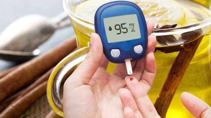
Diabetic Macular Edema: The Most Common Cause of Vision Loss
Diabetic macular edema (DME) — swelling of the macula (the central region of the retina responsible for sharp, detailed central vision) due to fluid leakage from damaged blood vessels — is the most common cause of vision loss in people with diabetic retinopathy. It can occur at any stage of retinopathy, including relatively early NPDR, and it produces the blurry, distorted, or washed-out central vision that significantly impairs reading, driving, and face recognition.
DME may be discovered incidentally on routine dilated eye examination before the person has noticed any visual symptoms — particularly in its early stages, when swelling is present but has not yet reached the fovea (the point of maximum visual acuity). This underscores the importance of regular dilated eye exams rather than relying on visual symptoms to prompt evaluation: by the time DME is causing noticeable vision loss, it is often in a more advanced state than if caught through screening. Treatment of DME has advanced dramatically over the past decade with the introduction of anti-VEGF injections (ranibizumab, aflibercept, bevacizumab, faricimab), which reduce retinal swelling and preserve or improve vision in the majority of treated eyes. The earlier treatment is initiated, the more vision can be preserved — making annual eye exams a critically important component of diabetes management.
Other Eye Problems Associated With High Blood Sugar
Beyond osmotic blurry vision and diabetic retinopathy, several other ocular conditions are associated with elevated blood sugar and diabetes:
Cataracts develop earlier and progress faster in people with diabetes, driven by the same sorbitol accumulation and oxidative stress that contribute to lens osmotic changes. The relationship between blood sugar and cataract risk is dose-dependent: higher glucose levels over longer time periods produce earlier and more rapid lens opacification. Cataracts are surgically treatable — cataract surgery is one of the most effective and commonly performed surgical procedures in medicine — but their earlier occurrence in people with diabetes adds to the burden of eye disease that good glucose control can help prevent.
Glaucoma occurs at approximately twice the rate in people with diabetes compared to the general population. The mechanism is not fully understood but likely involves microvascular damage to the optic nerve and increased intraocular pressure related to vascular changes. Glaucoma typically produces peripheral vision loss that is not noticed until significant damage has occurred, making regular pressure measurement and optic nerve assessment as part of the annual dilated eye exam important for early detection.
Fluctuating prescription needs are common in people with poorly controlled diabetes, as described in the osmotic mechanism section above. People with diabetes may find that their vision prescription changes frequently, that their current glasses don’t seem right even shortly after being fitted, or that vision seems clearest at certain times of day. All of these experiences reflect blood glucose-driven osmotic changes and should prompt blood sugar evaluation rather than repeated prescription adjustments. For a comprehensive overview of symptoms of type 2 diabetes including eye changes, and for guidance on the full range of early signs of high blood sugar that often accompany visual changes, see our dedicated guides.
Eye Exam Recommendations and Treatment Approaches
The American Diabetes Association recommends that all people with Type 2 diabetes receive a comprehensive dilated eye examination at the time of diagnosis, because many people with Type 2 diabetes have had elevated blood sugar for years before diagnosis and may already have retinopathy present. People with Type 1 diabetes should have their first dilated eye exam within five years of diagnosis (since the onset of Type 1 is typically precisely known), after which annual exams are recommended for both types. People with any detected retinopathy may need more frequent monitoring.
Treatment of diabetic retinopathy depends on the stage. Mild to moderate NPDR is typically managed with intensive blood sugar control and annual monitoring, as many cases stabilize or partially regress with glucose improvement. Severe NPDR and PDR are treated with laser photocoagulation (which destroys abnormal blood vessel growth in the peripheral retina) or anti-VEGF injections (which suppress the VEGF-driven neovascularization). Diabetic macular edema is treated primarily with anti-VEGF injections, which have largely replaced laser therapy for this indication due to their superior visual outcomes.
The most effective intervention for preventing retinopathy and slowing its progression remains glucose control. The DCCT and UKPDS trials both demonstrated that intensive glucose management — keeping A1C below 7 percent — dramatically reduced the development and progression of diabetic retinopathy: by 76 percent in Type 1 diabetes (DCCT) and by 25 percent in Type 2 diabetes (UKPDS). Understanding and achieving your A1C target — with your healthcare provider’s guidance — is the single most important intervention for long-term vision preservation. See our guide on the A1C test for a detailed explanation of what the A1C measures, what targets are appropriate, and how to interpret your results. And for practical monitoring tools to track glucose between clinic visits and understand how your daily choices affect your glucose control, see our guide on home blood sugar monitoring. For people who have not yet been formally diagnosed but recognize visual changes alongside other early metabolic symptoms, evaluating diabetes risk factors and seeking blood glucose testing is the appropriate first step — vision symptoms that accompany glucose dysregulation often resolve early in treatment but signal underlying metabolic changes that warrant attention.
How Blood Sugar Control Protects Vision Over the Long Term
The most powerful and evidence-supported intervention for preventing diabetic eye disease — including both retinopathy and DME — is sustained blood glucose control. The Diabetes Control and Complications Trial (DCCT) in Type 1 diabetes and the UK Prospective Diabetes Study (UKPDS) in Type 2 diabetes both demonstrated conclusively that reducing A1C through intensive glucose management dramatically reduces the risk of developing retinopathy and slows its progression in people who already have it. In the DCCT, intensive glucose control reduced the development of new retinopathy by 76 percent and slowed progression of existing retinopathy by 54 percent compared to conventional treatment. These reductions persisted long after the trial ended — a phenomenon called “metabolic memory” — suggesting that early, sustained glucose control imprints long-lasting benefits on retinal vascular health that continue even after glucose management becomes less intensive.
The practical implication is that every point of A1C reduction matters for vision. An A1C reduction from 9 percent to 7 percent, for instance, approximately halves the annual rate of retinopathy progression. This dose-response relationship means there is no threshold below which glucose improvement stops protecting the retina — benefit is cumulative and continuous, providing ongoing motivation for maintaining the best possible glucose control even when perfection is unachievable. Blood pressure control is the second most important intervention for retinopathy: hypertension accelerates retinal vascular damage through mechanisms independent of glucose, and treating blood pressure to below 130/80 mmHg in people with diabetes significantly reduces retinopathy progression. The combination of good glucose control, blood pressure control, and lipid management provides the most robust protection for long-term vision health. For comprehensive guidance on understanding your glucose control targets and what the A1C test tells you about your metabolic trajectory, see our guide on the A1C test.
Recognizing Visual Symptoms That Need Same-Day Evaluation
While blurry vision from osmotic lens changes is a non-urgent symptom that warrants medical evaluation but not emergency care, certain visual symptoms in people with diabetes require same-day ophthalmological evaluation to rule out serious complications that can cause permanent vision loss if untreated within hours.
Seek urgent eye evaluation the same day (not next week at a scheduled appointment) if you experience: sudden floaters — new black spots, cobwebs, or streaks moving in your visual field, which may represent vitreous hemorrhage (bleeding into the eye from fragile new blood vessels); flashes of light, particularly in one eye; a curtain or shadow appearing to drop over a portion of your vision (potential retinal detachment); sudden significant vision loss in one or both eyes; or double vision that has not previously been present. These symptoms, in people with diabetes, may represent advanced diabetic retinopathy complications that are ophthalmic emergencies — early treatment significantly improves outcomes while delayed treatment may mean permanent vision loss that cannot be recovered.
In contrast, gradually worsening blurry vision that fluctuates through the day, vision that changes when blood sugar is measured to be high versus lower, or difficulty reading that has developed over weeks to months represents the osmotic mechanism and warrants medical evaluation for blood sugar control within the next one to two weeks rather than same-day emergency care. The distinction between sudden and gradual, one-eye and both-eyes, and total versus partial visual change helps determine the appropriate urgency of evaluation. When in doubt, err toward earlier evaluation — an ophthalmologist who examines your eye and finds only early, treatable changes provides far better outcomes than one who sees you after damage has become irreversible. For a complete picture of all the symptoms and early signs associated with elevated blood sugar, including visual changes, see our guide on early signs of high blood sugar and our guide on symptoms of type 2 diabetes. People newly diagnosed with diabetes who want to understand how to monitor their blood sugar at home between clinic visits — and how this monitoring relates to both symptom management and long-term complication prevention — will find our guide on home blood sugar monitoring an essential practical resource.
The Importance of Annual Dilated Eye Exams
Diabetic retinopathy produces no symptoms in its early stages. Microaneurysms, dot hemorrhages, and early retinal edema can all be present and causing measurable damage without any awareness from the person affected — because these changes occur in the peripheral retina initially, outside the central field of vision that is used for most activities. By the time vision change is noticed, retinopathy is often in a moderate or advanced stage where treatment can still help but cannot recover the vision that has already been lost.
This is why annual dilated eye examinations — in which the pupils are pharmacologically widened to allow the ophthalmologist or optometrist to directly visualize the retinal blood vessels — are a non-negotiable component of diabetes care, regardless of whether any visual symptoms are present. The examination can detect early retinopathy, DME, and other changes when they are most responsive to intervention. Fundus photography (taking a photograph of the retina) and optical coherence tomography (OCT, a cross-sectional scan of retinal tissue that can detect even very early edema) extend the sensitivity of the examination beyond what clinical examination alone can detect.
Missing annual eye exams is unfortunately common among people with diabetes — surveys suggest that fewer than 60 percent of people with diabetes receive annual dilated eye exams, with access barriers, cost, competing health priorities, and lack of awareness of the recommendation all contributing. The consequences of missed exams are measured in degrees of preventable vision loss. Every annual exam that detects early retinopathy and leads to intensive glucose and blood pressure management is an opportunity to prevent progression to the stages where vision loss becomes inevitable without invasive treatment. For people who have not been regularly screened and are concerned about their retinal status, discussing the most appropriate evaluation with their diabetes care team or a retinal specialist is the first step toward understanding and protecting their long-term vision health. Understanding how managing other aspects of blood sugar affects complications like vision is part of the broader story of why blood sugar matters for long-term health — a comprehensive guide to the full spectrum of complications that sustained glucose elevation produces over years and decades.
Practical Steps After Noticing Blood Sugar-Related Vision Changes
If you notice that your vision has become blurry or that your previously adequate glasses no longer seem to work well, and you have known diabetes, prediabetes, or risk factors for elevated blood sugar, the recommended steps are: first, check your blood glucose if you have a meter — high readings confirm the likely osmotic cause; second, do not immediately schedule a new glasses or contacts fitting, as the prescription obtained during hyperglycemia will be inaccurate once blood sugar normalizes; third, contact your diabetes care provider within the next one to two weeks to review your glucose control and discuss whether treatment adjustment is needed; and fourth, ensure you have an annual dilated retinal examination scheduled, because visual symptoms from osmotic changes — while reassuring in their reversibility — are also signals that blood sugar has been elevated enough to affect eye tissues and that retinal evaluation for early retinopathy is warranted. For people without a prior diabetes diagnosis, new unexplained visual changes alongside increased thirst, fatigue, or frequent urination should prompt blood glucose testing as a priority — the combination of symptoms points strongly toward a metabolic basis that warrants evaluation before other explanations are pursued. For the practical tools to support ongoing glucose management and vision protection through good blood sugar control, our guide on home blood sugar monitoring covers both the tools and the decision-making framework for using them effectively at home. Understanding where diabetes fits in a comprehensive view of diabetes risk factors helps identify the other conditions that contribute to eye disease risk and that compound the effects of elevated blood sugar on long-term vision health.
Sources: American Diabetes Association. Standards of Medical Care in Diabetes — 2024. Diabetes Care. 2024;47(Suppl 1):S20–S42. • The Diabetes Control and Complications Trial Research Group. The Effect of Intensive Treatment of Diabetes on the Development and Progression of Long-Term Complications. N Engl J Med. 1993;329(14):977–986. • National Eye Institute. Diabetic Retinopathy. NEI; 2023.
This is one of the clearest explanations of blurry vision and high blood sugar: I have found. I appreciate that the article is careful about distinguishing between what is known and what is still being researched. This is exactly why I prefer this website over generic health platforms.
Really well-written article on blurry vision and high blood sugar:. The section on managing this condition day-to-day was especially useful for planning. Will definitely be coming back to this site for more health information.
Bookmarked this article on blurry vision and high blood sugar: immediately — going to reference it regularly. The specific numbers and thresholds mentioned are exactly what I needed to understand my results. Appreciate the effort that went into researching and writing this — it shows.