
Most adults know that the kidneys clean the blood. Fewer understand the actual mechanism — what structures do the filtering, how 180 liters of fluid gets processed into 1–2 liters of urine daily, or how the kidneys regulate blood pressure, produce hormones, and activate vitamin D entirely outside their filtration function. Understanding how the kidneys work is what makes kidney health concepts concrete. When you know what a nephron is and what glomerular filtration rate (GFR) measures, an eGFR result becomes a meaningful number rather than an abstract lab value. This guide explains the structure, function, and physiology of the kidneys in accessible terms — with enough clinical depth to make the health implications clear.
Where Are the Kidneys?
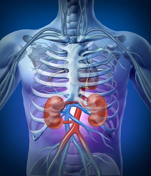
The kidneys are located retroperitoneally — behind the abdominal cavity’s peritoneal lining, on either side of the spine at approximately T12 to L3 vertebrae. In practical terms: the back of the abdomen, roughly at the level of the lowest ribs. Each kidney is about the size of a fist — approximately 10–12 cm long, 5–6 cm wide, weighing 120–170 grams. The right kidney sits slightly lower because the liver displaces it downward. When a kidney infection causes flank pain, the pain location corresponds directly to where the kidneys sit.
The kidneys receive an extraordinary volume of blood for their size: approximately 20–25% of total cardiac output at rest — roughly 1.0–1.2 liters per minute. This high blood flow reflects the scale of the filtration task. The kidneys process all the blood in the body approximately every 4–5 minutes.
The Structure of a Kidney
The Renal Cortex, Medulla, and Pelvis
Three distinct regions are visible inside a kidney. The outer layer — the renal cortex — is where filtration units (glomeruli) and most tubular processing occur. Beneath it lies the renal medulla, composed of triangular renal pyramids (bundles of collecting ducts). At the center is the renal pelvis, a funnel-shaped cavity that collects urine and channels it into the ureter. The hilum — the medial indentation on the inner curve — is where the renal artery, vein, lymphatics, and ureter enter and exit.
Blood Supply — From Aorta to Renal Vein
The renal artery branches from the abdominal aorta → segmental arteries → interlobar arteries → arcuate arteries → interlobular arteries → afferent arterioles feeding the glomerular capillary tuft. Blood exits via the efferent arteriole (higher resistance than afferent → maintains glomerular filtration pressure) → peritubular capillaries or vasa recta (where reabsorbed substances re-enter circulation) → renal vein → inferior vena cava.
The Nephron — The Kidney’s Working Unit
The functional unit of the kidney is the nephron. Each kidney contains approximately 1 million nephrons — though this varies (700,000 to 1.2 million) and declines with age, which is part of why kidney function decreases gradually over a lifetime.
The Glomerulus and Bowman’s Capsule
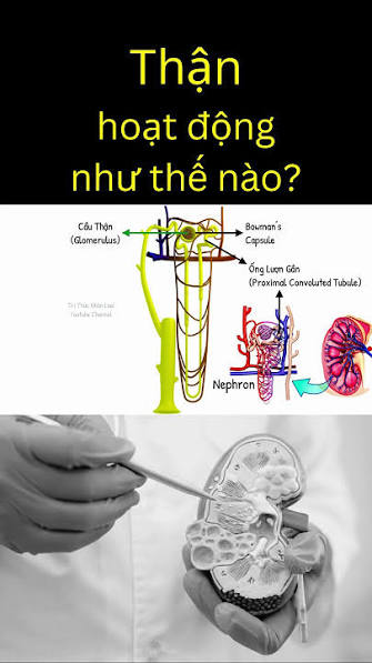
At the start of each nephron, the glomerulus — a tuft of capillaries enclosed in Bowman’s capsule — is where filtration occurs. Blood pressure forces fluid through a three-layer filtration barrier:
- Fenestrated endothelium: pores allow water and small solutes through; block red blood cells and platelets
- Glomerular basement membrane: size and charge selectivity — blocks albumin and large proteins
- Podocytes with filtration slits: specialized cells providing the final selective barrier
What crosses into Bowman’s capsule (the filtrate): water, glucose, amino acids, electrolytes, urea, creatinine. What stays in blood: red blood cells, platelets, albumin. When albumin appears in urine, it signals that this barrier has been damaged — the basis of the UACR test as an early kidney injury marker.
The Proximal Convoluted Tubule
The filtrate enters the proximal convoluted tubule (PCT) in the renal cortex — the highest-activity reabsorption segment. The PCT reclaims approximately 65–70% of filtered water and sodium, essentially all filtered glucose (via SGLT2 cotransporters — the same transporters targeted by SGLT-2 inhibitor medications used in diabetes and CKD), and all amino acids. PCT cells have a dense brush border of microvilli maximizing surface area for transport. The PCT also secretes hydrogen ions, uric acid, and many drugs into the tubule for excretion — which is why kidney function affects medication dosing.
The Loop of Henle
The filtrate descends into the Loop of Henle — a hairpin structure extending from the cortex into the medulla, critical for the kidney’s ability to concentrate urine. The mechanism works as a countercurrent multiplier:
- Thin descending limb: permeable to water, not salt → water exits by osmosis → tubular fluid concentrates
- Thick ascending limb: actively pumps NaCl out (Na/K/2Cl cotransporter) without allowing water to follow → tubular fluid dilutes; medullary interstitium concentrates (up to ~1,200 mOsm/kg)
This steep osmotic gradient in the medulla is what allows the collecting duct to produce concentrated urine when ADH is present. Loop diuretics like furosemide work by blocking this Na/K/2Cl pump.
The Distal Convoluted Tubule and Collecting Duct
The distal convoluted tubule (DCT) handles final electrolyte adjustments — sodium reabsorption regulated by aldosterone, calcium reabsorption by PTH. Thiazide diuretics block sodium transport here. The collecting duct is where final urine concentration is set: ADH (vasopressin) from the posterior pituitary inserts aquaporin water channels → water flows out → concentrated urine. Without ADH (diabetes insipidus), up to 15–20 L of dilute urine can be produced daily. Aldosterone (from adrenal cortex) reabsorbs sodium and excretes potassium here — the final step of the RAAS response.
How the Kidneys Filter Blood — Step by Step
The complete process from blood to urine passes through five stages:
- Blood enters glomerulus via afferent arteriole → hydrostatic pressure drives filtrate through the three-layer barrier into Bowman’s capsule (~125 mL/min = 180 L/day)
- Filtrate enters PCT → 65–70% of water, all glucose, all amino acids, ~65% of sodium reabsorbed
- Loop of Henle → NaCl removed, medullary osmotic gradient built, tubular fluid leaves ascending limb dilute
- DCT → fine-tuning of sodium, potassium, calcium under aldosterone and PTH
- Collecting duct → final water reabsorption under ADH → urine drains into renal pelvis → ureter → bladder
GFR (glomerular filtration rate) measures the rate of Step 1. At full function: ~125 mL/min. eGFR (estimated GFR from serum creatinine) below 60 mL/min/1.73m² for ≥3 months defines chronic kidney disease.
Beyond Filtration — Hormonal Functions
The RAAS System and Blood Pressure
The kidneys actively regulate blood pressure through the renin-angiotensin-aldosterone system. When blood pressure or sodium delivery drops, juxtaglomerular cells release renin → cleaves angiotensinogen (liver) → angiotensin I → converted by ACE (lungs) → angiotensin II: a potent vasoconstrictor that raises blood pressure, triggers aldosterone release, ADH release, and thirst. ACE inhibitors and ARBs block this cascade — which is why they are kidney-protective in CKD: they reduce intraglomerular pressure and proteinuria beyond their blood pressure effect.
Erythropoietin — How Kidneys Drive Red Blood Cell Production
Peritubular interstitial cells in the renal cortex detect low oxygen and produce erythropoietin (EPO), signaling the bone marrow to make more red blood cells. When CKD progresses (typically Stage 3b onward), EPO production falls → normocytic normochromic anemia develops. This “anemia of CKD” is managed with erythropoiesis-stimulating agents (ESAs) and iron supplementation.
Vitamin D Activation
Vitamin D from sun or diet is inactive until processed in two steps: liver produces 25-hydroxyvitamin D [25(OH)D] (the serum form measured on standard blood tests); then kidneys convert it to 1,25-dihydroxyvitamin D (calcitriol) — the biologically active form required for calcium absorption and bone mineralization. CKD impairs this conversion → functional vitamin D deficiency → secondary hyperparathyroidism → bone disease. This becomes clinically significant from CKD Stage 3 onward.
Prostaglandins and Why NSAIDs Affect the Kidneys
Kidneys produce local prostaglandins that dilate the afferent arteriole, maintaining GFR when blood pressure or volume drops. NSAIDs (ibuprofen, naproxen) block prostaglandin synthesis → afferent arteriole constricts → GFR falls. In healthy, well-hydrated adults this is usually reversible. In volume-depleted patients, the elderly, or those with existing CKD, NSAID use can precipitate acute kidney injury. Chronic NSAID use is associated with progressive kidney damage.
How the Kidneys Regulate Acid-Base Balance
Normal blood pH (7.35–7.45) is maintained long-term by the kidneys through three mechanisms:
- Bicarbonate reabsorption: the PCT reclaims most filtered HCO3−, preventing its loss in urine
- Hydrogen ion secretion: PCT and collecting duct actively secrete H+ into the tubule
- Ammonium buffering: PCT cells produce NH3 → combines with H+ → NH4+ excreted in urine, carrying away acid without further lowering urine pH
CKD impairs these mechanisms → serum bicarbonate falls → metabolic acidosis develops. Low serum bicarbonate is itself associated with faster CKD progression and is tracked alongside eGFR in CKD management.
What Healthy Urine Looks Like
Normal urine: pale yellow to amber (from urochrome, a hemoglobin breakdown pigment); 1–2 liters per day; transparent; slightly acidic (pH 5.5–7.0); mildly ammonia-scented. Abnormal signs worth knowing:
- Foamy or bubbly: excess albumin (proteinuria) — indicates glomerular damage
- Pink, red, or cola-colored: blood (hematuria) — warrants evaluation for UTI, stones, glomerulonephritis, or malignancy
- Cloudy: white blood cells (pyuria) — suggests urinary tract infection
- Dark amber or tea-colored: severe dehydration, or myoglobin from muscle breakdown (rhabdomyolysis)
- Glucose in urine: blood glucose exceeds the renal threshold (~180 mg/dL) — the mechanism SGLT-2 inhibitors exploit therapeutically
What Happens When the Kidneys Stop Working Properly
As GFR declines, each lost kidney function produces measurable consequences:
- Waste accumulation (uremia): urea, creatinine, and other metabolites build up → fatigue, nausea, cognitive changes
- Anemia: EPO production falls → reduced red blood cell production
- Bone disease: impaired calcitriol production → calcium-phosphorus dysregulation → secondary hyperparathyroidism
- Metabolic acidosis: impaired H+ excretion → serum bicarbonate falls
- Fluid overload and edema: reduced sodium and water excretion capacity
- Hyperkalemia: reduced potassium excretion → cardiac arrhythmia risk
These complications develop progressively and are tracked via routine blood tests: eGFR, serum bicarbonate, hemoglobin, potassium. This is the core reason regular lab monitoring in at-risk adults matters — the labs show what’s happening before symptoms develop.
How the Kidneys Relate to Urinary Health
The kidneys produce urine. The urinary tract — ureters, bladder, urethra — stores and eliminates it. These are closely related but distinct systems with different disease patterns. Kidney disease (CKD, glomerulonephritis) presents with lab abnormalities: elevated creatinine, low eGFR, proteinuria, electrolyte changes. Urinary tract disorders (UTIs, bladder control problems) present with symptoms: dysuria, urgency, frequency, incomplete emptying. Some conditions affect both: kidney stones form in the kidney but cause symptoms as they pass through the urinary tract; ascending UTIs can cause kidney infection (pyelonephritis) and scarring if untreated.
Our what is urinary health guide explains how the transport and storage system works. Our kidney health vs urinary health comparison guide covers how these two systems differ clinically.
Frequently Asked Questions
How much blood do the kidneys filter per day?
The kidneys filter approximately 180 liters of blood per day — about 125 mL per minute. Of that, over 99% is reabsorbed through the tubules; only 1–2 liters becomes urine. The kidneys process all the blood in the body roughly every 4–5 minutes.
Can you live with one kidney?
Yes. The remaining kidney undergoes compensatory hypertrophy, eventually compensating for approximately 70–75% of combined two-kidney GFR. Most people with a single kidney live normal lives. The remaining kidney warrants more careful protection — avoiding routine NSAID use, controlling blood pressure, staying hydrated — since there is no backup.
Do kidneys regenerate?
Kidney function can partially recover after acute kidney injury (AKI) if the cause is identified and treated quickly. But nephrons — the filtration units — do not regenerate once permanently damaged. Scarred glomeruli and tubules are not replaced. This is why CKD is progressive and why slowing progression is the clinical goal.
What does protein in urine mean?
Protein in the urine (specifically albumin, detected by UACR) indicates that the glomerular filtration barrier has been damaged. Healthy glomeruli keep albumin in the blood; damaged ones allow it to leak. A UACR above 30 mg/g on at least two of three measurements indicates persistent proteinuria — an early and sensitive marker of kidney injury, often appearing before eGFR declines. In adults with diabetes, rising UACR is the earliest signal of diabetic kidney disease.
How does age affect kidney function?
Kidney function naturally declines with age. The number of functional nephrons decreases approximately 10% per decade after age 40. GFR typically declines at about 1 mL/min/1.73m² per year after age 40, faster after age 70. An eGFR of 65 in a 75-year-old may be normal age-related decline; the same value in a 45-year-old warrants investigation. Older adults are also more vulnerable to acute kidney injury from dehydration, NSAID use, or illness because their renal reserve is lower.
For the broader clinical picture — what kidney health means, key lab values, and how to protect kidney function — our what is kidney health guide covers the practical detail. For the age-specific risks and screening that matter from midlife onward, our why kidney health matters after age 40 guide addresses the specific interventions and screening schedule relevant to adults over 40.
Understanding how the kidneys work transforms kidney health from abstract concepts into something concrete. Every metric used to assess kidney function — eGFR, UACR, serum creatinine, bicarbonate, hemoglobin — corresponds directly to a specific function the kidneys perform. When those numbers change, they reflect real changes in nephron filtration efficiency, hormonal output, and acid-base regulation. That connection between measurement and mechanism is what makes kidney lab results interpretable, and what makes the case for regular screening when those functions are at risk.
Clinical Implications: What This Physiology Means for Your Health
Understanding how the kidneys work makes a practical difference when you receive lab results or a new diagnosis. Here are the direct connections between kidney physiology and common clinical situations.
Why eGFR Uses Creatinine
Creatinine is a by-product of muscle metabolism, produced at a relatively constant rate by the body and excreted almost entirely by the kidneys through glomerular filtration with a small contribution from tubular secretion. Because its production is relatively stable and its clearance depends almost entirely on GFR, a rising serum creatinine is a reliable signal of falling GFR. The eGFR formula adjusts for age and sex (because muscle mass — and therefore creatinine production — differs systematically by these factors) to produce a more accurate estimate of actual filtration rate. This is why a 75-year-old woman with serum creatinine of 1.1 mg/dL may have an eGFR of 55, while a 35-year-old man with the same creatinine value has an eGFR of 80 — the kidneys are doing very different amounts of work in each case.
Why Potassium Becomes Dangerous in CKD
Potassium is handled primarily in the collecting duct, where aldosterone drives sodium reabsorption in exchange for potassium excretion. In healthy kidneys, potassium homeostasis is tightly maintained by increasing or decreasing its tubular secretion in response to dietary intake and aldosterone levels. As GFR falls and fewer functional nephrons remain, the kidney’s ability to excrete potassium on a per-nephron basis increases as a compensatory measure — but this compensation eventually reaches its limit, particularly in Stage 4–5 CKD. Hyperkalemia (elevated serum potassium) is dangerous because potassium controls cardiac muscle excitability: severely elevated potassium causes the cardiac conduction system to misfire, producing arrhythmias that can be fatal. This is why potassium is closely monitored in CKD, and why patients on ACE inhibitors or ARBs (which also raise potassium by blocking aldosterone) require more careful monitoring as GFR declines.
Why Loop Diuretics Are More Powerful Than Thiazides
The tubular segments targeted by different diuretics explain their relative potency. Thiazide diuretics block sodium transport in the distal convoluted tubule — a segment that handles roughly 5–10% of filtered sodium. Loop diuretics (furosemide, bumetanide) block the Na/K/2Cl pump in the thick ascending limb of the Loop of Henle — which handles approximately 25–30% of filtered sodium. Blocking the loop also collapses the medullary osmotic gradient, preventing the collecting duct from concentrating urine regardless of ADH levels — producing a more potent and faster diuresis. This is why loop diuretics are used in acute fluid overload and pulmonary edema, while thiazides are adequate for chronic blood pressure management in patients with normal or near-normal kidney function.
Why Kidney Disease Causes Bone Loss
The bone disease of CKD — called CKD-mineral and bone disorder (CKD-MBD) — develops through a sequence directly traceable to kidney physiology. Impaired calcitriol production → intestinal calcium absorption falls → blood calcium begins to drop → parathyroid glands increase PTH secretion (secondary hyperparathyroidism) → elevated PTH mobilizes calcium from bone to maintain serum calcium levels → progressive bone resorption and demineralization. Simultaneously, failing kidneys cannot excrete phosphorus adequately → hyperphosphatemia → phosphorus further suppresses calcitriol production and stimulates PTH. Treatment involves calcitriol supplementation (active vitamin D), phosphate binders taken with meals, and managing PTH levels — all targeting specific steps in this physiology-driven cascade.
Why SGLT-2 Inhibitors Protect the Kidneys
SGLT-2 (sodium-glucose cotransporter-2) is the primary transporter reclaiming filtered glucose in the proximal convoluted tubule. Blocking it with SGLT-2 inhibitors causes glucose (and sodium) to spill into the urine rather than being reabsorbed. The sodium that is not reabsorbed in the PCT reaches the macula densa — a specialized cluster of cells at the end of the thick ascending limb, adjacent to the glomerulus — in higher than normal concentrations. The macula densa responds by triggering afferent arteriole constriction (tubuloglomerular feedback), reducing intraglomerular pressure. This reduction in glomerular hyperfiltration is the primary kidney-protective mechanism of SGLT-2 inhibitors, operating entirely independently of blood glucose lowering. It is why the ADA Standards of Care 2024 recommend SGLT-2 inhibitors for adults with diabetes and CKD regardless of whether additional glycemic control is needed.
For authoritative patient-facing explanations of kidney anatomy and function, the NIDDK’s how your kidneys work resource is the primary reference. The National Kidney Foundation’s kidney function overview covers the same material in accessible patient terms. The Merck Manual’s kidney biology overview provides more detailed clinical physiology for readers who want the next level of depth.
Tracking Kidney Health Over Time
Because kidney disease is largely asymptomatic in its early and middle stages, the kidneys’ functional state is visible only through lab measurements. Keeping a record of your own eGFR and UACR results over time — noting the date of each test and the value — creates a personal trend line that is more useful than any single result. A slow, steady decline in eGFR over five years tells a different story than a sudden drop over three months. A UACR that rises from 15 to 45 mg/g over two years signals early damage even though both values might be reported without comment on a standard lab result. Asking your provider at each visit for both values, and whether the trend has changed, is the most practical application of understanding how the kidneys work.
Sources: National Institute of Diabetes and Digestive and Kidney Diseases (2023); National Kidney Foundation (2023); Guyton and Hall Medical Physiology 14th Edition (Chapters 26–30); KDIGO Clinical Practice Guidelines (2012); Merck Manual — Kidney Biology Overview (2023).
Pingback: Common Kidney Problems in Adults
Pingback: Signs of Healthy Kidney Function
Really well-written article on how the kidneys work: structure, function,. I especially valued the explanation of why these recommendations exist, not just what they are. I wish I had found this article earlier — would have saved a lot of confusion.
Really well-written article on how the kidneys work: structure, function,. I have tried following advice from several sources but this is most consistent with what my specialist told me. This is going into my health folder that I bring to every doctor’s visit.
My doctor recommended I look into how the kidneys work: structure, function, and this article covered it perfectly. I appreciated how the article addressed both the clinical side and the practical adjustments. Thank you for making complex medical information accessible without dumbing it down.