
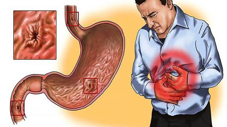
Gastritis is one of the most frequently self-diagnosed conditions in modern medicine — and one of the most frequently misunderstood. Many people use the word to describe any upper abdominal discomfort, nausea, or indigestion. Gastritis, as a medical diagnosis, means something specific: inflammation of the gastric mucosa, confirmed by biopsy. Most people with true gastritis have no symptoms whatsoever. And the most important cause — a bacterial infection called Helicobacter pylori — is entirely treatable, with eradication reducing not just the gastritis but the long-term risk of stomach cancer that untreated chronic gastritis carries.
What Is Gastritis?
Gastritis is inflammation of the gastric mucosa — the specialised inner lining of the stomach. It is a histological diagnosis, meaning it is established by examining stomach tissue under a microscope rather than by symptoms or imaging. The endoscopic appearance of the stomach lining correlates poorly with histological severity: gastritis can appear normal at endoscopy while showing significant inflammation on biopsy.
Acute gastritis is sudden-onset inflammation, typically caused by NSAIDs, alcohol, H. pylori initial infection, or severe physiological stress. It may cause mucosal erosions and can result in bleeding. When the causative factor is removed, acute gastritis generally resolves.
Chronic gastritis is long-standing mucosal inflammation. The two major forms are H. pylori-associated gastritis (Type B), affecting the antrum predominantly and the most common form worldwide, and autoimmune gastritis (Type A), affecting the corpus and fundus and caused by autoantibodies against parietal cells.
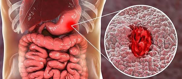
Atrophic gastritis is the end-stage: normal gastric glandular architecture is replaced by intestinal-type epithelium or fibrous tissue. Acid production is reduced or absent. Atrophic gastritis with intestinal metaplasia significantly elevates the risk of gastric cancer.
Types of Gastritis
H. pylori-associated gastritis (Type B): The dominant form globally. H. pylori infects approximately 44% of the world’s population, with prevalence much higher in developing countries. It causes a chronic active gastritis that, without treatment, persists indefinitely and in a subset of people progresses to atrophic gastritis, intestinal metaplasia, and ultimately gastric cancer. H. pylori is also responsible for most peptic ulcers and is causally linked to gastric adenocarcinoma and primary gastric MALT lymphoma.
Autoimmune gastritis (Type A): An underdiagnosed condition in which the immune system attacks parietal cells in the stomach’s corpus. Parietal cells produce both hydrochloric acid and intrinsic factor — the protein required for vitamin B12 absorption. Their destruction leads to achlorhydria, hypergastrinaemia, and ultimately pernicious anaemia from vitamin B12 deficiency. Autoimmune gastritis is strongly associated with Hashimoto’s thyroiditis, type 1 diabetes, and Addison’s disease.
Erosive and haemorrhagic gastritis: Caused by NSAIDs, aspirin, alcohol, and critical illness (stress ulceration). This form is characterised by mucosal erosions and, in severe cases, frank haemorrhage — the most common cause of upper GI bleeding in hospital inpatients.
Bile reflux gastritis: Occurs when bile refluxes from the duodenum into the stomach, most commonly after gastric surgery. Bile salts disrupt the mucosal barrier, causing chemical gastritis. Rare forms include eosinophilic gastritis (immune-mediated, associated with food allergy), granulomatous gastritis (Crohn’s disease, sarcoidosis, tuberculosis), and CMV gastritis in immunocompromised patients.
What Causes Gastritis?
Helicobacter pylori
H. pylori is a Gram-negative spiral bacterium that has evolved uniquely to survive in the stomach. It does so primarily through the production of urease, an enzyme that splits urea into ammonia and carbon dioxide — the ammonia neutralising stomach acid in the bacterium’s immediate microenvironment. Once established, H. pylori causes persistent mucosal inflammation through several mechanisms: its lipopolysaccharide activates innate immune receptors; its CagA virulence protein (in more virulent strains) is injected directly into gastric epithelial cells where it disrupts signalling and promotes oncogenic changes; and its VacA vacuolating cytotoxin causes cell damage and immunosuppression.
H. pylori is transmitted primarily by the faecal-oral route — contaminated water or food — with acquisition typically occurring in childhood. Once established, it persists lifelong unless treated. For more on H. pylori specifically, see our dedicated article on H. pylori infection and stomach health.
NSAIDs and Aspirin
Non-steroidal anti-inflammatory drugs — ibuprofen, naproxen, diclofenac, aspirin — are among the most widely used medications in the world and among the most common causes of gastric injury. Their mechanism involves inhibition of cyclooxygenase-1 (COX-1), an enzyme constitutively expressed in the gastric mucosa that produces prostaglandins essential for mucosal defence: mucus secretion, bicarbonate production, mucosal blood flow, and epithelial cell proliferation. When COX-1 is inhibited, this defensive cascade is disrupted, leaving the mucosa vulnerable to acid-mediated damage.
Risk factors for NSAID-associated gastric injury include: older age (over 60), high NSAID dose or combination use, concurrent corticosteroid or anticoagulant therapy, previous peptic ulcer, H. pylori co-infection. For people with multiple risk factors, PPI co-prescription is strongly recommended.
Alcohol and Stress
Alcohol is a direct mucosal irritant. Ethanol disrupts the hydrophobic mucus layer, increases mucosal permeability to hydrogen ions, and impairs the mucosal microcirculation. Acute heavy alcohol intake can cause acute erosive gastritis with haemorrhage.
In patients who are critically ill — on mechanical ventilation, with major burns, severe sepsis, or multi-organ failure — reduced splanchnic blood flow leads to mucosal ischaemia. This impairs energy-dependent mucosal defence, leading to erosion formation and significant upper GI haemorrhage in severe cases. Stress ulcer prophylaxis with PPIs is standard practice in high-risk ICU patients. Psychological stress does not directly cause gastritis in the histological sense — this is an important and commonly misunderstood distinction.
Autoimmune Mechanisms
In autoimmune gastritis, CD4+ T-cells sensitised against parietal cell antigens drive chronic inflammation and progressive destruction of the acid- and intrinsic factor-producing cells of the gastric corpus. The trigger is incompletely understood but may involve molecular mimicry from prior H. pylori infection, genetic susceptibility, and a broader autoimmune diathesis with co-existing thyroid and adrenal autoimmunity.
Gastritis Symptoms and Causes: Recognising the Signs
Perhaps the most clinically important point about gastritis symptoms and causes is that most people with gastritis have no symptoms. The degree of histological inflammation correlates poorly — sometimes not at all — with the presence or severity of symptoms. A person can have severe atrophic gastritis on biopsy and feel entirely well. Conversely, someone with severe epigastric pain may have a completely normal stomach lining on biopsy.
When gastritis does cause symptoms, they include:
- Epigastric pain or discomfort: upper central abdominal pain, which may be burning, aching, or gnawing; may be worsened by food or present on an empty stomach
- Nausea: particularly common in acute gastritis and erosive gastritis; may be accompanied by vomiting
- Bloating and early satiety: a sense of fullness rapidly after beginning a meal; upper abdominal distension
- Belching: from aerophagia associated with nausea and discomfort
- Upper GI bleeding: haematemesis (fresh red or coffee-ground vomiting) or melaena (black, tarry, offensive stools) — indicates erosive or haemorrhagic gastritis and requires urgent endoscopic assessment
Pernicious anaemia (autoimmune gastritis): fatigue, pallor, breathlessness from B12-deficiency anaemia; neurological symptoms — peripheral neuropathy (tingling, numbness, reduced vibration sense), subacute combined degeneration of the spinal cord affecting the dorsal columns (impaired proprioception, positive Romberg sign); glossitis; angular cheilitis. These neurological features, once present, may not fully reverse with B12 supplementation — making early detection critical.
Diagnosing Gastritis
Upper GI endoscopy (OGD) with biopsy is the gold standard. Endoscopy allows direct visualisation of the gastric mucosa and collection of biopsies for histological analysis. The Sydney Protocol recommends 5 biopsies: 2 from the antrum, 2 from the corpus, and 1 from the incisura angularis — providing the most accurate assessment of H. pylori status, gastritis distribution, and degree of atrophy and intestinal metaplasia. Histology reports should describe: degree of chronic inflammation, activity (neutrophil infiltration), atrophy, intestinal metaplasia (and subtype — complete/type I vs incomplete/type II–III, with type III carrying the highest cancer risk), H. pylori presence, and any dysplasia.
H. pylori testing options:
- Urea breath test (UBT): non-invasive, high sensitivity (~95%) and specificity (~96%); used for initial diagnosis and confirming eradication; must be performed ≥4 weeks after completing antibiotics and ≥2 weeks after stopping PPIs
- Stool antigen test: non-invasive; sensitivity and specificity comparable to UBT for both diagnosis and post-eradication testing
- Serology: detects IgG antibodies; cannot distinguish active from past infection; not useful for confirming eradication
- Rapid urease test (CLO test): biopsy-based; quick in-procedure result during endoscopy
Blood tests: Full blood count (iron-deficiency anaemia from gastric bleeding; macrocytic anaemia from B12 deficiency); B12 levels; parietal cell antibodies (present in ~90% of autoimmune gastritis); intrinsic factor antibodies (more specific for pernicious anaemia); fasting serum gastrin (markedly elevated in autoimmune gastritis with achlorhydria).
Treatment for Gastritis
H. pylori eradication: Standard first-line regimen: 7–14 days of triple therapy — a PPI (twice daily) + amoxicillin 1g (twice daily) + clarithromycin 500mg (twice daily). Longer courses (14 days) achieve higher eradication rates. Where clarithromycin resistance is above 15%, bismuth quadruple therapy is preferred: bismuth subcitrate + PPI + metronidazole + tetracycline for 10–14 days. Eradication must be confirmed 4–8 weeks after completing treatment using the urea breath test or stool antigen test — off PPI for 2 weeks before testing.
Successful H. pylori eradication resolves the associated gastritis in most cases, heals peptic ulcers, prevents ulcer recurrence, and significantly reduces the risk of gastric cancer in populations where gastritis has not yet progressed to high-grade atrophy or intestinal metaplasia. See our article on stomach ulcers: what adults should know for more on ulcer management.
Acid suppression: PPIs are the most effective medications for reducing acid, healing gastric erosions, and preventing further mucosal damage — used for eradication regimens, NSAID-associated gastritis, and co-prescription with NSAIDs in high-risk patients.
NSAID management: Stopping or reducing NSAIDs is the most important step when they are the causative factor. Where NSAIDs must continue, PPI co-prescription reduces gastric injury by approximately 80%. COX-2 selective inhibitors have lower GI toxicity than non-selective NSAIDs but are not free of gastric risk.
Autoimmune gastritis — B12 replacement: Intramuscular hydroxocobalamin (1mg every 3 months, lifelong) bypasses absent intrinsic factor and corrects B12 deficiency. Oral high-dose B12 (1000–2000mcg daily) can also achieve adequate serum levels through passive absorption, but intramuscular supplementation is standard for pernicious anaemia with neurological involvement. Iron supplementation addresses co-existing iron-deficiency anaemia, since achlorhydria impairs non-haem iron absorption.
Gastritis and Gastric Cancer Risk
The relationship between chronic gastritis and gastric cancer is one of the most well-studied sequences in oncology — the Correa cascade:
Normal mucosa → H. pylori gastritis → Chronic active gastritis → Atrophic gastritis → Intestinal metaplasia → Dysplasia → Gastric cancer (intestinal type)
This cascade unfolds over decades and is not inevitable — the majority of people with H. pylori gastritis never develop gastric cancer. However, certain stages carry substantially higher risk: atrophic gastritis carries 3–6× higher cancer risk than normal mucosa; incomplete intestinal metaplasia (type II–III) carries 10–15× higher risk in some studies; high-grade dysplasia requires endoscopic resection.
The OLGA (Operative Link on Gastritis Assessment) and OLGIM (Operative Link on Gastric Intestinal Metaplasia) staging systems provide a standardised way to classify gastric cancer risk from biopsy findings. Patients at OLGA/OLGIM Stage III–IV are classified as high-risk and require endoscopic surveillance every 3 years.
Autoimmune gastritis independently elevates gastric cancer risk (3–6× vs general population) through the corpus atrophy and achlorhydria pathway, and additionally elevates gastric neuroendocrine tumour risk from chronic hypergastrinaemia. H. pylori eradication reduces gastric cancer risk — particularly in younger patients and those who have not yet developed intestinal metaplasia. According to the World Health Organization, H. pylori is classified as a Group 1 carcinogen — a definite cause of gastric cancer in humans.
Gastritis vs Dyspepsia — Understanding the Difference
Gastritis and dyspepsia are frequently confused, both by patients and in clinical practice. The distinction matters because they are not the same thing and are managed differently.
Gastritis is a histological diagnosis — it means inflammation of the gastric mucosa confirmed on biopsy. It may or may not cause symptoms. Dyspepsia is a symptom complex — upper abdominal discomfort, pain, bloating, nausea, early satiety — with multiple possible causes. The majority of patients with dyspepsia in primary care do not have gastritis on biopsy. The most common endoscopic finding in dyspepsia is a normal stomach — a finding that leads to the diagnosis of functional dyspepsia, which involves disordered gut-brain signalling and visceral hypersensitivity rather than mucosal inflammation.
For a direct comparison, see our article on GERD vs gastritis: what is the difference? — including how these conditions overlap and diverge in practice.
Prevention
H. pylori prevention: There is no approved vaccine for H. pylori. Prevention relies on food and water hygiene — safe drinking water, adequate sanitation, and hand hygiene (particularly after toileting) are the primary interventions. In high-risk settings, test-and-treat programmes identify and eradicate infection before complications develop. According to the NHS Gastritis guidance, most cases of H. pylori-related gastritis can be resolved with a short course of antibiotics.
NSAID-associated gastritis prevention: Use the lowest effective NSAID dose for the shortest necessary duration. In patients requiring regular NSAIDs with risk factors for GI complications, PPI co-prescription is recommended. Substituting paracetamol for NSAIDs where the anti-inflammatory effect is not critical reduces GI risk substantially.
Lifestyle factors: Limiting alcohol consumption reduces acute erosive gastritis risk. Smoking impairs gastric mucosal healing, reduces mucosal blood flow, and is independently associated with H. pylori treatment failure — smoking cessation is an important component of gastritis prevention. According to a Maastricht V/Florence consensus report published in Gut, H. pylori eradication should be considered in all infected individuals, regardless of symptoms, given its role in cancer prevention.
Long-Term Management and Follow-Up
The long-term management of gastritis depends entirely on its underlying cause. For H. pylori-associated gastritis, the priority is confirming eradication and ensuring the patient does not have high-risk precancerous lesions that require ongoing surveillance. For autoimmune gastritis, lifelong B12 supplementation is the cornerstone of treatment, alongside periodic monitoring for pernicious anaemia, iron deficiency, and the elevated risk of gastric neuroendocrine tumours that arises from sustained hypergastrinaemia.
Patients with confirmed atrophic gastritis or intestinal metaplasia — regardless of cause — benefit from OLGA/OLGIM staging to risk-stratify their need for endoscopic surveillance. Current European Society of Gastrointestinal Endoscopy (ESGE) guidelines recommend endoscopic surveillance every 3 years for patients at OLGA/OLGIM Stage III–IV, while patients at Stage I–II with no additional risk factors (family history of gastric cancer, incomplete intestinal metaplasia, male sex, smoking, Eastern European or East Asian origin) may not require intensive surveillance.
For patients with erosive gastritis caused by NSAIDs or alcohol, the follow-up is largely centred on removing or mitigating the causative factor. In patients in whom long-term NSAID use is unavoidable (rheumatoid arthritis, cardiovascular secondary prevention with aspirin), a treatment plan combining PPI co-prescription and annual symptom review is standard. For patients with autoimmune gastritis who are asymptomatic and have normal B12 levels, a surveillance endoscopy every 3–5 years is recommended to screen for gastric neuroendocrine tumours and adenocarcinoma.
Dietary modification plays a supportive but not curative role in long-term management. Patients with atrophic gastritis and achlorhydria have impaired absorption not only of vitamin B12 but also of non-haem iron, calcium (particularly relevant for fracture risk), and zinc. These deficiencies should be screened for and supplemented accordingly. Patients with eosinophilic gastritis may benefit from dietary elimination of identified trigger foods in consultation with a dietitian and allergist.
Frequently Asked Questions
Acute gastritis caused by a specific trigger — a heavy drinking session, a short course of NSAIDs, or a viral illness — typically resolves within days to a few weeks once the trigger is removed. H. pylori gastritis does not resolve without antibiotic treatment — the bacteria persist indefinitely and the gastritis continues as long as H. pylori remains present. Autoimmune gastritis is progressive and does not resolve, though B12 supplementation prevents and treats the consequences of pernicious anaemia. Chronic atrophic gastritis does not reverse even after H. pylori eradication, though eradication prevents further progression.
No — though they are closely related. Gastritis is inflammation of the stomach lining, whereas a peptic ulcer is a deeper breach in the mucosal lining extending through the submucosa. Gastritis, particularly H. pylori gastritis, is a major predisposing cause of peptic ulcers — but not everyone with gastritis develops an ulcer. Both conditions share the same risk factors and many of the same treatments. For a comprehensive breakdown, see our article on stomach ulcers: what adults should know.
During symptomatic episodes, foods that increase acid production or directly irritate the gastric mucosa often worsen symptoms: alcohol, spicy foods, coffee and caffeine (which stimulate gastric acid secretion), acidic foods (citrus, tomatoes), carbonated drinks, and fatty or fried foods. There is no evidence-based “gastritis diet” — dietary modifications are supportive rather than curative. The underlying cause of the gastritis (H. pylori, NSAIDs, alcohol) is what requires treatment. Small, frequent meals may reduce epigastric discomfort.
H. pylori infection cannot be diagnosed from symptoms alone. The recommended non-invasive tests are the urea breath test (most accurate for active infection) and the stool antigen test. Serology can indicate exposure but cannot confirm active infection. In patients undergoing endoscopy, biopsies and a rapid urease test provide in-procedure diagnosis. If you are experiencing persistent dyspeptic symptoms — particularly under age 60 without alarm features — a test-and-treat approach for H. pylori is recommended before endoscopy in most guidelines.
It depends on the type and stage. Acute gastritis and uncomplicated H. pylori gastritis without atrophy carry a low absolute cancer risk. Chronic atrophic gastritis and intestinal metaplasia — particularly incomplete (type II–III) — carry significantly elevated gastric cancer risk. Autoimmune gastritis elevates risk 3–6 fold. H. pylori eradication in patients without established atrophy significantly reduces cancer risk. The degree of cancer risk can be stratified using OLGA/OLGIM staging on biopsy; high-risk patients (Stage III–IV) should undergo endoscopic surveillance every 3 years.
Physiological stress in the context of critical illness (major surgery, burns, severe sepsis, mechanical ventilation) causes stress ulceration — a form of erosive gastritis from mucosal ischaemia, which is a recognised medical emergency. Psychological stress (life stress, anxiety) does not directly cause gastritis in the histological sense, though it can exacerbate symptoms in people with existing gastric conditions, particularly functional dyspepsia, through gut-brain signalling pathways. The common belief that “stress causes ulcers” is largely a misconception — H. pylori and NSAIDs cause most ulcers.
Gastritis is inflammation of the stomach lining. GERD (gastro-oesophageal reflux disease) is a condition where stomach acid refluxes upward into the oesophagus, causing symptoms (heartburn, regurgitation) and potentially oesophageal inflammation (oesophagitis). Gastritis affects the stomach; GERD primarily affects the oesophagus. They can coexist, and both may be associated with H. pylori. The key distinguishing feature clinically is symptom location: gastritis typically causes epigastric discomfort; GERD typically causes burning behind the breastbone and acid taste in the throat. For a detailed comparison, see our article on GERD vs gastritis.
Medical Disclaimer: This article is for educational purposes only and does not constitute medical advice. Persistent upper abdominal symptoms, especially with rectal bleeding, unexplained weight loss, or difficulty swallowing, should be evaluated promptly by a healthcare professional.
References
- Rugge M et al. Gastritis: The histology report. Dig Liver Dis. 2011;43 Suppl 4:S373–84.
- Malfertheiner P et al. Management of Helicobacter pylori infection — the Maastricht V/Florence Consensus Report. Gut. 2017;66(1):6–30. Available at: PubMed.
- Correa P. Human gastric carcinogenesis: a multistep and multifactorial process. Cancer Res. 1992;52(24):6735–40.
- Lahner E et al. Autoimmune gastritis. Best Pract Res Clin Gastroenterol. 2018;32–33:55–62.
- World Health Organization. Helicobacter pylori. Available at: who.int.
- NHS. Gastritis. Available at: nhs.uk.
I was diagnosed with H. pylori gastritis three months ago after finally getting an endoscopy for epigastric pain I’d had on and off for two years. My GP had attributed the symptoms to stress and given me antacids repeatedly, but the endoscopy found active gastritis with H. pylori on the CLO test. I completed the 14-day triple therapy course and had the urea breath test six weeks later — it was negative. The pain resolved within about two weeks of finishing the antibiotics. What this article explained that nobody told me at the time is that eradication also reduces long-term cancer risk — I had no idea there was a progression from H. pylori gastritis to atrophic changes to potential malignancy. My gastroenterologist didn’t mention any of that. I’ve now asked for a follow-up letter confirming whether I had any atrophic changes on my biopsy results, which I didn’t think to ask about at the time.
Rachel — your experience of a two-year delay before endoscopy, with symptoms attributed to stress and managed with antacids alone, is unfortunately very common. The challenge with H. pylori-associated gastritis is that symptoms are non-specific and often indistinguishable from functional dyspepsia — and the test-and-treat approach (non-invasive H. pylori testing before endoscopy) is recommended in most guidelines for patients under 60 without alarm features, precisely to identify and treat H. pylori without necessarily requiring endoscopy first. The key issue in your case, as you have now identified, is understanding what your biopsy showed beyond the H. pylori result itself. Specifically, whether there was any atrophy or intestinal metaplasia on the Sydney Protocol biopsies — and if so, what the OLGA/OLGIM stage was. If your biopsies showed no atrophy and no metaplasia (which is common in straightforward antral H. pylori gastritis in younger adults), your cancer risk after successful eradication is low and no endoscopic surveillance is typically recommended. If there was early atrophic change, that changes the follow-up plan. The follow-up letter you have requested is entirely appropriate, and asking specifically about these histological findings — and whether any surveillance is recommended — is the right question to put to your gastroenterologist. James — the decision to move to intramuscular B12 injections at a higher frequency than the standard 3-monthly schedule is a reasonable and well-evidenced approach in patients with active neurological symptoms. The evidence for oral B12 supplementation through passive absorption is adequate for maintaining serum levels once stores are repleted, but the intramuscular route remains standard for pernicious anaemia with neurological involvement because it guarantees absorption and allows higher tissue delivery in the early repletion phase. The co-existence of iron deficiency with autoimmune gastritis is underrecognised and worth highlighting: achlorhydria significantly impairs non-haem iron absorption, and patients with autoimmune gastritis should have ferritin, haemoglobin, and mean cell volume checked alongside B12 at their monitoring appointments. If you have not already, discussing with your haematologist or gastroenterologist whether your current monitoring schedule includes ferritin would be worthwhile.
I have autoimmune gastritis diagnosed four years ago alongside Hashimoto’s thyroiditis — both confirmed on the same set of blood tests. I was initially started on oral B12 supplements, but my neurologist recommended switching to intramuscular injections after I developed some mild peripheral neuropathy (tingling in both feet). The injections have been every 8 weeks rather than the standard 12 weeks because my symptoms were more prominent. The article’s point about neurological features potentially not fully reversing even with treatment is correct and something that I think needs to be said more clearly to patients at diagnosis — the earlier B12 replacement starts, the better the neurological outcome. My tingling has improved significantly but not fully resolved after two years on injections. I also appreciated the mention of iron deficiency alongside B12 — achlorhydria affecting iron absorption is something I wasn’t told about and I was iron deficient for over a year before it was identified.