
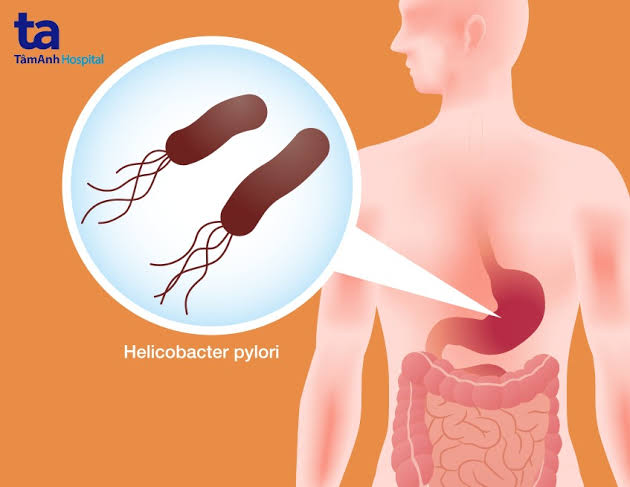
In 1984, an Australian physician named Barry Marshall drank a solution containing Helicobacter pylori — a bacterium he had isolated from a patient with stomach inflammation — to prove it was causing the disease. He developed gastritis within days. The experiment was unorthodox. The conclusion was transformative. The bacterium Marshall and his colleague Robin Warren had identified turned out to be the cause of most peptic ulcers, a major driver of gastric cancer, and one of the most successful human pathogens in history — infecting approximately 2.1 billion people globally. In 2005, they received the Nobel Prize in Physiology or Medicine. H. pylori infection remains the single most important preventable cause of gastric cancer in the world today, and it is entirely curable with a course of antibiotics.
What Is H. pylori?
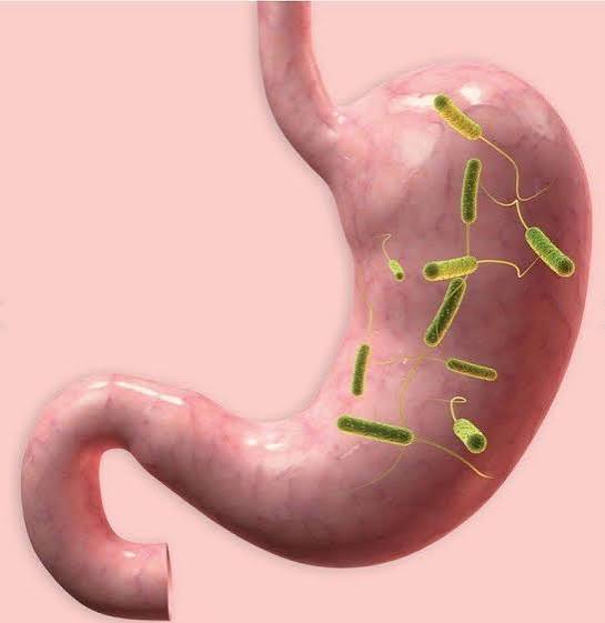
Helicobacter pylori is a Gram-negative, spiral-shaped bacterium that has evolved over thousands of years to colonise one of the most hostile environments in the human body — the stomach. With a pH typically between 1.5 and 3.5, the stomach kills most bacteria within minutes. H. pylori survives through a series of evolved mechanisms, primarily the production of urease, an enzyme that creates a localised alkaline microenvironment by converting urea (present in gastric secretions) to ammonia and carbon dioxide. Once established, it colonises the mucous layer overlying the gastric epithelium and persists indefinitely unless treated.
The World Health Organization classifies H. pylori as a Group 1 carcinogen — meaning there is definitive evidence that it causes cancer in humans, specifically gastric adenocarcinoma. It is the only bacterium known to cause cancer. Global prevalence is approximately 44% of the world’s population — roughly 2.1 billion people — making it one of the most prevalent chronic bacterial infections in human history. Prevalence is much higher in developing countries (Africa, South-East Asia, South and Central America: 70–90%) than in developed countries (Western Europe, North America, Australia: 20–40%).
How H. pylori Spreads
H. pylori is transmitted primarily by the faecal-oral route — through water or food contaminated with the bacterium. Poor sanitation infrastructure, inadequate clean water supply, and crowded living conditions are the most important risk factors for acquisition. The bacterium can also be transmitted by the oral-oral route — it has been cultured from saliva and dental plaque — likely explaining some person-to-person spread through shared utensils and close contact.
Acquisition occurs predominantly in early childhood, typically before age five in high-prevalence settings. Once acquired, H. pylori persists lifelong without treatment — it does not spontaneously clear in adults. This means adults carrying H. pylori acquired it decades earlier, usually during childhood, and may have been infected throughout their entire adult life without knowing it.
How H. pylori Damages the Stomach
H. pylori does not directly destroy gastric tissue — instead, it induces a chronic inflammatory response that, over years to decades, causes cumulative mucosal damage. The key mechanisms are:
Urease and ammonia: The ammonia produced to buffer stomach acid is directly toxic to gastric epithelial cells. While it protects H. pylori from acid, it contributes to mucosal cell injury in the bacterium’s immediate vicinity.
CagA protein (cytotoxin-associated gene A): CagA-positive strains inject the CagA protein directly into gastric epithelial cells via a Type IV secretion system — essentially a molecular syringe. Inside the cell, CagA disrupts signalling pathways that regulate cell growth, differentiation, and survival, promoting uncontrolled division and inhibiting normal apoptosis. CagA has been called a “bacterial oncoprotein” — a bacterial product that directly promotes cancer-associated cellular changes. CagA-positive strains are more strongly associated with peptic ulcer disease, gastric cancer, and MALT lymphoma.
VacA (vacuolating cytotoxin A): VacA forms pores in cell membranes, causing vacuolation, mitochondrial damage, and apoptosis. It also inhibits T-cell function, helping H. pylori evade immune clearance — a key reason why the immune system cannot eliminate the infection without antibiotic help.
Chronic active gastritis: The net result is chronic active gastritis — persistent mucosal inflammation that drives the sequential changes of the Correa cascade: atrophic gastritis → intestinal metaplasia → dysplasia → gastric cancer, in a subset of infected individuals over decades. For more on this process, see our article on gastritis symptoms and causes.
Symptoms of H. pylori Infection
Approximately 70–80% of people infected with H. pylori are entirely asymptomatic. They carry the bacterium, have histological gastritis on biopsy, and feel nothing. The infection is discovered incidentally — through targeted testing, population screening, or only when a complication develops.
When H. pylori does cause symptoms, they are non-specific and indistinguishable from functional dyspepsia:
- Epigastric discomfort or pain — upper central abdominal pain, often burning, gnawing, or aching
- Nausea
- Bloating and abdominal fullness
- Belching
- Early satiety (feeling full quickly when eating)
When H. pylori has caused a peptic ulcer, the symptom picture becomes more specific: duodenal ulcers produce epigastric pain relieved by food and classic nocturnal pain (waking at 1–3am); gastric ulcer pain may worsen with food and be associated with weight loss. Upper GI bleeding — haematemesis or melaena — indicates a bleeding complication and requires urgent assessment. For a full breakdown of peptic ulcer symptoms and complications, see our article on stomach ulcers: what adults should know.
Conditions Caused by H. pylori Infection in the Stomach
Peptic ulcer disease: H. pylori is responsible for approximately 90–95% of duodenal ulcers and 70–80% of gastric ulcers globally. Eradication heals ulcers and reduces recurrence from ~70–80%/year (untreated) to less than 5%/year after confirmed eradication.
Gastric adenocarcinoma: The most important preventable risk factor for gastric cancer globally. Approximately 75% of gastric adenocarcinomas are attributable to H. pylori. The absolute lifetime risk for any infected individual is approximately 1–3%, but given 2.1 billion infected people, H. pylori accounts for nearly 800,000 gastric cancer deaths annually. According to the World Health Organization, H. pylori infection is the leading known risk factor for gastric cancer.
Gastric MALT lymphoma: Primary gastric MALT lymphoma — a B-cell lymphoma arising from lymphoid tissue accumulated in response to H. pylori — is approximately 90% H. pylori-associated. In early-stage localised disease, H. pylori eradication alone achieves complete histological remission in approximately 75% of patients — without chemotherapy or radiotherapy. This is one of very few examples in oncology where a cancer can be cured by treating its bacterial trigger.
Functional dyspepsia: H. pylori is found in ~30–40% of patients with functional dyspepsia. A test-and-treat approach is recommended in most guidelines. The benefit of eradication is modest — a genuine but small effect, with approximately 10% of treated patients achieving long-term relief attributable to eradication (NNT approximately 10).
Iron deficiency anaemia: H. pylori-associated gastritis impairs iron absorption. In refractory iron deficiency anaemia with no other identifiable cause, H. pylori testing and eradication are recommended — eradication often improves haemoglobin without additional iron supplementation.
Immune thrombocytopaenia (ITP): H. pylori eradication is associated with platelet count improvement in approximately 50% of H. pylori-positive ITP patients in some series — likely through resolution of molecular mimicry between H. pylori antigens and platelet surface proteins.
Diagnosing H. pylori Infection
Urea breath test (UBT): The gold standard non-invasive test. The patient drinks labelled urea (13C or 14C); H. pylori urease cleaves it, releasing labelled CO₂ detected in expired breath. Sensitivity ~94%, specificity ~95%. Used for both initial diagnosis and post-eradication confirmation. Critical timing: at least 4 weeks after completing antibiotics and at least 2 weeks after stopping PPIs (both suppress H. pylori and cause false-negatives). According to the Maastricht V/Florence Consensus, the UBT is the preferred test for confirming eradication.
Stool antigen test (SAT): Detects H. pylori antigens in stool using monoclonal antibody ELISA. Sensitivity ~94%, specificity ~97%. Comparable to UBT; the same timing requirements apply. Preferred where UBT equipment is unavailable.
Serology: Detects IgG antibodies. Cannot distinguish active from past infection — antibodies remain positive for months to years after eradication. Do not use to confirm eradication. Limited utility where UBT or SAT are accessible.
Endoscopy-based tests: Rapid urease test (CLO test) gives a quick in-procedure result from biopsy; histology is the most sensitive individual test and provides full mucosal assessment; culture allows antibiotic susceptibility testing for second-line therapy selection.
Treatment: Eradicating H. pylori
Standard triple therapy (where clarithromycin resistance is <15%):
- PPI (twice daily) + amoxicillin 1g (twice daily) + clarithromycin 500mg (twice daily)
- Duration: 14 days (eradication rates ~80%)
Bismuth quadruple therapy (where clarithromycin resistance >15%, penicillin allergy, or first-line failure):
- Bismuth subcitrate 120mg (four times daily) + PPI (twice daily) + metronidazole 400mg (three times daily) + tetracycline 500mg (four times daily)
- Duration: 10–14 days; eradication rates ~85–90%
After two treatment failures, culture-guided therapy — selecting antibiotics based on sensitivity testing of a biopsied H. pylori isolate — is the recommended approach. A levofloxacin-based triple regimen (PPI + amoxicillin + levofloxacin × 10 days) is a common second-line option in settings where clarithromycin-based triple therapy was used first-line. For full details on peptic ulcer management following eradication, see our article on peptic ulcer disease explained.
H. pylori and Gastric Cancer Risk
The connection between H. pylori and gastric cancer operates through the Correa cascade — a sequence of mucosal changes beginning with H. pylori gastritis and progressing, over decades, to gastric cancer in a subset of patients. Not everyone with H. pylori follows this cascade; factors influencing risk include: CagA-positive strain (higher risk), corpus-predominant or pangastric gastritis, smoking, high-salt diet, low fruit and vegetable intake, male sex, and family history of gastric cancer.
OLGA and OLGIM staging assigns cancer risk based on the degree and distribution of atrophy and intestinal metaplasia on biopsy. Patients at Stage III–IV require endoscopic surveillance every 3 years.
H. pylori eradication reduces gastric cancer risk — confirmed in multiple randomised controlled trials. The reduction is greatest in patients treated before atrophic gastritis or intestinal metaplasia has developed. Eradicating H. pylori in younger adults with no atrophic changes prevents the cancer cascade from initiating; eradicating it in patients with established atrophy slows but does not fully reverse pre-existing changes. According to a landmark meta-analysis, H. pylori eradication reduces the incidence of gastric cancer by approximately 34–45% compared to no treatment.
Prevention of H. pylori Infection
No approved vaccine currently exists, though several candidates are in development. Primary prevention relies on improving the infrastructure determinants of transmission: safe drinking water, adequate sanitation, and hygiene education. These public health measures have driven the declining H. pylori prevalence in developed countries over recent decades.
The following groups should be offered H. pylori testing and treatment if positive:
- First-degree relatives of patients with gastric cancer
- Patients with established atrophic gastritis or intestinal metaplasia
- Patients with previous gastric MALT lymphoma
- Patients planning long-term NSAID therapy (prior eradication reduces NSAID GI risk)
- Patients with unexplained iron deficiency anaemia refractory to oral iron
Screen-and-treat programmes targeting H. pylori in high-prevalence populations have been shown to reduce gastric cancer incidence over 10–20 year follow-up periods. For how H. pylori relates to GERD and overlapping upper GI conditions, see our article on GERD vs gastritis: what is the difference?
After H. pylori Treatment: What to Expect
Most patients who complete a full course of H. pylori eradication therapy and confirm successful eradication with a urea breath test can expect their H. pylori-related gastritis to resolve over the following weeks to months. In patients whose symptoms were caused by active H. pylori gastritis — rather than by a co-existing functional dyspepsia — symptom improvement typically begins within 1–2 weeks of starting treatment and continues after completion. In patients with H. pylori-associated peptic ulcer disease, ulcer healing follows eradication on continued acid suppression, and the lifetime risk of ulcer recurrence is dramatically reduced.
For patients found to have atrophic gastritis or intestinal metaplasia on biopsies taken at the time of H. pylori diagnosis, eradication is still important — but the follow-up plan changes. These patients require ongoing OLGA/OLGIM staging and endoscopic surveillance, because their gastric cancer risk remains elevated even after successful eradication. The key question is how far along the Correa cascade their mucosal changes are: patients with atrophic gastritis and extensive incomplete intestinal metaplasia (type II or III) in the corpus are in the higher-risk surveillance category regardless of H. pylori status post-eradication.
Understanding the distinction between the bacterial infection being cured (H. pylori eradicated, confirmed by breath test) and the mucosal consequences of the infection being resolved (gastric atrophy and intestinal metaplasia, which do not reverse) is important for setting patient expectations. Eradication is curative for the infection; it is not fully curative for pre-existing high-grade mucosal changes that confer independent cancer risk. This is why follow-up endoscopy is recommended in high-risk patients even after confirmed eradication — the infection is gone, but the structural changes in the stomach lining that it caused over decades persist and require monitoring.
Patients who have been treated for H. pylori-associated MALT lymphoma follow a separate and closely monitored surveillance pathway. After confirmed H. pylori eradication, endoscopic re-staging is performed at 3–6 months to assess histological remission. Approximately 75% of patients with early-stage (Stage I–II) gastric MALT lymphoma achieve complete remission with eradication therapy alone; those with residual disease at restaging may require additional treatment with radiotherapy or rituximab-based immunochemotherapy. Long-term endoscopic surveillance is recommended for all gastric MALT lymphoma patients after initial remission, given the risk of relapse, particularly if H. pylori re-infection occurs.
Finally, for patients with H. pylori-associated immune thrombocytopaenia who achieve platelet count improvement after eradication, the mechanism is not fully understood but the effect is durable in most responders. Periodic platelet count monitoring is recommended after eradication in this group to confirm sustained response and to detect any later relapse that might indicate H. pylori re-infection or an alternative cause of the ITP requiring separate management.
H. pylori Testing in Special Populations
In certain clinical contexts, H. pylori testing and treatment decisions require specific consideration. Patients undergoing long-term NSAID therapy for conditions such as rheumatoid arthritis or osteoarthritis benefit from pre-treatment H. pylori testing and eradication: the combination of H. pylori and NSAIDs increases peptic ulcer risk multiplicatively, and eliminating H. pylori before commencing NSAIDs substantially reduces this combined risk. This is a recommended strategy in both the Maastricht V guidelines and the American College of Gastroenterology’s H. pylori management guidelines.
Patients with a first-degree family history of gastric cancer — a parent, sibling, or child with gastric cancer — have an approximately 2–3 fold higher baseline gastric cancer risk compared to the general population. In this group, H. pylori testing and eradication is recommended regardless of symptoms, since the background cancer risk makes the incremental benefit of eradication more significant. Endoscopic surveillance of first-degree relatives of gastric cancer patients, with OLGA/OLGIM staging of any biopsies taken, is recommended in many specialist guidelines.
Frequently Asked Questions
No — H. pylori does not spontaneously clear in adults. Once established in the gastric mucosa, it persists indefinitely. The bacterium has evolved sophisticated immune evasion mechanisms — including VacA-mediated T-cell suppression — that prevent the normal immune response from clearing the infection. Spontaneous clearance has been documented in rare cases in some studies, but cannot be relied upon. Treatment with antibiotics is the only way to achieve eradication.
Since most H. pylori infections are asymptomatic, the infection is often found on targeted testing rather than symptom investigation. The best non-invasive tests are the urea breath test and the stool antigen test — both require being off PPIs for at least 2 weeks and off antibiotics for at least 4 weeks to avoid false-negative results. Blood serology tests can indicate exposure but cannot confirm active infection. If you have upper abdominal symptoms and are under 55 without alarm features, the test-and-treat approach (test non-invasively, treat if positive, reassess if symptoms persist) is recommended in most guidelines before proceeding to endoscopy.
H. pylori is transmissible — primarily through contaminated water and food (faecal-oral route) and possibly through close contact such as shared utensils (oral-oral route). However, it does not spread easily between adults in typical household settings in countries with good sanitation. Transmission is most efficient in early childhood in settings with poor water sanitation and crowded living conditions. Adults living with an infected partner have somewhat higher H. pylori rates, but the absolute additional risk is modest in developed-country settings.
Most people with untreated H. pylori never develop serious complications. The bacterium persists for life, chronic gastritis continues, and in approximately 10–15% a peptic ulcer will develop over a lifetime. In approximately 1–3%, the Correa cascade progresses to gastric cancer over decades. In patients also taking NSAIDs, the combined GI risk is multiplicative. For patients with established atrophic gastritis, continuing without treatment allows the cancer cascade to progress unchecked. The risk-benefit calculus strongly favours eradication in all H. pylori-positive individuals.
Re-infection after successful eradication occurs at approximately 1–2% per year in developed countries — substantially lower than the original infection risk. In high-prevalence settings with poor sanitation, re-infection rates are higher (up to 10–15% over 3 years). After confirmed eradication in a developed-country adult, re-infection is relatively uncommon. If symptoms recur after confirmed eradication, repeat testing with UBT or SAT is appropriate to distinguish re-infection from other causes.
No — the vast majority of infected people never develop gastric cancer. H. pylori is a necessary but not sufficient cause of H. pylori-associated gastric cancer. The lifetime risk in an H. pylori-positive individual is approximately 1–3% — meaningfully elevated compared to uninfected individuals, but not a near-certainty. Risk is higher with CagA-positive strains, corpus-predominant gastritis, smoking, high-salt diet, male sex, and family history of gastric cancer. H. pylori eradication, particularly before atrophic changes develop, substantially reduces this risk.
There is no specific evidence-based H. pylori diet. During treatment, completing the full antibiotic and PPI course is the priority. Alcohol and spicy foods may worsen gastritis symptoms and are best avoided during treatment. Probiotic supplementation (particularly Lactobacillus rhamnosus) has been shown in some trials to improve H. pylori eradication rates and reduce antibiotic side effects when taken alongside eradication therapy. High-salt diets are associated with increased gastric cancer risk in the context of H. pylori infection. A diet rich in fruit, vegetables, and antioxidants — particularly vitamin C — has been associated with reduced gastric cancer risk in observational studies.
Medical Disclaimer: This article is for educational purposes only and does not constitute medical advice. Persistent upper abdominal symptoms, unexplained weight loss, or blood in vomit or stools require prompt medical evaluation.
References
- Hooi JKY et al. Global Prevalence of Helicobacter pylori Infection. Gastroenterology. 2017;153(2):420–429. Available at: PubMed.
- Malfertheiner P et al. Maastricht V/Florence Consensus Report. Gut. 2017;66(1):6–30. Available at: PubMed.
- Sugano K et al. Kyoto global consensus report on H. pylori gastritis. Gut. 2015;64(9):1353–1367.
- Wroblewski LE et al. Helicobacter pylori and gastric cancer: factors that modulate disease risk. Clin Microbiol Rev. 2010;23(4):713–739.
- World Health Organization. Helicobacter pylori. Available at: who.int.
I had H. pylori diagnosed six months ago during an endoscopy for a duodenal ulcer. I was CagA-positive on biopsy, which my gastroenterologist mentioned briefly without explaining what it meant. I found your explanation of CagA — the Type IV secretion system, the bacterial oncoprotein concept, the higher cancer risk association — genuinely illuminating. Nobody told me any of this. I completed 14-day bismuth quadruple therapy (chosen because I was tested for antibiotic resistance and was clarithromycin-resistant) and had a negative UBT at 6 weeks post-antibiotics. The ulcer was confirmed healed on repeat endoscopy at 8 weeks. My biopsies showed mild atrophic changes in the antrum — nothing in the corpus. I’ve been told no surveillance is needed. Is this correct for mild antral atrophy only (OLGA Stage I–II)?
Michael — the combination of CagA-positivity, confirmed eradication, healed ulcer, and biopsies showing only mild antral atrophy (no corpus atrophy, no intestinal metaplasia) is actually a reassuring constellation of findings. Under OLGA staging, mild atrophic changes confined to the antrum (no atrophy in the corpus) typically places a patient at Stage I, and current European guidelines (ESGE 2019) do not recommend endoscopic surveillance for OLGA Stage I–II patients in the absence of additional high-risk features such as family history of gastric cancer, incomplete intestinal metaplasia in the corpus, or Eastern European/East Asian origin. The absence of intestinal metaplasia — particularly the absence of incomplete (type II/III) metaplasia — is the most reassuring feature of your biopsy result. CagA positivity is a strain-specific risk factor for the initial development of these mucosal changes; once the infection is eradicated and the current mucosal status is staged, the ongoing cancer risk is assessed from the mucosal findings themselves rather than from the bacterial virulence factor. Your gastroenterologist’s recommendation that no surveillance is needed for mild antral atrophy only is consistent with current ESGE guidelines. You would be appropriately reviewed at any point if symptoms develop or if you develop features that shift your risk profile (e.g., new weight loss, iron deficiency anaemia, family history update).
Priya — your decision to proceed with H. pylori testing and treatment after your mother’s gastric cancer diagnosis is exactly the right one, and your question about endoscopic surveillance after eradication confirmation is the right next question. The recommendation in most specialist guidelines for first-degree relatives of gastric cancer patients is to test for H. pylori and treat if positive — which you are doing — and then to consider endoscopic assessment, particularly to obtain biopsies for OLGA/OLGIM staging to determine your personal mucosal cancer risk. Given that you are 41 and asymptomatic, and that you are treating the H. pylori proactively, your cancer risk from H. pylori is likely to be substantially reduced by eradication. The question of whether endoscopy and mucosal staging is warranted in your specific case depends on additional factors your gastroenterologist can assess: your ethnicity, whether there are further first-degree relatives with gastric cancer, and whether your mother had any identifiable hereditary gastric cancer syndrome. At a minimum, confirming eradication and having a single staging endoscopy to assess your mucosal baseline after eradication — ideally 12 months post-treatment when the mucosa has had time to respond to the absence of H. pylori — would be a reasonable and well-supported approach to ask your gastroenterologist about.
My mother was diagnosed with gastric cancer at age 67 — intestinal-type adenocarcinoma of the antrum. She is currently in remission after surgery and chemotherapy. Following her diagnosis, our family was told to consider H. pylori testing. I’m 41 and was tested using a stool antigen test — positive. I’ve just started the triple therapy course. The article’s explanation of the Correa cascade is the most coherent explanation I’ve found of why treating a bacterial infection in an asymptomatic person (me, I have no symptoms at all) reduces cancer risk. I’d been a bit sceptical about why I needed antibiotics for something I couldn’t feel. I also appreciated the section on first-degree relatives of gastric cancer patients and the recommendation for endoscopic surveillance with OLGA staging — my GP hadn’t mentioned this possibility and I will be asking my gastroenterologist about it at my next appointment after I confirm eradication.