Why Melanoma Symptoms Demand Immediate Attention
Melanoma symptoms are unlike the symptoms of most cancers — they are visible to the naked eye, they appear on the skin surface, and they can be detected by the patient themselves months or even years before melanoma penetrates deeply enough to metastasize. This makes melanoma one of the most detectable cancers in existence, and it makes early symptom recognition one of the most impactful self-care behaviors for cancer prevention.
The catch is that melanoma symptoms are frequently dismissed. A changing mole is attributed to normal variation. A new dark spot is assumed to be a freckle. A nodule that does not fit any known benign pattern goes unexamined for months. These delays — between first symptom and first physician evaluation — are the most modifiable variable in melanoma outcomes. For comprehensive background on the disease itself, see our guide to melanoma.
The ABCDE Framework for Melanoma Symptoms
The ABCDE criteria are the most widely used and validated framework for recognizing early melanoma symptoms. They were developed to give both clinicians and patients a systematic vocabulary for describing suspicious pigmented lesions.
A — Asymmetry: Draw an imaginary line through the center of the lesion in any direction. If one half does not mirror the other, the lesion is asymmetric. Benign common moles are almost always symmetric; melanoma lesions, which grow at variable rates in different directions, commonly are not.
B — Border: Benign moles have smooth, well-defined borders. Melanoma borders are irregular — notched, scalloped, feathered, or indistinct, fading gradually into surrounding skin rather than ending sharply.
C — Color: A normal mole is one uniform shade of brown or tan. Melanoma characteristically displays multiple colors within a single lesion: mixtures of brown, dark brown, black, red (inflammation or vascularity), white (regression), and blue-gray (deep pigment). Any lesion with more than two distinct colors warrants evaluation.
D — Diameter: Melanomas are often larger than 6mm (the diameter of a pencil eraser) when diagnosed. However, this criterion has the weakest specificity — many melanomas are smaller than 6mm, and many benign lesions are larger. D should never be used as a standalone criterion to provide false reassurance.
E — Evolution: Evolution is the most sensitive of the ABCDE criteria. Any change in a lesion — growth, darkening, lightening, shape change, or a new symptom such as itching, bleeding, or crusting — over a period of weeks to months is a melanoma symptom until proven otherwise. A mole that has been completely stable for decades and then begins to change requires evaluation.
For a detailed exploration of how to apply ABCDE criteria during skin self-examination, see our dedicated guide to ABCDE melanoma warning signs.
Superficial Spreading Melanoma Symptoms
Superficial spreading melanoma (SSM) is the most common subtype, accounting for approximately 70% of all melanomas. Its typical presentation gives patients and clinicians the best opportunity for early recognition because SSM has an extended radial (horizontal) growth phase during which it remains confined to the epidermis and is virtually always curable with excision.
SSM symptoms include:
- A new or changing flat to slightly raised pigmented plaque
- Irregular borders with notching or indistinct edges
- Multiple colors: combinations of tan, brown, dark brown, black, pink, white (regression), or blue-gray
- Gradual enlargement over months to years
- Occasionally itching or surface irregularity; rarely bleeding in the radial phase
The classic location is the back in men and the legs in women, but SSM can arise on any skin surface including non-sun-exposed areas. Because of the extended radial phase, SSM caught before vertical growth begins is nearly universally curable.
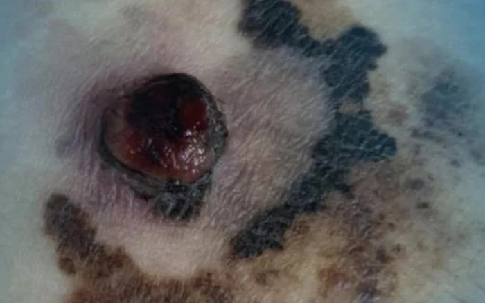
Nodular Melanoma Symptoms
Nodular melanoma (NM) is the second most common subtype and the most lethal per case because it has no recognizable radial growth phase — it begins with vertical (downward) invasion of the dermis immediately. By the time NM is noticed, it is frequently already thick.
Nodular melanoma symptoms are distinct and should trigger urgent evaluation:
- A rapidly growing raised nodule — visible growth over weeks, not months
- Dark blue-black or occasionally amelanotic (flesh-colored, pink, or red) surface
- Smooth, dome-shaped, or irregular surface
- Spontaneous bleeding without trauma — a common early symptom of nodular melanoma
- May be symptomatic: tender, itchy, or burning
The rapid growth and atypical (often amelanotic) color of nodular melanoma means it does not always satisfy the ABCDE criteria, particularly for color (which may be uniform) and evolution may not yet be recognized as concerning. Any rapidly growing new skin nodule that bleeds should be biopsied promptly.
Lentigo Maligna Melanoma Symptoms
Lentigo maligna melanoma (LMM) develops on chronically sun-damaged skin — most often the face, ears, scalp, or neck of older adults — and has the most protracted natural history of any melanoma subtype, often remaining in situ (lentigo maligna stage) for years or decades before invasive transformation occurs.
Symptoms of LMM include:
- A large (often several centimeters), slowly enlarging flat patch on sun-damaged facial skin
- Irregular, mottled pigmentation: variable shades of tan, brown, and dark brown
- Islands of regression within the lesion (pale, hypopigmented areas)
- Ill-defined edges blending with surrounding skin
- Occasional focal raised papule or nodule within a flat lesion — indicates transition from in situ to invasive disease
Acral Lentiginous Melanoma Symptoms
Acral lentiginous melanoma (ALM) is unique among melanoma subtypes in its anatomical location (palms, soles, and nail apparatus) and its independence from UV exposure. ALM is the subtype most commonly diagnosed in Black, Asian, and Hispanic patients — not because these groups have higher risk, but because UV-independent ALM represents a larger proportion of melanomas in darker-skinned individuals.
ALM symptoms by location:
Palmar and plantar ALM: An irregular, slowly expanding pigmented patch on the palm of the hand or the sole of the foot. The lesion is often initially light tan or brown, with progressive darkening and border irregularity as it enlarges. Because these sites are not routinely examined, ALM at this location is frequently discovered late — often when patients notice a new dark area they cannot explain, or when it becomes symptomatic (bleeding, pain, or discharge).
Subungual (nail) melanoma: Presents as longitudinal melanonychia — a dark streak running the full length of the nail from the proximal nail fold (where the nail is generated) to the free edge. Key distinguishing feature: Hutchinson’s sign — extension of the pigment from the nail bed onto the surrounding skin of the proximal nail fold or lateral nail folds. Hutchinson’s sign has high specificity for subungual melanoma and should trigger urgent biopsy. Advanced subungual melanoma may cause nail destruction, lifting of the nail plate (onycholysis), or a nodular growth under the nail.
Amelanotic Melanoma Symptoms
Amelanotic melanoma is one of the most diagnostically challenging presentations because it lacks the pigmentation that defines most melanoma warning sign frameworks. Without color variation to alert the patient or physician, amelanotic melanoma relies entirely on structural features and behavioral changes for recognition.
Amelanotic melanoma symptoms include:
- A new pink, red, skin-colored, or lightly pigmented raised lesion
- May be nodular (amelanotic nodular melanoma) or flat/plaque-like
- Spontaneous bleeding without trauma — a red flag for any unexplained vascular skin lesion
- Rapid growth over weeks to months
- May mimic: pyogenic granuloma, Spitz nevus, amelanotic BCC, squamous cell carcinoma, inflamed seborrheic keratosis, or granulation tissue
Because amelanotic melanoma defies the standard color-based ABCDE recognition framework, physicians and patients must rely on the E (evolution) and B (border) criteria alongside clinical suspicion based on growth rate. Any rapidly growing, bleeding skin lesion that does not resolve or respond to standard wound care within 4–6 weeks should be biopsied.
Symptoms of Metastatic Melanoma
When melanoma spreads beyond the primary skin site, it generates symptoms that reflect the affected organ system. Recognizing these patterns can guide evaluation for patients with a known history of melanoma who develop new symptoms.
Regional lymph node metastasis: The first symptom of nodal spread is often a painless, rubbery lymph node enlargement in the drainage basin of the primary tumor (axillary nodes for upper extremity/trunk melanoma; inguinal nodes for lower extremity melanoma; cervical nodes for head/neck primaries). In-transit metastases — tumor deposits between the primary site and the regional nodes — appear as skin-colored or pigmented papules or nodules along the dermis of the limb.
Pulmonary metastasis: The lungs are the most common visceral site for melanoma metastasis. Symptoms include persistent cough, hemoptysis (coughing blood), and progressive dyspnea. These symptoms develop after sufficient tumor burden accumulates in the pulmonary parenchyma or pleura.
CNS metastasis: Brain metastases are common in advanced melanoma — melanoma has one of the highest rates of CNS tropism of any solid tumor. Symptoms include new-onset headache (particularly early morning), seizures, focal neurological deficits (weakness, speech or vision changes), and personality or behavioral changes. Any new neurological symptom in a melanoma patient with known or suspected metastatic disease requires urgent brain MRI.
Hepatic metastasis: Right upper quadrant pain, nausea, early satiety, and jaundice can develop when melanoma metastasizes to the liver. Elevated liver enzymes on routine blood work may precede symptoms.
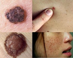
Melanoma Symptoms vs. Benign Skin Lesions: Common Mimickers
Several benign skin conditions can closely resemble melanoma symptoms, and distinguishing them requires dermatological expertise and often dermoscopy or biopsy.
Seborrheic keratosis: The most common mimicker of melanoma. Can be darkly pigmented, multi-colored, and irregular. Distinguishing features: a “stuck-on” waxy or greasy surface, cerebriform (brain-like) warty texture, clearly pasted-on appearance, and comedone-like openings on dermoscopy. However, inflamed, irritated, or atypical seborrheic keratoses can be genuinely difficult to distinguish clinically, and biopsy is appropriate when there is genuine uncertainty.
Blue nevus: A uniformly blue-gray, well-defined, stable papule. Most blue nevi are benign and represent deeply situated melanocytes. Malignant blue nevus is extremely rare but does occur; any blue-gray lesion that grows or changes warrants evaluation.
Spitz nevus: Most common in children and young adults; typically a pink or red dome-shaped papule that grows rapidly over months and then stabilizes. Spitz nevi can be histologically very difficult to distinguish from melanoma (“Spitzoid melanoma”). Any rapidly growing pink nodule in a child or adolescent warrants dermatological evaluation.
Pigmented basal cell carcinoma: Can appear dark brown or blue-black, sometimes resembling melanoma or pigmented nevus. On dermoscopy, pigmented BCC shows leaf-like pigment areas, arborizing (branching tree-like) vessels, and spokes — features that differ from melanoma. Biopsy provides definitive diagnosis.
See a dermatologist within days — not weeks — for any of these melanoma symptoms: A new mole that appeared in adulthood; any existing mole that has grown, changed color, or changed shape within the past 2–4 weeks; a mole or skin spot that bleeds without injury; a dark streak under a fingernail or toenail (especially if it has appeared recently or is widening); any raised, fast-growing nodule that bleeds easily; or any skin lesion that your doctor previously described as stable that has now changed. Evolution — change — is the red flag that overrides all other considerations.
Frequently Asked Questions
Can melanoma develop in a place that doesn’t get sun?
Yes. Approximately 70–80% of melanomas arise de novo on skin that may never have been examined or considered at risk. Acral lentiginous melanoma occurs on palms, soles, and under nails — UV-independent sites. Mucosal melanoma occurs inside the mouth, nose, genitalia, and anorectal region. Uveal (eye) melanoma develops within the choroid. Any melanocyte in the body can transform into melanoma regardless of UV history.
What is the difference between a benign mole and a melanoma?
A benign common mole (melanocytic nevus) is symmetric, has a well-defined smooth border, is a single uniform shade of brown or tan, is usually smaller than 6mm, and has been stable for years. Melanoma is asymmetric, has irregular or poorly defined borders, displays multiple colors, may be larger than 6mm, and crucially — is changing. Stable lesions are reassuring; changing lesions are not. For a complete guide to recognizing changes, see our article on skin cancer symptoms.
Do all melanomas start as moles?
No. The majority of melanomas — approximately 70–80% — arise on apparently normal-looking skin, not within a pre-existing mole. This is one reason why monthly total-body skin self-examination is more informative than simply monitoring known moles.
When should I go to the emergency room for melanoma symptoms?
Emergency department evaluation is appropriate for new neurological symptoms in a patient with known advanced melanoma (suggesting possible brain metastasis requiring urgent imaging), hemorrhage from a skin lesion that cannot be controlled with direct pressure, or signs of systemic illness with a newly discovered suspicious skin lesion in an immunocompromised patient. For asymptomatic suspicious lesions, a dermatology appointment within days is appropriate — not an emergency visit, but not a 3-month wait either.
Sources
- American Academy of Dermatology. Melanoma: Signs and Symptoms (ABCDE Criteria).
- National Comprehensive Cancer Network. NCCN Guidelines: Melanoma v2.2024.
- National Cancer Institute. Melanoma Treatment (PDQ) — Patient Version.
- Sloot S et al. Amelanotic melanoma: distinctive clinical and dermoscopic features. JAAD. 2018.
- Rigel DS et al. ABCDE — an evolving concept in the early detection of melanoma. JAAD. 2005.
The “Ugly Duckling” Sign: A Complementary Symptom Recognition Tool
The ugly duckling sign is a complementary approach to melanoma symptom recognition based on the observation that in most people, moles look similar to one another — they form a “flock” of similar-appearing lesions. A melanoma often looks different from a patient’s other moles: it is the outlier, the lesion that does not fit the pattern of everything else on the skin. This approach is particularly useful for people with many moles, where the ABCDE criteria can be challenging to apply uniformly.
Clinically, the ugly duckling sign works like this: when examining a patient with 50 or more moles, the dermatologist is not comparing each individual lesion against an abstract standard — they are looking for the one mole that stands out as different from the rest. That mole may be larger, darker, more elevated, or differently shaped than its neighbors. It may simply feel “wrong” against the background of the patient’s usual mole pattern. This outlier deserves closer inspection and often biopsy.
Patients can apply this concept during monthly self-examination. After becoming familiar with your own mole pattern, look for any lesion that seems out of place — that appears different from everything else — as well as any lesion that changes relative to how it looked during the previous month’s examination.
How to Recognize Melanoma Symptoms During Skin Self-Examination
Effective recognition of melanoma symptoms requires a systematic, monthly skin self-examination using good lighting, a full-length mirror, and a hand mirror for areas not directly visible. The steps described below are aimed specifically at melanoma symptom recognition, not just general skin awareness:
- Face and neck: Check for any new pigmented spot, particularly on sun-exposed areas. Lentigo maligna — the precursor to LMM — appears on the face of older adults as an irregular, slowly enlarging tan patch. Any such lesion in this distribution that has changed over 6–12 months warrants evaluation.
- Scalp: Use a blow-dryer or comb to part the hair systematically. Scalp melanoma is frequently detected late because it is covered by hair. Ask a partner to examine the posterior scalp and crown.
- Back: The back is one of the most common sites for melanoma in men. Use a full-length mirror and a hand mirror together. Any lesion on the back that is changing, asymmetric, or multi-colored deserves a photograph for documentation and a dermatology visit.
- Palms, soles, and nails: Inspect the palms and soles for any irregular dark patch. Check all 10 fingernails and 10 toenails for dark longitudinal streaks. A new dark streak under a nail — particularly one that is widening or that extends onto the skin around the nail (Hutchinson’s sign) — requires prompt evaluation regardless of skin tone.
- Genitalia: Melanoma and mucosal melanoma can arise on genital skin or mucosa. Any new pigmented lesion or patch in this area that has appeared recently or changed warrants evaluation.
Taking monthly photographs of suspicious or borderline lesions allows direct comparison over time and substantially improves the reliability of the E (evolution) criterion. A mole that appeared to be unchanged in memory alone is often documented to have grown meaningfully when a photograph from 3 months earlier is available for comparison. For guidance on how often professional skin examinations should occur based on individual risk level, see our guide to skin cancer screening.
When Melanoma Symptoms Need Biopsy vs. Monitoring
Not every suspicious lesion requires immediate biopsy. Dermatologists use clinical and dermoscopic assessment to stratify lesions into three management pathways:
Biopsy now: Lesions with dermoscopic features highly specific for melanoma (blue-white veil, irregular network, peripheral streaks, multiple regression structures), lesions that have grown rapidly, lesions on the palms/soles/nails fitting the ALM pattern, any lesion that bleeds spontaneously, or amelanotic nodular lesions with rapid growth. The threshold for biopsy should be low when the consequences of a missed early melanoma are considered against the minimal risk of an excisional biopsy of a benign lesion.
Short-interval monitoring (3 months): Lesions with some concerning features but no definitive dermoscopic red flags may be followed with digital dermoscopy at 3-month intervals. This approach is applied to stable but borderline lesions in patients who are reliable for follow-up. Any growth or change at the 3-month visit triggers biopsy.
Routine annual monitoring: Lesions assessed as low-risk with no ABCDE features, stable on dermoscopy, and fitting a patient’s usual mole pattern — these are photographed and added to the baseline surveillance record for annual review.
Patients should advocate for biopsy when a lesion is genuinely concerning to them, even if a clinician initially suggests monitoring. A negative biopsy result provides definitive reassurance; continued uncertainty does not. For a comprehensive overview of the full spectrum of concerning lesions and symptoms beyond melanoma, see our article on skin cancer symptoms.