What Is Melanoma?
Melanoma is cancer of the melanocytes — the specialized cells that produce melanin, the pigment responsible for skin, hair, and eye color. Melanocytes are most concentrated in the skin, but they are also present in the eyes, ears, nasal cavity, mouth, gastrointestinal tract, and meninges, which is why melanoma can arise at sites far removed from sun-exposed skin.
Among all skin cancers, melanoma accounts for only about 1% of cases but causes approximately 80% of skin cancer deaths. This disproportionate lethality reflects melanoma’s capacity for early lymphatic and hematogenous spread — a single melanoma cell can metastasize to the lymph nodes, lungs, liver, brain, or bone before the primary lesion on the skin surface has reached 1 millimeter in thickness.
In the United States, approximately 100,000 new melanomas are diagnosed each year, with around 8,300 deaths. When detected at Stage I, the 5-year survival rate exceeds 97%. When detected at Stage IV, survival — even with modern immunotherapy — is approximately 30–35% at 5 years. Early detection is not merely advantageous; for melanoma, it is the determining variable in survival. For guidance on how and when to be examined, see our overview of skin cancer screening.
Melanoma Subtypes
Not all melanomas look alike, and recognizing the different presentations prevents delays in diagnosis.
Superficial spreading melanoma (SSM) accounts for approximately 70% of all melanomas. It begins with a radial (horizontal) growth phase along the epidermis before transitioning to a vertical growth phase that penetrates deeper into the dermis. This radial phase can last months to years and is the window during which SSM is most likely to display classic ABCDE features.
Nodular melanoma accounts for 15–30% of cases and is the most aggressive subtype by behavior. Unlike SSM, nodular melanoma begins with vertical growth immediately, meaning it invades the dermis from the outset. It often presents as a rapidly enlarging blue-black or amelanotic (flesh-colored or reddish) raised nodule. By the time patients notice it, nodular melanoma has frequently reached a Breslow thickness of 2mm or greater.
Lentigo maligna melanoma (LMM) develops on chronically sun-damaged skin, typically the face, neck, or dorsal forearms of older adults. It begins as a lentigo maligna — an in situ precursor — which can persist for years before invasive transformation. LMM classically appears as a large, irregularly pigmented flat patch that slowly expands over the course of decades.
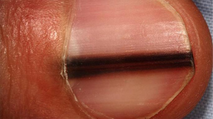
Acral lentiginous melanoma (ALM) develops on the palms, soles, and beneath the fingernails or toenails. Unlike other subtypes, ALM is not associated with UV exposure — it occurs at equal rates across skin tones, making it proportionally the most common melanoma subtype in Black, Asian, and Hispanic patients. Subungual ALM often presents as a dark streak running the length of the nail (Hutchinson’s sign when the pigment extends onto the periungual skin).
Amelanotic melanoma lacks pigment and can present as a pink, red, or flesh-colored papule or nodule. Because it does not display the expected dark pigmentation, amelanotic melanoma is frequently misdiagnosed as a benign growth.
Desmoplastic melanoma is a rare variant characterized by spindle cells and strong perineural invasion tendency. It often appears as a firm, flesh-colored plaque on the head or neck, associated with a background of lentigo maligna, and has a high local recurrence rate requiring wider surgical margins than other subtypes.
Who Is at Risk for Melanoma
Ultraviolet exposure is the primary environmental driver of cutaneous melanoma. Intermittent intense sun exposure, particularly blistering sunburns in childhood and adolescence, increases risk substantially. Tanning bed use increases melanoma risk by 59–75% for users who start before age 35.
Skin type and pigmentation: People with Fitzpatrick skin types I and II (very fair skin that always burns, often with red or blonde hair and light eyes) have the highest baseline risk. However, people of all skin tones can and do develop melanoma — particularly ALM, which is not UV-related.
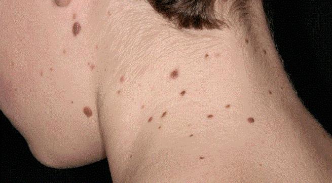
Mole count and atypical nevi: Having more than 50 ordinary moles doubles melanoma risk. Having multiple atypical (dysplastic) nevi and a family history of melanoma constitutes the familial atypical multiple mole melanoma (FAMMM) syndrome, which carries a lifetime melanoma risk exceeding 50%.
Family history and genetics: Approximately 10% of melanomas occur in familial clusters. Mutations in the CDKN2A gene confer a 76% lifetime melanoma risk. A patient who has had one melanoma has a 10-fold higher risk of developing a second primary melanoma. For an understanding of the full spectrum of skin cancer risk factors including genetic predispositions, see our overview of skin cancer causes and types.
Immunosuppression: Organ transplant recipients, people on long-term biologic therapies, and those with HIV and low CD4 counts all have elevated melanoma risk.
ABCDE Warning Signs of Melanoma
The ABCDE criteria represent the most widely validated framework for identifying early melanoma. Any skin lesion meeting one or more of these criteria warrants dermatological evaluation.
- A — Asymmetry: One half of the lesion does not match the other half. Benign moles are symmetric; melanoma lesions commonly are not.
- B — Border: Irregular, notched, scalloped, or poorly defined borders distinguish melanoma from the smooth, well-defined edges of benign nevi.
- C — Color: Multiple colors within a single lesion — mixtures of tan, brown, dark brown, black, red, white, or blue — indicate variable melanocyte activity consistent with malignant transformation.
- D — Diameter: A lesion larger than 6mm (approximately the diameter of a pencil eraser) warrants evaluation, though melanomas can be smaller.
- E — Evolution: Any change in a lesion — new appearance, growth, color change, shape change, or new symptoms such as bleeding or itching — is the most sensitive indicator of melanoma.
For a more detailed exploration of these features and how to perform a systematic self-examination, see our dedicated guide to ABCDE melanoma warning signs and our overview of skin cancer symptoms.
Melanoma Staging: The AJCC 8th Edition System
Melanoma staging uses the AJCC 8th edition TNM system, integrating tumor characteristics, nodal status, and metastatic spread. The single most important prognostic factor for localized melanoma is Breslow thickness — the depth of invasion measured from the granular cell layer of the epidermis to the deepest tumor cell.
T classification (primary tumor depth):
- T1a: less than 0.8mm, no ulceration
- T1b: less than 0.8mm with ulceration, OR 0.8–1.0mm (any)
- T2a: 1.01–2.0mm, no ulceration
- T2b: 1.01–2.0mm, with ulceration
- T3a: 2.01–4.0mm, no ulceration
- T3b: 2.01–4.0mm, with ulceration
- T4a: greater than 4.0mm, no ulceration
- T4b: greater than 4.0mm, with ulceration
Ulceration — microscopic loss of the overlying epidermis — is an independent adverse prognostic factor that upstages tumors within each thickness category. N classification captures lymph node involvement and satellite/in-transit metastases; M classification captures distant spread by site (skin and soft tissue, lung, visceral, CNS) and LDH level.
Surgery: Wide Local Excision and Sentinel Lymph Node Biopsy
Wide local excision (WLE) is the definitive surgical treatment for primary melanoma. Excision margins are determined by Breslow thickness:
- Melanoma in situ: 0.5cm margin
- Breslow ≤1.0mm: 1.0cm margin
- Breslow 1.01–2.0mm: 1.0–2.0cm margin
- Breslow >2.0mm: 2.0cm margin
Sentinel lymph node biopsy (SLNB) is the standard staging procedure for melanomas T1b and thicker. The procedure identifies whether melanoma cells have spread to the first-echelon regional lymph node. A positive sentinel node indicates regional micrometastasis, upgrades the patient to Stage III, and qualifies them for adjuvant systemic therapy.
Following the MSLT-II trial (2017), complete lymph node dissection (CLND) is no longer routinely recommended after a positive sentinel node. The trial demonstrated no overall survival benefit from CLND versus active nodal surveillance with ultrasound, while CLND added substantially higher lymphedema and complication rates.
Immunotherapy for Melanoma
Immune checkpoint inhibitors fundamentally transformed the treatment of advanced melanoma, converting a disease with a historical Stage IV 5-year survival under 10% to one achieving approximately 30–52% at 5 years depending on regimen.
Anti-PD-1 monotherapy: Pembrolizumab (Keytruda) is FDA-approved for adjuvant treatment of Stage IIB/IIC and Stage III/IV resected melanoma — the Stage IIB indication was added following the KEYNOTE-716 trial. Nivolumab (Opdivo) is approved for adjuvant Stage III/IV and metastatic disease.
Combination anti-CTLA-4 + anti-PD-1: The CheckMate 067 trial (Larkin J et al., NEJM 2015) demonstrated 5-year overall survival of 52% for ipilimumab + nivolumab, compared to 44% for nivolumab alone and 26% for ipilimumab alone, in previously untreated metastatic melanoma. The combination achieves superior response rates but with grade 3–4 immune toxicity in ~59% of patients.
LAG-3 inhibition: Relatlimab + nivolumab (Opdualag), FDA-approved March 2022, demonstrated superior progression-free survival versus nivolumab alone in the RELATIVITY-047 trial, adding a new first-line option for unresectable or metastatic melanoma.
Targeted Therapy: BRAF and MEK Inhibitors
Approximately 45–50% of cutaneous melanomas harbor an activating BRAF V600E mutation, which constitutively activates the MAPK signaling pathway driving uncontrolled proliferation. Targeted BRAF+MEK inhibitor combinations produce rapid, high response rates in BRAF-mutated tumors:
- Dabrafenib + trametinib (Tafinlar + Mekinist)
- Vemurafenib + cobimetinib (Zelboraf + Cotellic)
- Encorafenib + binimetinib (Braftovi + Mektovi)
The pivotal trametinib trial (Flaherty KT et al., NEJM 2012) demonstrated the power of MEK inhibition in BRAF-mutated melanoma. Combined BRAF+MEK blockade achieves response rates of 60–70%, but acquired resistance develops in most patients within 12–18 months. In BRAF-mutated advanced melanoma, the choice between immunotherapy and targeted therapy as first-line treatment requires individualization based on disease burden, LDH level, and the urgency of response needed.
Survival Rates by Stage
Five-year survival rates from NCI SEER data (AJCC 8th edition):
- Stage IA: ~99% | Stage IB: ~97%
- Stage IIA: ~94% | Stage IIB: ~87% | Stage IIC: ~82%
- Stage IIIA: ~93% | Stage IIIB: ~83% | Stage IIIC: ~69% | Stage IIID: ~32%
- Stage IV: ~30–35% (modern immunotherapy era)
A notable feature of melanoma staging is that Stage IIIA — low-volume nodal disease from thin primaries — has better survival than Stage IIC (thick tumors without nodal involvement), illustrating that stage number alone does not linearly reflect prognosis. Each patient’s staging profile must be interpreted individually.
See a doctor promptly if you notice: A new dark spot, mole, or lesion that appeared within the past few months; any existing mole that has changed in color, shape, or size; a mole that bleeds without injury; a dark streak under a fingernail or toenail that was not caused by trauma; or any lesion on the palm or sole that is new, pigmented, or slowly enlarging. Do not apply a “wait and see” approach to suspicious skin lesions — melanoma diagnosed at Stage I is almost always curable; the same lesion discovered at Stage III carries substantially worse odds.
Melanoma vs. Other Skin Cancers
Understanding how melanoma differs from the other common skin cancers — basal cell carcinoma (BCC) and squamous cell carcinoma (SCC) — clarifies why early detection matters so much more for melanoma specifically.
BCC arises from basal cells in the deepest layer of the epidermis. It grows slowly and almost never metastasizes — the metastasis rate for BCC is estimated at less than 0.1%. SCC arises from squamous (flat) epidermal cells and metastasizes in approximately 2–5% of cases. Melanoma, by contrast, can metastasize at Breslow thicknesses as thin as 1.0mm, and the metastatic potential increases substantially with thickness and ulceration.
This difference in metastatic biology is why melanoma accounts for the overwhelming majority of skin cancer deaths despite being far less common than BCC and SCC. For comprehensive information on how BCC and SCC present and are treated, see our articles on basal cell carcinoma and squamous cell carcinoma.
Frequently Asked Questions
What is the difference between melanoma and other skin cancers?
Basal cell and squamous cell carcinomas arise from keratinocytes and rarely metastasize. Melanoma arises from melanocytes and has a substantially higher metastatic potential. A melanoma that has reached 4mm Breslow thickness has approximately a 40–50% chance of nodal involvement — which is why melanoma causes the vast majority of skin cancer deaths despite being less common than BCC or SCC.
Can melanoma develop in someone with dark skin?
Yes. While the absolute incidence is lower in people with darker skin tones, melanoma does occur and is disproportionately diagnosed at later stages. Acral lentiginous melanoma — occurring on palms, soles, and under the nails — is not sun-related and occurs across all racial and ethnic backgrounds. For more on how melanoma symptoms present across skin tones, see our guide to melanoma symptoms.
Is melanoma always associated with a mole?
No. Only approximately 20–30% of melanomas arise within a pre-existing mole. The majority arise de novo on apparently normal skin. This is why total skin examination is important — waiting for an existing mole to change misses most melanomas.
What does a BRAF test result mean for treatment?
BRAF mutation testing is performed on all advanced melanoma specimens. A BRAF V600E or V600K mutation means the patient is eligible for BRAF+MEK targeted therapy, which can achieve rapid tumor shrinkage (response rate ~60–70%) and is often preferred when disease burden is high. BRAF wild-type patients receive immunotherapy as first-line treatment. Immunotherapy is effective regardless of BRAF status.
Sources
- National Cancer Institute SEER. Cancer Stat Facts: Melanoma of the Skin.
- National Comprehensive Cancer Network. NCCN Guidelines: Melanoma v2.2024.
- Larkin J et al. Combined nivolumab and ipilimumab or monotherapy in untreated melanoma. N Engl J Med. 2015.
- Flaherty KT et al. Improved survival with MEK inhibition in BRAF-mutated melanoma. N Engl J Med. 2012.
- American Society of Clinical Oncology. Melanoma: Types of Treatment.
Melanoma Prevention: What the Evidence Supports
While not all melanomas are preventable — acral lentiginous and mucosal subtypes have no UV link — the majority of cutaneous melanomas are causally related to UV exposure, making primary prevention a meaningful intervention.
Sunscreen: Broad-spectrum SPF 30 or higher, applied daily to sun-exposed skin, reduces UV damage. The landmark randomized controlled trial (Green AC et al., Journal of Clinical Oncology, 2011) demonstrated that daily application of SPF 16 sunscreen reduced melanoma incidence by 50% and halved melanoma mortality over a 10-year follow-up in an Australian cohort. Higher SPF sunscreens applied in adequate quantities (2mg/cm² of skin surface) provide greater protection. Sunscreen should be reapplied every 2 hours during extended outdoor exposure.
Protective clothing and shade: UPF (ultraviolet protection factor) clothing, wide-brimmed hats, and UV-blocking sunglasses provide physical protection that is not subject to the application inconsistencies that reduce sunscreen effectiveness in practice. Seeking shade during peak UV intensity hours (10am–4pm) substantially reduces cumulative UV dose.
Avoiding tanning beds: Given the clear dose-response relationship between tanning bed use and melanoma risk — particularly for users who begin before age 35 — avoiding tanning beds is one of the most effective specific prevention actions for young adults. Many countries have banned commercial tanning bed use under age 18 on this basis.
Vitamin D: A common concern about sun avoidance is vitamin D deficiency. The evidence supports that adequate vitamin D can be maintained through dietary sources and supplementation without ultraviolet skin exposure. Intentional sun exposure for vitamin D synthesis is not recommended as a cancer prevention strategy, and the doses of UV required for significant vitamin D synthesis also confer meaningful skin cancer risk.
Melanoma Surveillance After Treatment
Following definitive treatment of melanoma — whether surgical alone for early-stage disease, or combined with adjuvant immunotherapy or targeted therapy for higher-stage disease — ongoing surveillance is essential for two reasons: to detect recurrence as early as possible, and to identify new primary melanomas.
Surveillance visits typically include a complete skin examination, palpation of regional lymph nodes, and a structured review of symptoms. Imaging (CT, PET-CT, or MRI) is added according to stage-based NCCN protocols, most intensively during the first 2–3 years when recurrence risk is highest.
Patients with a history of melanoma should continue annual dermatology examination for life, regardless of time since diagnosis. The 10-fold increased risk of a second primary melanoma does not diminish over time — it is a permanent feature of the risk profile following any melanoma diagnosis. Patients should also perform monthly skin self-examination between professional visits. For the schedule of follow-up examinations by melanoma stage, see our guide to skin cancer screening after a melanoma diagnosis.
Clinical Trials and Emerging Treatments
Melanoma has been one of the most active areas of oncology research over the past 15 years, and clinical trials continue to advance treatment options for both early and advanced disease. Current areas of active investigation include:
- Personalized mRNA cancer vaccines: A Phase 2b trial (Moderna/Merck mRNA-4157 + pembrolizumab) demonstrated a significant reduction in melanoma recurrence and distant metastasis in high-risk resected Stage IIB–IV patients compared to pembrolizumab alone (KEYNOTE-942, 2023). This represents the first meaningful signal for individualized neoantigen cancer vaccines in any solid tumor.
- Neoadjuvant immunotherapy: Treating Stage III melanoma with immunotherapy before surgery (neoadjuvant) has shown pathological complete response rates of 30–60% in early trials, potentially sparing some patients more extensive surgical procedures and providing powerful prognostic information.
- Intralesional therapies: T-VEC (talimogene laherparepvec) is already FDA-approved for unresectable cutaneous/subcutaneous melanoma. Newer oncolytic viruses and intralesional agents are in clinical trials.
- NRAS-targeted therapy: Approximately 20% of melanomas harbor NRAS mutations. Effective targeted therapy for NRAS-mutated melanoma remains an unmet need; MEK inhibitor trials and combinations are ongoing.
Patients with advanced melanoma who are not responding to or who have progressed on standard therapies should be referred to a melanoma center with active clinical trial programs. The rapid pace of melanoma therapeutic development means that clinical trial access represents a meaningful treatment option, not a last resort.
Melanoma outcomes have improved dramatically over the past decade, and the trajectory of progress continues. Patients diagnosed today have access to treatments — checkpoint immunotherapy, targeted BRAF+MEK inhibition, and emerging mRNA vaccine approaches — that did not exist 15 years ago. For patients at any stage, the combination of regular skin self-examination, professional dermatological surveillance, and prompt evaluation of any changing or new skin lesion remains the most reliable pathway to catching melanoma when treatment is most effective. The biology of melanoma is unforgiving once metastatic spread occurs; the biology of early melanoma is among the most favorable of any human cancer, with cure rates approaching 99% for Stage IA disease. The difference between those two outcomes, for most patients, is detection.