What Is Squamous Cell Carcinoma?
Squamous cell carcinoma (SCC) is the second most common skin cancer, with approximately 1.8 million new cases diagnosed in the United States each year. SCC arises from squamous cells — the flat, scale-like keratinocytes that make up the majority of the epidermis. Unlike basal cell carcinoma (BCC), which almost never spreads, SCC has a genuine metastatic potential: approximately 2–5% of cutaneous SCCs spread to regional lymph nodes or distant organs, and this rate rises substantially in high-risk presentations.
Cutaneous SCC causes an estimated 15,000 deaths per year in the United States — more deaths than melanoma in some analyses, reflecting the sheer volume of SCC cases and the population of immunosuppressed patients in whom SCC behaves with particular aggression. For most otherwise healthy individuals with a localized, low-risk SCC, treatment is highly effective and curative. The challenge lies in identifying and treating high-risk SCC before it metastasizes, and in managing the heavily immunosuppressed patient population in whom SCC is a major cause of mortality. For context on where SCC falls in the skin cancer spectrum, see our overviews of skin cancer types, basal cell carcinoma, and melanoma.
From Sun Damage to Cancer: How SCC Develops
The development of cutaneous SCC follows a well-characterized progression from UV-damaged skin to premalignant change to invasive carcinoma. Understanding this continuum guides both prevention and early intervention.
UV damage and TP53 mutations: Ultraviolet radiation — particularly UVB — causes characteristic mutations in the TP53 tumor suppressor gene, which normally triggers apoptosis (programmed cell death) in cells with damaged DNA. When TP53 is mutated, cells with UV-damaged DNA survive and continue to replicate, accumulating additional mutations over time. This process underlies the field cancerization concept — the entire chronically sun-exposed skin surface (the “field”) sustains UV damage and is at elevated risk for developing multiple AKs and SCCs over time.
Actinic keratoses (AKs): The premalignant precursor to SCC. AKs appear as rough, scaly, erythematous papules or plaques on sun-exposed skin — the face, scalp, ears, dorsal hands, and forearms. Each individual AK has an approximately 1–3% annual risk of progressing to invasive SCC, and a 10% lifetime risk. A patient with dozens of AKs distributed across sun-damaged skin (field cancerization) has a substantially elevated cumulative SCC risk, justifying field therapy rather than individual lesion treatment.
EGFR and other molecular drivers: Epidermal growth factor receptor (EGFR) is overexpressed in the majority of cutaneous SCCs and represents a therapeutic target. HPV infection, particularly HPV types 16 and 18, contributes to SCC carcinogenesis at mucosal sites (anogenital, oral, oropharyngeal) and is implicated in a subset of cutaneous SCCs in immunosuppressed patients.
Actinic Keratoses: Treating the Precursor to Prevent SCC
Because AKs are the recognized precursor to SCC, treating AKs represents a form of primary prevention for invasive SCC. For patients with isolated AKs (1–5 lesions), cryotherapy — liquid nitrogen applied to each lesion individually — is the most common approach. For patients with field cancerization and many AKs distributed across a skin area, field therapy is preferred:
- Topical 5-fluorouracil (5-FU, Efudex, Carac): Applied twice daily for 2–4 weeks to the affected field. 5-FU preferentially disrupts rapidly dividing dysplastic cells, causing significant local inflammation (redness, erosion, crusting) that resolves over 2–4 weeks post-treatment. The intense local reaction is a sign that the treatment is working, not a reason to discontinue.
- Imiquimod 3% or 5%: Applied 2–3 times weekly for 4–16 weeks; stimulates local immune response to eliminate dysplastic cells. Slower than 5-FU with less intense inflammatory reaction; appropriate for patients who require a milder regimen.
- Photodynamic therapy (PDT): ALA or MAL photosensitizer is applied to the field; after incubation, illumination with red light activates the photosensitizer to generate reactive oxygen species that selectively destroy dysplastic cells. Excellent cosmetic results; requires clinic visit; highly effective for AK clearance.
SCC Subtypes and Special Presentations
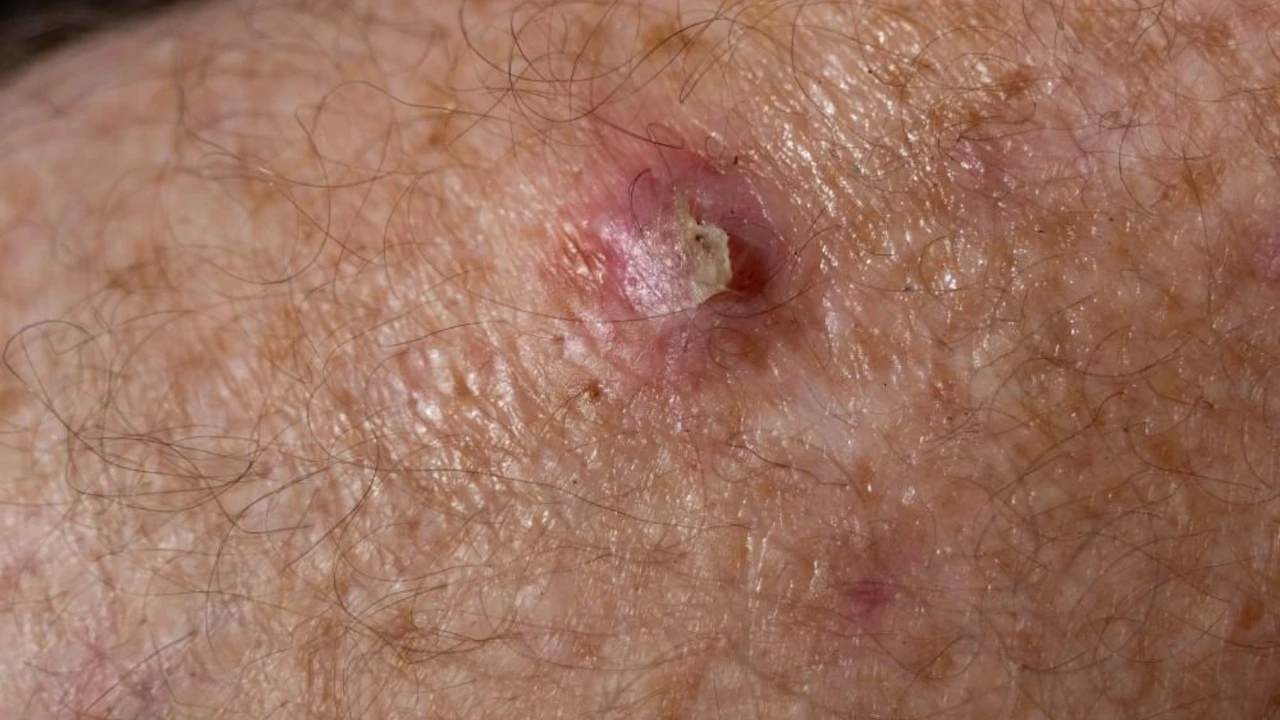
Well-differentiated SCC is the most common form: a firm, keratinized papule, plaque, or nodule, often with a rough or warty surface, on sun-exposed skin. It grows steadily but usually more slowly than poorly differentiated SCC.
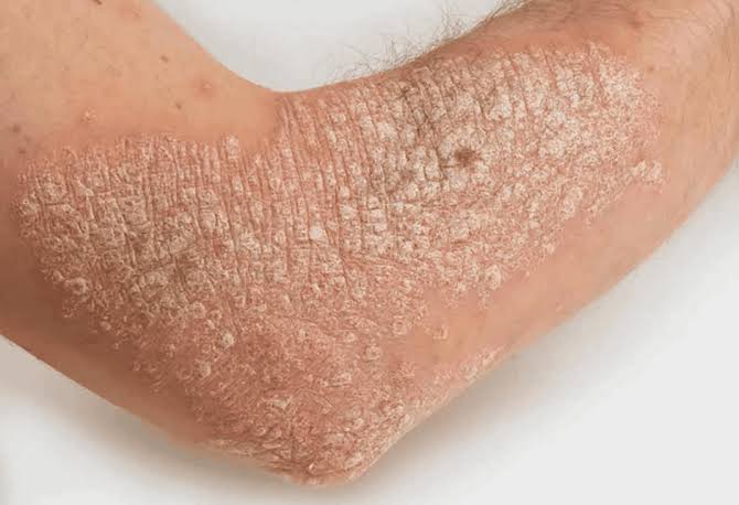
SCC in situ (Bowen’s disease) is intraepidermal SCC that has not yet breached the basement membrane. It presents as a red, scaly, sharply demarcated patch that can resemble eczema or psoriasis. Without treatment, approximately 3–5% of Bowen’s disease lesions progress to invasive SCC. Treatment options include 5-FU, imiquimod, PDT, cryotherapy, or excision.
Poorly differentiated SCC is less keratinized, grows more rapidly, and has a substantially higher metastatic potential than well-differentiated SCC. Perineural invasion — tumor cells tracking along nerve sheaths — is more common in poorly differentiated SCC and is an independent predictor of metastatic risk.
Marjolin’s ulcer is SCC arising in a chronic wound, burn scar, or area of chronic inflammation. It is notably aggressive — metastasis rates for Marjolin’s ulcer SCC range from 20–30%, far exceeding those of UV-associated SCC. Any non-healing wound or persistently ulcerated scar that has been present for many years should be biopsied to exclude SCC.
Keratoacanthoma (KA) is a rapidly growing, crater-shaped, keratinous papule that classically appears within 6–8 weeks and may spontaneously involute over months. It is histologically related to SCC and some authorities classify it as a variant of well-differentiated SCC. The clinical behavior (spontaneous regression) differs from classic SCC, but biopsy is required to distinguish KA from invasive SCC, and most dermatologists treat KA with excision rather than observation due to inability to predict which lesions will involute.
Risk Factors for Squamous Cell Carcinoma
Cumulative UV exposure is the dominant risk factor, particularly for SCC arising in the typical distribution — face, scalp, ears, dorsal hands. Unlike melanoma (where intermittent intense sunburns are a key driver), SCC is more directly linked to total lifetime UV dose, making it a particular concern for people with significant occupational sun exposure.
HPV infection: Human papillomavirus types 16 and 18 are established causes of SCC at mucosal sites — cervical, vaginal, vulvar, penile, anal, and oropharyngeal SCCs are largely HPV-driven. For cutaneous SCC, HPV plays a supporting role particularly in immunosuppressed patients. HPV vaccination (Gardasil 9) prevents HPV 16/18 infection and substantially reduces HPV-associated SCC risk at mucosal sites.
Immunosuppression: This is the most critical risk factor modifier. Organ transplant recipients develop SCC at 65–250 times the rate of immunocompetent individuals — the highest relative risk increase of any skin cancer in transplant patients. Transplant-associated SCC is more aggressive, more frequently multifocal, and more often lethal than UV-associated SCC in immunocompetent individuals. HIV infection (particularly with low CD4 counts), long-term biologic therapy, and chronic corticosteroid use all elevate SCC risk.
Chronic wounds and scars: Marjolin’s ulcer — SCC arising in burn scars, chronic wounds, or areas of chronic osteomyelitis — represents a particularly aggressive SCC phenotype. Any wound that has been present for many years without healing should be evaluated for malignant transformation.
High-Risk SCC: Who Needs More Aggressive Treatment
NCCN guidelines stratify SCC into low-risk and high-risk categories based on clinical and pathological features. High-risk SCC requires more aggressive treatment — typically Mohs surgery — and closer follow-up:
High-risk anatomical locations: Ear, lip, temple, scalp, hands, feet, anogenital region, and areas of prior radiotherapy. These sites have higher metastatic potential and/or more limited surgical margin options.
High-risk histological features:
- Poorly differentiated or undifferentiated histology
- Perineural invasion (PNI) — one of the most important high-risk features; associated with local recurrence and nodal metastasis
- Lymphovascular invasion
- Tumor thickness >2mm or Breslow depth >4mm
- Clark level IV–V invasion
High-risk host and tumor characteristics:
- Size >2cm
- Recurrent SCC (previously incompletely excised)
- Immunosuppressed patient (especially organ transplant recipient)
- SCC arising in chronic wound, scar, or radiation field
Surgical Treatment: Excision and Mohs Surgery
For low-risk SCC, standard excision with 4–6mm margins achieves cure in the majority of cases. Permanent section margin evaluation is recommended.
Mohs micrographic surgery is indicated for high-risk SCC — providing the same advantage of complete margin evaluation as for BCC, with the ability to spare normal tissue on cosmetically and functionally critical areas. Mohs is particularly important for SCC with perineural invasion, recurrent SCC, and SCC in immunosuppressed patients where any residual tumor represents significant recurrence risk.
Sentinel lymph node biopsy (SLNB) is not standard for all SCCs, but is considered for high-risk SCC (thick tumor, perineural invasion, immunosuppressed patient) where the risk of nodal micrometastasis is high enough to justify the procedure. When nodes are clinically positive, lymph node dissection is performed (unlike melanoma, where CLND after positive SLNB is no longer standard, lymph node dissection for clinically positive SCC nodal disease remains standard of care).
Radiation therapy is an alternative for patients who cannot undergo surgery and as adjuvant therapy when perineural invasion or positive margins are identified after surgical excision.
Advanced SCC: Immunotherapy with Cemiplimab and Pembrolizumab
For SCC that is locally advanced (unresectable) or has metastasized, checkpoint immunotherapy has become the standard of care:
Cemiplimab (Libtayo), an anti-PD-1 antibody, was FDA-approved in 2018 as the first systemic therapy specifically approved for cutaneous SCC. The pivotal trial (Migden MR et al., NEJM 2018) demonstrated an objective response rate of approximately 47% in metastatic cSCC — a remarkable result for a disease that previously had no approved systemic treatment.
Pembrolizumab (Keytruda) received FDA approval in 2020 for recurrent or metastatic cutaneous SCC not curable by surgery or radiation, providing a second anti-PD-1 option.
EGFR inhibitors (cetuximab, erlotinib) had been used before checkpoint inhibitor approval and remain an option for patients who cannot receive or progress on immunotherapy. Platinum-based chemotherapy (cisplatin ± 5-FU) may be used in combination with cetuximab for head/neck SCC or in patients not suitable for checkpoint inhibitors.
See a dermatologist promptly for these SCC warning signs: A firm, rough, scaly papule or nodule that is growing on sun-exposed skin; a sore on the lip, ear, or scalp that is not healing; any non-healing wound or ulcer that has been present for more than 6 weeks; a rapidly growing skin growth (especially dome-shaped with central crater); any lesion on a burn scar or old wound that has changed or started to bleed; new numbness or tingling along the course of a facial nerve (suggesting perineural invasion). SCC caught early at the primary site is highly curable; SCC with nodal spread requires more extensive treatment with less favorable outcomes.
SCC in Organ Transplant Recipients: Special Considerations
Organ transplant recipients represent the highest-risk population for SCC and deserve specific mention. The 65–250-fold elevated SCC risk in transplant patients — driven by chronic immunosuppression, often decades of pre-transplant UV accumulation, and HPV-driven carcinogenesis — means that SCC is a leading cause of morbidity and mortality in this population, not a minor dermatological concern.
All organ transplant recipients should:
- Begin dermatological surveillance 6–12 months after transplantation
- Be seen by a dermatologist at least annually, and quarterly if skin cancer has developed
- Practice rigorous sun protection (SPF 50+, UPF clothing, hat) as a mandatory post-transplant lifestyle modification
- Have any new or changing skin lesion promptly evaluated — the threshold for biopsy should be extremely low
Systemic retinoids (acitretin) are used in some high-risk transplant recipients with multiple SCCs to reduce new SCC formation. Modification of the immunosuppressive regimen — for example, switching from calcineurin inhibitors to mTOR inhibitors (sirolimus, everolimus) — may reduce SCC risk and is discussed with transplant nephrology when the SCC burden is significant.
Frequently Asked Questions
Can squamous cell carcinoma spread?
Yes. Unlike BCC, SCC has genuine metastatic potential. Low-risk SCC (small, well-differentiated, no perineural invasion, immunocompetent host) metastasizes in approximately 2–5% of cases. High-risk SCC — particularly in transplant recipients, or with perineural invasion, poorly differentiated histology, or arising in a chronic wound — metastasizes at substantially higher rates (10–30%). Prompt treatment of primary SCC before nodal spread is the most important factor in avoiding metastatic disease. For a comparison with the metastatic potential of other skin cancers, see our guide to skin cancer symptoms.
What is the difference between squamous cell carcinoma and basal cell carcinoma?
Both arise from the epidermis, but from different cell types. BCC arises from basal cells and almost never metastasizes; SCC arises from squamous cells and has real metastatic potential. SCC also has a broader range of risk factors — including HPV and chronic wounds — beyond UV exposure. SCC tends to present on sun-exposed skin as a scaly, keratinizing lesion rather than the pearly, translucent nodule of BCC. When in doubt, biopsy distinguishes them definitively.
What does a squamous cell carcinoma look like?
SCC typically presents as a firm, scaly or keratinized, pink or red papule, plaque, or nodule on sun-exposed skin. The surface may be rough, crusted, or ulcerated. It is often tender to touch. SCC in situ (Bowen’s disease) appears as a flat, scaly, red plaque that can resemble eczema. Rapidly growing SCCs or keratoacanthomas may appear as a dome-shaped nodule with a central keratotic crater. Any new or growing scaly, rough lesion on sun-damaged skin warrants evaluation. See our guide to skin cancer screening for how often to be professionally evaluated.
Can actinic keratoses be left untreated?
Individual AKs may spontaneously resolve — some studies suggest 20–25% of AKs regress without treatment. However, the same UV-damaged skin that produces one AK continues to accumulate UV damage and can produce new AKs. A patient who waits for AKs to self-resolve while continuing UV exposure will likely develop new lesions as old ones disappear. Field therapy — treating all AKs and the surrounding UV-damaged field simultaneously — is more effective than treating isolated lesions and is associated with lower subsequent SCC risk.
Sources
- American Cancer Society. Squamous Cell Skin Cancer.
- National Comprehensive Cancer Network. NCCN Guidelines: Squamous Cell Skin Cancer.
- Migden MR et al. PD-1 Blockade with Cemiplimab in Advanced Cutaneous Squamous-Cell Carcinoma. N Engl J Med. 2018.
- American Academy of Dermatology. Squamous Cell Carcinoma: Overview.
- National Cancer Institute. Skin Cancer Treatment (PDQ) — Patient Version.
Prevention and Long-Term Management of SCC Risk
Given SCC’s strong dependence on cumulative UV damage and field cancerization, prevention and long-term risk reduction are as important as treating individual tumors. A patient who has had one SCC is at elevated risk for additional SCCs and for actinic keratosis recurrence regardless of whether the original lesion was treated successfully.
Daily sunscreen: Broad-spectrum SPF 50+ sunscreen applied daily to all sun-exposed skin — face, ears, dorsal hands, scalp if applicable — is the most impactful preventive behavior. The evidence base for sunscreen reducing cutaneous SCC specifically is strong: Green AC et al. (Journal of Clinical Oncology, 2011) demonstrated a 40% reduction in SCC incidence in the group randomized to daily sunscreen use. Daily application — not just on days with planned outdoor activity — is required because UV exposure from incidental activities (driving, walking to/from the car, window glass transmission) accumulates significantly over years.
Protective clothing and behavior: Wide-brimmed hats, UPF-rated long-sleeved shirts, and UV-blocking sunglasses provide physical protection not dependent on correct application. Behavior modification — seeking shade between 10am and 4pm, avoiding midday sun exposure for outdoor work — reduces UV dose substantially in high-exposure individuals.
Nicotinamide (vitamin B3) 500mg twice daily: Reduces new SCC formation by 30% in high-risk patients with prior multiple skin cancers (Chen AC et al., NEJM 2015). This is the same well-tolerated, inexpensive B vitamin supplement studied for BCC chemoprevention. It is appropriate for any patient with a history of multiple SCCs or BCCs and should be discussed with a dermatologist.
HPV vaccination: Gardasil 9, which covers HPV types 6, 11, 16, 18, 31, 33, 45, 52, and 58, prevents HPV infection at mucosal sites and substantially reduces HPV-associated SCC risk at cervical, anal, penile, vulvar, vaginal, and oropharyngeal sites. The vaccine is approved for males and females ages 9–45. For prevention of HPV-related cancers, vaccination before first sexual exposure is most effective, but vaccination at any age before HPV acquisition provides benefit.
Follow-Up After Squamous Cell Carcinoma
Surveillance after SCC treatment is individualized based on tumor risk category and patient immune status:
- Low-risk SCC, immunocompetent: Physical examination every 6 months for 2 years, then annually; annual total-body skin examination for life
- High-risk SCC features (perineural invasion, positive or close margins, large size): Every 3–6 months for 2 years; consider imaging (CT or PET-CT) of the regional nodal basin
- SCC in immunosuppressed patient (transplant, HIV): Every 3 months; lower threshold for imaging of regional nodes
- Regional lymph node involvement: Follow NCCN head/neck cancer surveillance protocols including serial imaging
Patients with SCC should continue monthly skin self-examination and be familiar with the scar from their treated SCC so they can identify local recurrence — typically a new papule or induration at or adjacent to the scar margin — which is most common within the first 2 years after treatment. Any new skin growth on photodamaged skin, any new rough or scaly lesion in a field of actinic damage, or any lesion that bleeds or fails to heal should be evaluated promptly. For complete guidance on how often professional skin examination should occur at different risk levels, including the specific intervals recommended after a history of skin cancer, see our comprehensive guide to skin cancer screening.
Squamous cell carcinoma sits at an important middle ground in the skin cancer spectrum — more dangerous than basal cell carcinoma by virtue of its metastatic potential, but far more manageable than metastatic melanoma when treated at the localized stage. The key clinical insight is that SCC risk is not uniform: a small, well-differentiated primary SCC in an immunocompetent patient is an entirely different management challenge from a poorly differentiated, perineural-invasive SCC in a transplant recipient. Recognizing the features that place any given SCC into the high-risk category — and ensuring those patients receive appropriately aggressive treatment and intensive follow-up — is the central challenge of SCC management. For anyone with a history of significant sun damage, multiple actinic keratoses, or prior skin cancer, the combination of consistent UV protection and regular professional dermatological evaluation represents the most effective long-term risk management strategy available. Understanding the signs and symptoms of skin cancer across all types remains an essential tool for early detection.