Cirrhosis and liver cancer are inseparable in clinical medicine. Approximately 80 to 90 percent of all hepatocellular carcinoma (HCC) — the most common form of primary liver cancer — arises in livers that have been damaged to the point of cirrhosis. This profound association is not coincidental: the biology of cirrhosis creates precisely the conditions that enable cancer to develop. Understanding why scarred liver tissue is so cancer-prone, how the severity of cirrhosis is measured and managed, and what this means for cancer surveillance and treatment is essential for the millions of people worldwide living with chronic liver disease.
Cirrhosis does not announce itself clearly in early stages. Many patients live for years with compensated cirrhosis — their liver still managing its functions adequately despite substantial architectural damage. It is often only when a complication arises, or when a routine surveillance scan finds a lesion, that the full weight of the diagnosis becomes apparent. For those with established cirrhosis, liver cancer surveillance is not optional; it is the most important ongoing commitment a patient can make to their own longevity.
What Is Cirrhosis and How Does It Develop
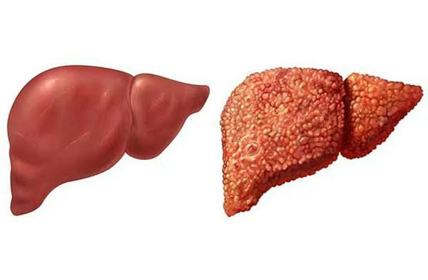
Cirrhosis is the final stage of progressive hepatic fibrosis — a process in which the liver’s normal cellular architecture is replaced by fibrous scar tissue and abnormal regenerative nodules. The normal liver is a highly organized organ with hepatocytes arranged in functional lobules, blood flowing through defined sinusoids, and bile flowing toward bile ducts. Cirrhosis disrupts this architecture entirely, replacing ordered structure with irregular nodules of hepatocytes surrounded by bands of fibrous tissue.
This structural replacement has profound functional consequences. Fibrous tissue increases resistance to blood flow through the liver, creating portal hypertension — the elevated blood pressure in the portal vein that drives many of cirrhosis’s most serious complications. The remaining hepatocytes must work harder to compensate for the loss of functional tissue, and the liver’s regenerative capacity is channeled into producing more nodules — maintaining the constant cycle of proliferation that, over time, leads to cancer.
Cirrhosis develops from virtually any chronic liver injury. Common causes include: chronic viral hepatitis (HBV and HCV — explored in depth in our article on liver cancer and hepatitis); alcohol-related liver disease; non-alcoholic fatty liver disease (NAFLD) and MASH; hereditary hemochromatosis; primary biliary cholangitis; autoimmune hepatitis; and Wilson’s disease. Approximately 4.5 million adults in the United States have cirrhosis, and an estimated 112 million people worldwide are affected.
80–90% of all hepatocellular carcinoma occurs in cirrhotic livers. Annual HCC incidence in cirrhosis ranges from 1–5% per year depending on etiology. Cirrhotic patients face TWO simultaneous diseases — the cancer and the liver failure — and treatment must address both.
Why Cirrhotic Livers Develop Cancer — The Biology
The relationship between cirrhosis and HCC is not simply correlational — cirrhosis creates a highly specific biological environment that promotes cancer development through multiple simultaneous mechanisms.
Chronic hepatocyte turnover and replication errors: In a healthy liver, hepatocytes are long-lived cells that rarely divide. In a cirrhotic liver, the ongoing cycle of cell death and regenerative proliferation means hepatocytes are constantly dividing to replace damaged neighbors. Every cell division is an opportunity for DNA replication errors. Over years, accumulated errors in repeatedly dividing hepatocytes create the genetic mutations that can transform a normal cell into a malignant one.
Persistent inflammatory signaling: Chronic liver inflammation maintains high levels of inflammatory cytokines — IL-6, TNF-α, TGF-β — that activate oncogenic signaling pathways (JAK-STAT, NF-κB, Wnt/β-catenin). These pathways promote cell survival, proliferation, and resistance to apoptosis — hallmarks of malignant behavior. Inflammation that would normally be a temporary healing response becomes a permanent feature of the cirrhotic liver.
Oxidative stress: Reactive oxygen species (ROS) generated by immune cells and damaged mitochondria cause direct DNA damage: strand breaks, base pair mismatches, and chromosomal rearrangements accumulate in hepatocytes exposed to chronic oxidative stress.
Telomere shortening: Repeated regeneration cycles shorten telomeres — the protective caps on chromosomes. In a cirrhotic liver, accelerated telomere shortening produces chromosomal instability that creates the abnormalities characterizing cancer cells.
The dysplastic nodule pathway: Carcinogenesis in the cirrhotic liver follows a multi-step sequence that imaging can sometimes track: Regenerative nodule (benign) → Low-grade dysplastic nodule → High-grade dysplastic nodule (HGDN) → Early HCC → Progressed HCC. High-grade dysplastic nodules have a 30 to 40 percent probability of progressing to HCC within 2 years — representing the last stage in the cancer development sequence before actual malignancy is established.
How Severe Is Your Cirrhosis? Child-Pugh and MELD Scores
The Child-Pugh Score
The Child-Pugh score quantifies liver function using five clinical and laboratory parameters, each scored 1 to 3 points: serum bilirubin, serum albumin, prothrombin time (INR), presence and severity of ascites, and degree of hepatic encephalopathy. Total scores classify cirrhosis severity:
- Child-Pugh A (5–6 pts): well-compensated; liver is functioning adequately
- Child-Pugh B (7–9 pts): significant functional impairment; some treatments are limited
- Child-Pugh C (10–15 pts): severely decompensated; liver transplantation is typically the only meaningful treatment
In liver cancer management, Child-Pugh class is integral to the BCLC staging system. Only Child-Pugh A or B patients are generally considered for active anti-cancer treatment; Child-Pugh C patients — unless they can receive a liver transplant — are managed with palliative and supportive care.
The MELD Score
The MELD score (Model for End-Stage Liver Disease) uses serum creatinine, serum bilirubin, and INR to produce a score from 6 to 40 that predicts 90-day mortality in liver disease patients. MELD-Na (incorporating serum sodium) is the current US standard for transplant allocation. Patients with HCC within Milan criteria receive additional MELD exception points to increase their transplant list priority, acknowledging that their cancer may progress while waiting. For patients with both cirrhosis and HCC, a patient with MELD 8 and a small single HCC may best be served by resection or ablation; a patient with MELD 18 and the same tumor may derive more benefit from liver transplantation.
Compensated vs. Decompensated Cirrhosis
The clinical course of cirrhosis divides into two phases with vastly different prognoses.
Compensated cirrhosis is when the liver retains enough functional capacity to maintain most of its essential roles. Patients may be asymptomatic or have only mild symptoms. Median survival in compensated cirrhosis exceeds 12 years — the primary reason HCC surveillance is so impactful, catching tumors before symptoms develop when curative treatment is still possible.
Decompensated cirrhosis occurs when the liver can no longer compensate, and major complications arise. The first episode of decompensation marks a critical turning point: median survival without liver transplantation drops to 1 to 2 years. Major complications include:
- Ascites: fluid accumulation from portal hypertension and salt/water retention; managed with sodium restriction, diuretics (spironolactone + furosemide), and large-volume paracentesis; TIPS for refractory ascites
- Spontaneous bacterial peritonitis (SBP): bacteria seed ascitic fluid (neutrophil count ≥250 cells/μL = diagnosis); treated with IV antibiotics (ceftriaxone) + long-term prophylaxis
- Variceal bleeding: portal hypertension ruptures esophageal or gastric varices; emergency management: IV octreotide + antibiotics + urgent endoscopy with band ligation; prevented with non-selective beta-blockers
- Hepatic encephalopathy: failure to clear ammonia → brain dysfunction from subtle cognitive changes to coma; treated with lactulose (acidifies colon, traps ammonia as ammonium) + rifaximin
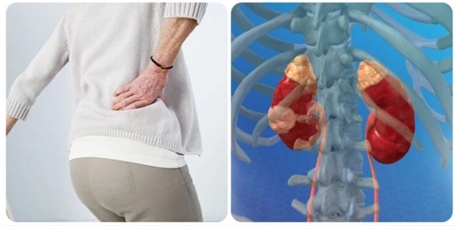
- Hepatorenal syndrome (HRS): renal vasoconstriction causing kidney failure; treated with terlipressin + albumin; liver transplantation is the only definitive treatment
HCC Surveillance in Cirrhosis — The Core Recommendation
Every major hepatology guideline recommends that all patients with cirrhosis, regardless of etiology, undergo HCC surveillance with abdominal ultrasound every 6 months, with or without AFP testing. This applies throughout the patient’s life with cirrhosis, including after HCV cure or during HBV antiviral therapy. The annual HCC incidence in cirrhosis across all etiologies is approximately 2 to 5 percent — well above the 1.5 percent threshold at which surveillance is cost-effective. Full details on the surveillance protocol and what happens when a nodule is found are covered in our complete guide to liver cancer screening.
HCC Risk by Type of Cirrhosis
While all cirrhosis warrants surveillance, annual HCC incidence varies by etiology:
- HCV cirrhosis (untreated): 3–5% per year; highest risk
- HCV cirrhosis post-SVR (cured): ~1–2% per year; surveillance continues indefinitely
- HBV cirrhosis: 2–5% per year; antiviral treatment reduces risk 50–70% but does not eliminate it
- Hereditary hemochromatosis + cirrhosis: ~200x background population risk
- NAFLD/MASH cirrhosis: ~2–3% per year; AFP may be normal even when HCC is present; ultrasound is especially important
- Alcohol-related cirrhosis: ~2–3% per year; ongoing alcohol use adds further risk
- Primary biliary cholangitis + cirrhosis: ~1–1.5% per year
When Cirrhosis and HCC Coexist — Treatment Decisions
When HCC is diagnosed in a cirrhotic patient, treatment must account for both the cancer and the underlying liver disease. This makes liver cancer treatment unique among solid tumor oncology — the organ in which the cancer resides is also diseased, and its functional status directly constrains what treatments are safe.
Surgical resection requires Child-Pugh A liver function without clinically significant portal hypertension. Patients with Child-Pugh B or C have too little functional reserve for major hepatectomy — the cirrhotic remnant does not regenerate as predictably as healthy liver, and hepatic failure after resection is a serious risk.
Liver transplantation is the only treatment that addresses both HCC and cirrhosis simultaneously. For patients who meet the Milan criteria, transplantation achieves five-year survival rates of 70 to 80 percent. It is particularly valuable when cirrhosis is severe enough to make resection dangerous. Locoregional therapies — TACE and ablation — serve as bridge therapy while patients wait for transplant, keeping tumors within Milan criteria and preventing progression beyond transplant eligibility.
TACE, TARE, and systemic therapy for intermediate and advanced HCC can be offered to Child-Pugh A and selected Child-Pugh B patients. Child-Pugh C patients are not candidates for these treatments because the risk of precipitating decompensation outweighs any potential benefit. A multidisciplinary liver tumor board — including a hepatologist, liver surgeon, interventional radiologist, and medical oncologist — must evaluate both tumor characteristics and liver functional reserve before any treatment decision is made.
Managing Cirrhosis to Reduce Cancer Risk
While established cirrhosis is generally irreversible, managing its underlying causes and complications can slow further progression, reduce HCC incidence, and preserve functional reserve. Curing HCV reduces HCC incidence by 70 to 80 percent even in cirrhotic patients. Suppressing HBV viral replication reduces HCC risk by approximately 50 to 70 percent. Alcohol abstinence halts ongoing alcoholic hepatitis and, in some patients, allows partial fibrosis regression. Weight loss of 7 to 10 percent in MASH patients improves liver histology and reduces inflammation.
Cirrhotic patients should avoid hepatotoxins: acetaminophen above 2 g/day, NSAIDs (which worsen fluid retention by reducing renal blood flow), herbal supplements with hepatotoxic potential, and alcohol. Adequate nutritional support — protein intake of 1.2 to 1.5 g/kg/day, small frequent meals, and late evening snacks to reduce the overnight fasting period — helps maintain muscle mass and hepatic encephalopathy tolerance.
Liver Transplantation — Treating Both Diseases at Once
Liver transplantation represents the only therapy that simultaneously cures cirrhosis and removes HCC. For eligible patients, it offers the best long-term outcome because it addresses the root cause of the cancer — the cirrhotic liver — rather than just the tumor. For comprehensive staging information, our guide to liver cancer covers BCLC staging and all treatment modalities in detail. Patients experiencing new symptoms should review our article on liver cancer symptoms.
Living with cirrhosis requires ongoing vigilance — not fatalism. Most people with cirrhosis, particularly compensated cirrhosis, live many years with manageable disease. The combination of treating the underlying cause, adhering to surveillance, staying engaged with a hepatology team, and acting promptly when complications or new lesions arise gives cirrhotic patients the best possible chance of catching liver cancer early — and the best possible quality of life in the years before that risk materializes.
Sources: National Cancer Institute — Liver Cancer | American Liver Foundation — Cirrhosis | AASLD HCC and Cirrhosis Guidelines
Lifestyle Factors That Affect Cirrhosis and HCC Progression
For patients living with cirrhosis, daily choices accumulate into meaningful differences in disease trajectory. Alcohol is the most damaging modifiable factor: any ongoing alcohol consumption in a cirrhotic patient accelerates portal hypertension, worsens hepatic function, and substantially increases the annual HCC incidence rate. Complete abstinence is not a recommendation — it is a clinical imperative. Even patients whose cirrhosis was caused by viral hepatitis or metabolic disease face additional harm from any alcohol use superimposed on their existing fibrosis.
Obesity and insulin resistance in patients with MASH cirrhosis are independently associated with higher HCC risk. Even in patients who have progressed to cirrhosis, weight reduction of 7 to 10 percent body weight — achieved through sustained dietary changes — has been shown to reduce liver inflammation scores and, in non-cirrhotic patients, to partially reverse fibrosis. While fibrosis reversal is less certain at the cirrhotic stage, reducing metabolic stress on the liver preserves functional reserve.
Smoking accelerates cirrhosis progression and is independently associated with higher HCC incidence in several cohort studies. Patients should be counseled toward tobacco cessation as part of comprehensive cirrhosis management.
Coffee consumption is associated with lower fibrosis progression rates and lower HCC incidence — one of the few dietary factors with consistent observational support across multiple large cohort studies. The mechanism likely involves inhibition of TGF-β signaling and reduction in hepatic inflammation. While it is not a treatment, patients need not avoid coffee and may benefit from regular moderate consumption.
Medications to Avoid in Cirrhosis
Cirrhotic patients have impaired drug metabolism and are highly sensitive to medications that the liver processes or that affect hepatic blood flow and renal perfusion. Medications that must be used carefully or avoided include:
- NSAIDs (ibuprofen, naproxen): reduce renal perfusion by inhibiting prostaglandin synthesis; can precipitate acute kidney injury or hepatorenal syndrome; should generally be avoided in patients with ascites or Child-Pugh B/C
- Sedatives and opioids: enhanced CNS sensitivity in cirrhosis; benzodiazepines and opioids can precipitate or worsen hepatic encephalopathy; if necessary, use lowest doses with careful monitoring
- Herbal supplements: multiple supplements — including kava, comfrey, green tea extract, germander, and pennyroyal — are directly hepatotoxic; avoid without hepatologist consultation
- Acetaminophen: generally safe at ≤2 g/day; higher doses deplete hepatic glutathione stores and can cause acute liver injury; preferred over NSAIDs for pain management when used at safe doses
- Statins: observational data suggest statin use is associated with 30–40% lower HCC incidence in cirrhotic patients; they appear safe in compensated cirrhosis and should not be withheld without reason
Monitoring Intervals and the Importance of Not Missing Surveillance
The 6-month surveillance interval for ultrasound was chosen based on the average tumor doubling time for early HCC — approximately 3 to 6 months. Extending the interval to 12 months reduces the detection rate for very early tumors; compressing to 3 months has not been shown to improve outcomes and increases costs and false-positive scans. The 6-month interval is therefore not arbitrary — it is calibrated to tumor biology.
Patients frequently miss surveillance appointments due to travel, cost, transportation difficulties, or feeling well and underestimating the risk. Each missed surveillance scan represents a window in which a tumor could grow from very early (T1, ≤2 cm, potentially curative) to BCLC intermediate stage (beyond curative therapies). Studies of HCC outcomes consistently show that surveillance-detected tumors present at earlier stages with longer survival times than symptom-detected tumors. The difference is measured in years, not months.
If an ultrasound is of poor quality — which happens in patients with morbid obesity or high echogenicity from fatty liver — the hepatologist may order CT or MRI of the liver as the surveillance modality. Patients who consistently have technically inadequate ultrasounds should discuss alternative imaging surveillance with their hepatologist.
Emotional and Psychological Dimensions of Living With Both Diagnoses
Patients who receive both a cirrhosis diagnosis and an HCC diagnosis — or who carry the ongoing uncertainty of cirrhosis knowing liver cancer is a real probability — face a distinct psychological burden. The constant awareness of cancer risk during surveillance, the anxiety before and after each 6-month scan, and the fear of decompensation create chronic psychological stress. Research shows that patients with cirrhosis have significantly higher rates of depression and anxiety than the general population, yet these conditions are under-diagnosed and under-treated in hepatology practice.
Validated screening tools for depression (PHQ-9) and anxiety (GAD-7) take less than 5 minutes to complete and are recommended at hepatology follow-up visits. Hepatic encephalopathy can mimic or amplify depression symptoms — treating subclinical encephalopathy with lactulose may itself improve cognitive function and mood. Support groups — in-person through the American Liver Foundation and online through patient communities — provide connection with others navigating the same dual diagnosis.
Understanding the surveillance process — what each scan looks for, what findings trigger what next steps, what “stable nodule” and “LI-RADS 2” mean — gives patients agency and reduces anxiety. A well-informed patient who understands their imaging reports is better equipped to participate in decisions and maintain adherence to surveillance than one who feels passive and uncertain between appointments.
What to Discuss With Your Hepatologist
Patients with cirrhosis should come to each appointment prepared to review and update the following with their care team:
- Current Child-Pugh score and whether it has changed since last visit
- MELD score and implications for transplant listing if applicable
- Upcoming surveillance ultrasound schedule and any results from the last scan
- Status of the underlying cause — HBV viral load, HCV SVR status, alcohol abstinence, weight management
- Current medications — any new prescriptions from other providers, over-the-counter use, supplements
- Any new symptoms: new or worsening abdominal swelling, ankle edema, confusion, jaundice, bleeding, unintentional weight loss, right upper quadrant pain
- Nutritional status — any difficulty maintaining weight or muscle mass
- Vaccination status — hepatitis A, hepatitis B (if applicable), pneumococcal, influenza
Building a Long-Term Plan With Cirrhosis
Cirrhosis is a chronic disease that demands a chronic management plan. Patients benefit most from a named hepatologist who knows their case, a clear schedule of surveillance ultrasounds, and an established relationship with a liver transplant center — not to imply transplantation is imminent, but because when listing decisions need to be made quickly, the prior relationship makes the process faster and less traumatic. Patients should understand their Child-Pugh class, their MELD score, and what changes would trigger a re-evaluation of their transplant status. They should have a clear plan for who to call and where to go in case of acute decompensation: sudden abdominal pain, vomiting blood, profound confusion, or jaundice. Cirrhosis managed proactively and systematically gives patients the longest window in which surveillance can catch liver cancer at a curable stage — and the best functional status for tolerating treatment when it is needed.