
Cardiac MRI: When It May Be Used and What It Shows
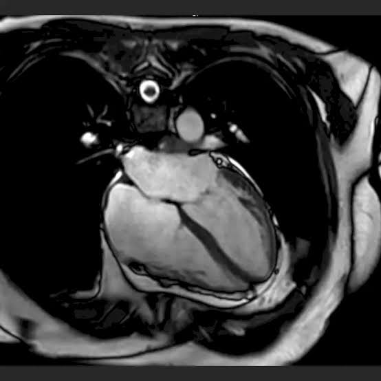
Cardiac MRI ??? also called cardiovascular magnetic resonance (CMR) ??? is the most comprehensive non-invasive cardiac imaging modality available, providing simultaneous assessment of cardiac structure, function, perfusion, viability, and tissue characterization that no other single test can replicate. Unlike the echocardiogram (limited to acoustic imaging of structure and function) or CT (limited to anatomy and coronary calcium), cardiac MRI uses magnetic fields and radiofrequency pulses to generate detailed images of cardiac tissue composition ??? distinguishing healthy myocardium from scar, fibrosis, inflammation, and infiltrative disease at a level of precision unmatched by any other non-invasive test.
The reason cardiac MRI is not the first-line cardiac test for every patient ??? despite its superior information content ??? is practical: it is more expensive than echocardiography, takes 45 to 75 minutes compared to 20 to 30 minutes for echo, is not available at the bedside or in emergency settings, requires patients to hold still for extended periods, and has contraindications related to implanted metallic devices. For most routine cardiac structural and functional assessments, echocardiography provides sufficient information at lower cost and with greater accessibility. Cardiac MRI is reserved for specific clinical questions where its unique tissue characterization capabilities are needed and no other test can provide equivalent information.
What Cardiac MRI Shows That Other Tests Cannot
The defining advantage of cardiac MRI over all other cardiac imaging modalities is tissue characterization ??? the ability to distinguish different types of cardiac tissue based on their intrinsic magnetic properties:
Late gadolinium enhancement (LGE): The cornerstone cardiac MRI technique. When intravenous gadolinium contrast is injected, it distributes into the extracellular space of the myocardium and is washed out from healthy (living) myocardial cells within 10 to 15 minutes, leaving the cells’ intracellular space gadolinium-free. In areas of myocardial infarction or fibrosis ??? where cell membranes are disrupted and the extracellular space is expanded ??? gadolinium accumulates and is retained, producing a bright signal (“enhancement”) on T1-weighted inversion recovery MRI sequences. This LGE pattern provides a precise map of myocardial scar and fibrosis at millimeter-level spatial resolution ??? impossible to achieve with any other imaging technique.
T1 and T2 mapping: Quantitative tissue mapping sequences measure intrinsic magnetic relaxation properties (T1 and T2 relaxation times) of the myocardium, detecting diffuse changes in myocardial tissue composition that focal LGE imaging cannot identify. Native T1 is elevated in myocardial edema (acute myocarditis, acute MI, Takotsubo cardiomyopathy), interstitial fibrosis, and amyloid infiltration; reduced in iron overload (hemochromatosis) and Fabry disease (lysosomal storage disorder causing glycolipid accumulation in the myocardium). T2 mapping quantifies myocardial water content, elevated during acute inflammation and edema.
Extracellular volume fraction (ECV): Calculated from pre- and post-contrast T1 maps, ECV directly quantifies the proportion of myocardial volume occupied by extracellular space ??? normally approximately 25 percent, elevated to 30 to 50 percent in diffuse interstitial fibrosis and amyloid infiltration. ECV is a quantitative biomarker of myocardial fibrosis burden ??? independently predictive of cardiovascular events and mortality in heart failure patients, beyond what LVEF or LGE alone can predict.
Specific Conditions Where Cardiac MRI Is Recommended
The major cardiology guidelines identify specific clinical scenarios where cardiac MRI provides unique and clinically essential information:
Cardiomyopathy diagnosis and classification: The LGE pattern is pathognomonic (specific enough to be diagnostic) for several cardiomyopathy subtypes. Ischemic cardiomyopathy produces subendocardial or transmural LGE in a coronary artery territory ??? starting at the subendocardium (farthest from the epicardial coronary artery blood supply, most vulnerable to ischemia) and extending outward with increasing severity of infarction. Non-ischemic dilated cardiomyopathy typically produces mid-wall septal LGE (a stripe of fibrosis through the middle of the septum, not following a coronary territory). Cardiac sarcoidosis produces patchy basal septal and lateral wall LGE with a distinct distribution pattern. Myocarditis produces epicardial or mid-wall LGE predominantly in the lateral wall ??? the site of maximal inflammation in the most common viral myocarditis patterns. Cardiac amyloidosis produces a global subendocardial LGE pattern with characteristic “nulling” difficulty (the diseased myocardium reaches null point at a different inversion time than normal myocardium, producing a striped or concentric pattern).
Hypertrophic cardiomyopathy (HCM): CMR provides more accurate LV wall thickness measurements than echocardiography (particularly in the anterolateral free wall and apex ??? segments that are difficult to image by TTE), identifies apical HCM (a variant that specifically involves the apical segments, often missed by echo), and quantifies LGE burden (the amount of myocardial fibrosis on LGE CMR is a predictor of arrhythmia risk and sudden cardiac death risk in HCM, informing decisions about implantable defibrillator implantation). ACC/AHA HCM guidelines recommend CMR for all patients with clinically suspected HCM where echocardiography does not provide definitive assessment.
Myocarditis: Acute or suspected myocarditis (inflammation of the heart muscle, most commonly from viral infection, autoimmune disease, or immune checkpoint inhibitor therapy) is definitively diagnosed on cardiac MRI using the updated Lake Louise Criteria ??? which incorporate T2 mapping (for myocardial edema quantification) and LGE (for myocardial injury mapping). The CMR findings confirm the diagnosis, assess the extent of myocardial involvement, and provide baseline data for monitoring recovery ??? avoiding the need for endomyocardial biopsy in most cases. CMR is the preferred test for suspected myocarditis in stable patients who do not require urgent biopsy.
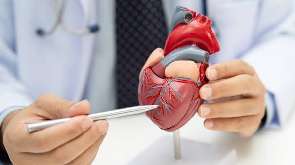
Cardiac MRI for Arrhythmogenic Right Ventricular Cardiomyopathy (ARVC)
Arrhythmogenic right ventricular cardiomyopathy (ARVC) ??? a genetic cardiomyopathy characterized by fibrofatty replacement of the right ventricular myocardium, causing life-threatening ventricular arrhythmias predominantly in young athletes ??? is one of the conditions for which cardiac MRI has become the diagnostic reference standard. The 2010 revised Task Force Criteria for ARVC diagnosis incorporate CMR findings as major criteria ??? RV dilation (RV end-diastolic volume index ???110 mL/m?? in men or ???100 mL/m?? in women), reduced RV ejection fraction (???40%), and regional RV wall motion abnormalities (akinesis, dyskinesis, or dyssynchrony) in specific segments (subtricuspid, RV inflow, RV outflow tract).
CMR is preferred over echocardiography for ARVC assessment because of its superior RV imaging capability: the right ventricle lies immediately behind the sternum and is difficult to image by transthoracic echo (poor acoustic window), while CMR’s field of view and spatial resolution make detailed RV assessment reliable. CMR also detects fatty infiltration (high-signal myocardium on T1-weighted fat-saturated sequences) and fibrosis (LGE in the RV free wall and outflow tract) ??? tissue changes that are pathognomonic of ARVC and invisible on echocardiography. For families of ARVC patients undergoing genetic cascade screening, CMR is the preferred imaging modality for identifying subclinical disease in gene-positive, phenotype-negative relatives.
Cardiac MRI in Congenital Heart Disease ??? The Lifelong Monitoring Role
Adults with repaired congenital heart defects ??? a growing population as pediatric cardiac surgery success rates have improved ??? require lifelong cardiac monitoring for late complications of their repair. Cardiac MRI has become the preferred imaging modality for adult congenital heart disease surveillance because of its ability to image complex three-dimensional cardiac anatomy, quantify ventricular volumes and function without geometric assumptions, and measure blood flow in any vessel by phase-contrast techniques.
The most common adult congenital heart disease application for CMR is monitoring patients with repaired tetralogy of Fallot (ToF) ??? the most common complex congenital heart defect. After surgical repair, most ToF patients have residual pulmonary regurgitation (PR) from the transannular patch used to relieve right ventricular outflow obstruction. Progressive PR causes progressive RV dilation and dysfunction over years to decades ??? eventually requiring pulmonary valve replacement (PVR) to prevent irreversible RV damage and ventricular arrhythmias. CMR is the reference standard for measuring RV volumes, quantifying PR fraction (the proportion of each RV stroke volume that regurgitates back through the pulmonic valve ??? measured by phase-contrast flow in the pulmonary artery), and determining the timing of PVR intervention (current guidelines recommend PVR when RV end-diastolic volume index exceeds 150 mL/m??, based largely on CMR data demonstrating improved RV remodeling after timely intervention).
What to Expect During a Cardiac MRI
Preparing for and undergoing a cardiac MRI is straightforward for most patients, though the longer scan time (compared to echo or CT) and the enclosed scanner environment require awareness:
Before the scan: You will be asked to complete a detailed safety screening questionnaire covering all implanted metallic devices, prior surgeries involving metallic implants, and any metallic foreign bodies (particularly in the eyes ??? relevant for metalworkers). Pacemakers and ICDs must be identified and their MRI conditional status verified before scheduling. Bring any device cards or implant documentation to the appointment. No fasting is required for a standard CMR without stress perfusion; fasting for 4 to 6 hours is recommended for stress perfusion CMR. Beta-blockers may be prescribed to lower resting heart rate below 75 to 80 beats per minute ??? a prerequisite for optimal image quality at most heart rates in CMR.
During the scan: You will lie flat on the MRI table, which slides into the scanner bore ??? a cylindrical tube approximately 60 to 70 cm in diameter and 1.5 to 2 meters long. ECG electrodes are attached to your chest for cardiac gating. You will hear loud knocking, clicking, and buzzing sounds throughout the scan ??? these are the gradient coils firing and are entirely normal. Earplugs or MRI-compatible headphones are provided. You will be coached through multiple breath-hold commands (typically 8 to 12 seconds each) for each imaging sequence. For gadolinium contrast sequences, you will receive an IV injection of gadolinium chelate (standard dose 0.1 mmol/kg) partway through the scan.
Gadolinium safety: Gadolinium contrast is generally very safe ??? serious allergic reactions are rare (approximately 0.03 to 0.1 percent). Patients with severely reduced kidney function (eGFR below 30 mL/min/1.73m??) have a very small risk of nephrogenic systemic fibrosis (NSF) from gadolinium deposition ??? this risk is essentially eliminated with modern macrocyclic gadolinium agents (gadobutrol, gadoteridol) even in severe renal impairment, but the decision to administer contrast in advanced renal failure should be made by the ordering physician after weighing the clinical necessity of the gadolinium sequences.
See our related articles on common heart tests explained, what is an echocardiogram, heart failure symptoms and monitoring, cardiomyopathy: a simple guide, and atrial fibrillation symptoms and risks. The American Heart Association cardiac MRI guide, NHLBI cardiac MRI overview, and Society for Cardiovascular Magnetic Resonance guidelines provide authoritative clinical standards for CMR practice.
- Schulz-Menger J, et al. Standardized image interpretation and post processing in cardiovascular magnetic resonance. J Cardiovasc Magn Reson. 2020;22(1):19.
- Ferreira VM, et al. Cardiovascular Magnetic Resonance Myocardial T1 Mapping (MOLLI). JACC Cardiovasc Imaging. 2018;11(11):1650-1655.
- Marcus FI, et al. Diagnosis of Arrhythmogenic Right Ventricular Cardiomyopathy/Dysplasia. Circulation. 2010;121(13):1533-1541.
- Ommen SR, et al. 2020 AHA/ACC Guideline for the Diagnosis and Treatment of Patients With Hypertrophic Cardiomyopathy. J Am Coll Cardiol. 2020;76(25):e159-e240.
- Fratz S, et al. Guidelines and protocols for cardiovascular magnetic resonance in children and adults with congenital heart disease. J Cardiovasc Magn Reson. 2013;15(1):51.
Cardiac MRI for Stress Perfusion Testing ??? Detecting Ischemia Without Exercise
Beyond tissue characterization, cardiac MRI can evaluate myocardial blood flow during stress ??? providing functional ischemia data that complements the anatomical information from LGE and T1/T2 mapping. Stress perfusion CMR has emerged as one of the most accurate non-invasive tests for detecting obstructive coronary artery disease (CAD), with diagnostic accuracy comparable to nuclear stress testing (myocardial perfusion imaging) but superior spatial resolution that allows detection of subendocardial perfusion abnormalities invisible on SPECT imaging.
The stress perfusion CMR protocol uses pharmacological stress ??? vasodilator stress with adenosine or regadenoson (not exercise stress, since patients must lie still in the scanner). Adenosine or regadenoson dilates the coronary microcirculation: in territories supplied by normal coronary arteries, blood flow increases four to five fold from baseline (hyperemic response). In territories supplied by significantly stenosed coronary arteries (hemodynamically significant stenosis ??? typically greater than 70 percent luminal narrowing), the microcirculation is already maximally dilated at rest to compensate for reduced pressure distal to the stenosis; vasodilator stress produces little or no further flow increase. This relative perfusion difference ??? normal territories lighting up brightly with gadolinium first-pass enhancement, ischemic territories remaining dark ??? identifies the ischemic zone and its coronary territory.
The diagnostic performance of stress perfusion CMR for obstructive CAD is 87 to 90 percent sensitivity and 83 to 87 percent specificity ??? superior to exercise stress ECG (approximately 68 percent sensitivity, 77 percent specificity) and comparable to stress nuclear imaging (SPECT) while avoiding radiation. Importantly, stress perfusion CMR combined with rest LGE in a single comprehensive examination can simultaneously identify ischemia (perfusion defect during stress), infarct (fixed perfusion defect and LGE at rest), myocardial viability (LGE transmural extent predicting recovery after revascularization), and biventricular function ??? providing the complete diagnostic picture in a single 75-minute examination that would otherwise require separate stress nuclear, echocardiogram, and viability imaging sessions.
Cardiac MRI vs. Echocardiogram ??? When to Choose Which
The echocardiogram and cardiac MRI assess overlapping but distinct aspects of cardiac structure and function. Understanding when each test adds unique value ??? rather than duplicating information the other can provide ??? is key to appropriate test selection:
Choose echocardiography first when: The clinical question is straightforward structural assessment (valve function, pericardial effusion, left atrial size, global LV function in a patient with good acoustic windows); the patient needs real-time bedside assessment (critical care, procedural guidance, emergencies); rapid results are required (echo can be performed and interpreted within 30 minutes at the bedside, CMR requires 45 to 75 minutes in a fixed scanner); serial monitoring of a known finding where echo quality is adequate (annual LVEF monitoring in a chemotherapy patient with good windows ??? if the echo LVEF is reproducible, CMR is unnecessary); or the patient has metallic implants contraindicated for MRI.
Choose cardiac MRI when: Echo image quality is inadequate due to poor acoustic windows (obese patients, severe COPD, post-cardiac surgery chest wall deformity); tissue characterization is the clinical question that echo cannot answer (distinguishing myocarditis from ischemic cardiomyopathy, identifying amyloid infiltration, characterizing a cardiac mass as thrombus vs. tumor vs. lipoma, quantifying diffuse myocardial fibrosis); RV structure and function need precise assessment (ARVC evaluation, post-tetralogy of Fallot monitoring, pulmonary hypertension RV assessment ??? CMR is the reference standard, echo is less accurate due to RV geometry); serial LVEF monitoring requires high reproducibility that echo cannot deliver (cardiotoxicity surveillance in high-risk chemotherapy patients where a real 5 percent LVEF change must be distinguished from measurement variability ??? CMR’s lower inter-observer variability makes it the preferred monitoring tool in cardiology guidelines for this indication); or the clinical question is one that specifically requires tissue characterization, flow quantification, or RV volumetry.
The Cardiac MRI Report ??? What Cardiologists Look For
Understanding the key elements of a cardiac MRI report helps patients and non-specialist clinicians interpret results and communicate effectively with the ordering cardiologist or cardiac MRI specialist:
Biventricular volumes and function: The report will state LV end-diastolic volume (LVEDV), LV end-systolic volume (LVESV), LV stroke volume (SV = LVEDV ??? LVESV), LV ejection fraction (LVEF = SV/LVEDV ?? 100%), and the same measurements for the right ventricle. Normal LVEF is ???55 percent; mildly reduced is 45 to 54 percent; moderately reduced is 35 to 44 percent; severely reduced is below 35 percent. LV mass (grams of myocardial tissue) is also reported ??? elevated in LV hypertrophy from hypertension, HCM, or aortic stenosis. RV volumes and EF are critical for ARVC, congenital heart disease, and pulmonary hypertension assessment.
Late gadolinium enhancement findings: The LGE section describes the pattern (subendocardial, transmural, mid-wall, epicardial, global subendocardial), location (which cardiac segments, expressed as AHA 17-segment model positions ??? e.g., “basal inferolateral” or “mid-septal”), and extent (percentage of myocardial wall thickness involved ??? less than 50 percent transmural extent = viable, greater than 50 percent = non-viable). The combination of pattern and location determines the diagnosis: subendocardial basal inferior LGE in the right coronary artery territory = ischemic; mid-wall septal LGE without coronary territory pattern = dilated cardiomyopathy; epicardial lateral LGE = myocarditis.
T1/T2 mapping and ECV values: If performed, native T1 values (milliseconds) and ECV fraction (percentage) are reported with reference ranges from the specific scanner and field strength. Elevated native T1 and ECV suggest diffuse myocardial fibrosis or infiltration; reduced native T1 suggests iron or fat deposition. These quantitative values are particularly important for amyloidosis monitoring, myocarditis follow-up, and cardiotoxicity surveillance.
Regional wall motion: Based on cine imaging, each of the 17 myocardial segments is described as normal, hypokinetic (reduced but present contraction), akinetic (absent contraction), or dyskinetic (paradoxical outward motion during systole ??? indicating scar). Wall motion abnormalities in a coronary territory suggest ischemic disease; global hypokinesis suggests diffuse cardiomyopathy.
Cardiac MRI and Implanted Devices ??? The Evolving Safety Landscape
The MRI contraindication issue ??? historically a significant barrier to cardiac MRI in a broad cardiac population where pacemakers and ICDs are common ??? has evolved substantially over the past decade. The practical implications for patients with cardiac implants:
MRI-conditional pacemakers and ICDs: Modern pacemakers and ICDs manufactured since approximately 2011 are increasingly labeled “MRI Conditional” ??? meaning they can be safely scanned under specific conditions (typically 1.5 Tesla field strength, specific SAR limits, no scanning over the device, and reprogramming before the scan under cardiac monitoring with a device specialist present). Before scheduling a cardiac MRI, patients with pacemakers or ICDs should ask their device clinic whether their specific device model is MRI conditional at the relevant field strength. The device is reprogrammed before the scan (often to an asynchronous pacing mode to prevent inhibition by gradient fields), the patient is monitored throughout the scan, and the device is reprogrammed back to the original settings immediately after. This process is safe and well-established at experienced centers.
Legacy pacemakers (MR Unsafe): Older pacemakers labeled “MR Unsafe” are genuinely contraindicated for MRI ??? the gradient magnetic fields can cause reed switch activation (disrupting pacing), generator heating, and lead tip heating from gradient currents. However, “legacy device MRI” protocols exist at some specialized centers that can safely scan selected patients with older devices under closely controlled conditions with cardiac monitoring and resuscitation equipment immediately available. These protocols are not universally available and require specialist consultation.
Other implants: Coronary stents (bare metal and drug-eluting), mechanical heart valves (most modern models), prosthetic rings, and cardiac occluder devices are generally MRI safe or conditional and do not represent absolute contraindications. Cochlear implants, cerebral aneurysm clips (ferromagnetic types), and certain older vascular clips require careful review of the specific implant model against published MRI safety databases before scanning.
Finally a resource that explains cardiac mri: when it may be in plain language. It is refreshing to see an article that acknowledges individual variation rather than giving one-size-fits-all advice. Will definitely be coming back to this site for more health information.
Came across this while researching cardiac mri: when it may be for a family member. The connection between diet, lifestyle, and long-term outcomes is explained clearly here. Keep up this kind of thorough health journalism — it genuinely helps patients like me.
I have been reading about cardiac mri: when it may be for weeks and this is the most thorough guide I have found. The practical tips at the end made this immediately actionable, not just theoretical. This is going into my health binder that I bring to every doctor’s visit.
Really well-written article on cardiac mri: when it may be. I especially valued the explanation of why these recommendations exist, not just what they are. Looking forward to reading more articles from this website.
I shared this article on cardiac mri: when it may be with my doctor and they appreciated the level of detail. This is the kind of evidence-based writing that actually changes how people approach their health. Looking forward to reading more articles from this website.
This breakdown of cardiac mri: when it may be is exactly what patients need before a specialist appointment. The specific numbers and thresholds mentioned are exactly what I needed to understand my results. This gave me real confidence going into my next specialist appointment.