
Blood Tests for Heart Health: What Each One Measures

Blood tests for heart health are among the most actionable tools available in cardiovascular medicine ??? providing quantitative data on cholesterol levels, inflammation, cardiac muscle damage, cardiac function, blood sugar control, and kidney health that directly guide treatment decisions. Unlike imaging tests that visualize anatomy or function, blood tests measure chemical concentrations and biomarkers that reflect the metabolic and structural state of the cardiovascular system ??? often identifying risk or disease years before symptoms develop or anatomy changes become visible.
Understanding what each cardiovascular blood test measures, what the results mean in clinical context, and when each test is appropriate helps patients participate meaningfully in their cardiac care decisions. This guide covers the major blood tests used in heart health assessment ??? organized by their primary clinical purpose: chronic risk assessment, acute cardiac event diagnosis, and heart failure evaluation.
The Fasting Lipid Panel ??? The Foundation of Cardiovascular Risk Assessment
The lipid panel is the cornerstone of cardiovascular blood testing and the most widely ordered cardiac blood test in primary care. A standard fasting lipid panel (drawn after 9 to 12 hours without food or caloric beverages) measures four values: total cholesterol, LDL cholesterol (low-density lipoprotein ??? the primary atherogenic lipoprotein), HDL cholesterol (high-density lipoprotein ??? associated with cardiovascular protection), and triglycerides (blood fats reflecting dietary carbohydrate and caloric intake, and a marker of insulin resistance in metabolic syndrome).
LDL cholesterol is the most clinically important lipid value for treatment decisions and monitoring. LDL carries cholesterol into arterial walls ??? the initiating step in atherosclerotic plaque formation. Every 38.7 mg/dL (1 mmol/L) reduction in LDL-C with statin therapy reduces major cardiovascular events by approximately 20 to 25 percent in randomized controlled trials ??? making LDL-C reduction the primary therapeutic target in cardiovascular prevention. Standard LDL-C is calculated (not directly measured) using the Friedewald equation: LDL-C = Total Cholesterol ??? HDL-C ??? (Triglycerides ?? 5), in mg/dL. This calculation becomes inaccurate when triglycerides exceed 400 mg/dL ??? at high triglyceride levels, direct LDL-C measurement by ultracentrifugation or the more accurate Martin-Hopkins equation should be used.
HDL cholesterol is an inverse risk factor ??? higher HDL is associated with lower cardiovascular risk in population studies, reflecting reverse cholesterol transport (HDL particles remove cholesterol from arterial walls and transport it back to the liver). However, clinical trials that raised HDL with pharmacological agents (niacin, CETP inhibitors) did not reduce cardiovascular events ??? indicating that HDL cholesterol as a number is a marker of metabolic health rather than a directly modifiable therapeutic target. Low HDL (<40 mg/dL in men, <50 mg/dL in women) is a risk factor identified in the ACC/AHA risk calculation, but raising HDL pharmacologically is not a treatment goal.
Triglycerides reflect dietary fat and carbohydrate intake in the fasting state. Severely elevated triglycerides (???500 mg/dL) cause acute pancreatitis ??? a medical emergency ??? and require urgent triglyceride-lowering therapy (fibrates, omega-3 fatty acids, dietary carbohydrate restriction). Moderately elevated triglycerides (150 to 499 mg/dL) indicate insulin resistance, metabolic syndrome, or familial hypertriglyceridemia ??? all associated with increased cardiovascular risk. Non-fasting triglycerides can be drawn for routine screening (the 2018 ACC/AHA guidelines allow non-fasting lipid panels for initial risk assessment) but fasting values are required for accurate LDL-C calculation and therapeutic decision-making.
High-Sensitivity C-Reactive Protein ??? The Inflammation Marker
Cardiovascular disease is fundamentally an inflammatory process ??? atherosclerotic plaque formation, growth, and rupture all involve inflammatory signaling. High-sensitivity C-reactive protein (hsCRP) is produced by the liver in response to systemic inflammation, and elevated hsCRP levels independently predict cardiovascular events beyond what LDL cholesterol alone predicts ??? a finding demonstrated in large prospective studies including the Physicians’ Health Study and the Women’s Health Study.
The landmark JUPITER trial (2008) demonstrated that adults with normal LDL-C (<130 mg/dL) but elevated hsCRP (???2 mg/L) had a significant reduction in major cardiovascular events (MI, stroke, revascularization, CV death) when treated with rosuvastatin compared to placebo ??? establishing that hsCRP elevation identifies statin-responsive cardiovascular risk even in the absence of elevated LDL cholesterol. This finding underpins the current ACC/AHA guideline recommendation to measure hsCRP as a “risk-enhancing factor” in adults with intermediate 10-year ASCVD risk (7.5 to 20 percent) who are uncertain about statin therapy ??? an elevated hsCRP in this group tips the benefit-risk calculation toward initiating statin treatment.
Important caveats about hsCRP interpretation: hsCRP is an acute-phase reactant that rises with any acute infection, inflammatory flare (rheumatoid arthritis, lupus, inflammatory bowel disease), physical injury, or recent surgery. An hsCRP measured during acute illness reflects that acute illness, not baseline cardiovascular risk ??? the test should be repeated 2 to 4 weeks after recovery for accurate cardiovascular risk assessment. hsCRP is also not a diagnostic test for any specific condition ??? it is elevated in dozens of inflammatory states. Its value is specifically in cardiovascular risk stratification in otherwise well patients with stable metabolic conditions.
Cardiac Troponin ??? Diagnosing Heart Muscle Damage
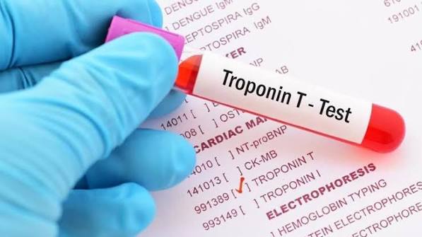
Cardiac troponins (troponin I and troponin T) are structural proteins in the cardiac muscle cell’s contractile apparatus. They are released into the bloodstream when cardiomyocytes are damaged or dying ??? making troponin the gold standard biomarker for detecting acute myocardial infarction (heart attack) and other forms of cardiac injury.
High-sensitivity troponin assays (hsTnI and hsTnT) ??? now the standard of care at most major hospitals ??? detect troponin concentrations in the low nanogram-per-liter range, far below the detection threshold of older conventional assays. This sensitivity allows rapid rule-out protocols: in patients presenting to the emergency department with chest pain, a normal hsTnI or hsTnT at 0 hours (initial draw) AND at 1 to 2 hours with no significant delta change effectively rules out acute MI with greater than 99 percent negative predictive value ??? allowing safe early discharge and avoiding prolonged observation admissions for the majority of chest pain patients.
BNP and NT-proBNP ??? The Heart Failure Biomarkers
B-type natriuretic peptide (BNP) and its inactive cleavage product NT-proBNP are neurohormones released by ventricular cardiomyocytes in response to increased wall stress ??? the stretching of heart muscle cells caused by elevated filling pressures (volume or pressure overload). They are the primary blood biomarkers for detecting and monitoring heart failure:
BNP above 100 pg/mL (or NT-proBNP above age-stratified thresholds: 450 pg/mL for age under 50, 900 pg/mL for age 50 to 75, 1800 pg/mL for age over 75) has high sensitivity for heart failure as a cause of acute dyspnea (difficulty breathing). The most important clinical use is differentiating cardiac dyspnea (heart failure) from pulmonary dyspnea (COPD exacerbation, pneumonia, pulmonary embolism) in the acute setting ??? a distinction that is clinically difficult without biomarker support because both present with similar symptoms.
BNP and NT-proBNP are also used for chronic heart failure management: serial measurements track treatment response (successful diuresis and neurohormonal blockade reduce BNP/NT-proBNP levels; rising levels despite therapy suggest decompensation), and a low discharge BNP/NT-proBNP is associated with reduced 30-day readmission risk. The biomarker is elevated in conditions beyond decompensated heart failure ??? pulmonary hypertension, right heart strain from PE, AF, and severe renal failure all cause BNP/NT-proBNP elevation without primary left heart failure ??? requiring clinical context for interpretation.
Hemoglobin A1c ??? The Diabetes-Cardiovascular Risk Connection
Hemoglobin A1c (HbA1c or A1c) measures the percentage of hemoglobin molecules in red blood cells that have glucose attached ??? reflecting average blood glucose over the preceding 2 to 3 months (the lifespan of a red blood cell). Diabetes is defined as A1c ???6.5 percent; prediabetes as 5.7 to 6.4 percent; normal as below 5.7 percent.
The cardiovascular connection is direct and powerful: diabetes is an independent cardiovascular risk factor that approximately doubles the risk of coronary artery disease, stroke, and peripheral arterial disease. Prediabetes (A1c 5.7 to 6.4 percent) is also associated with increased cardiovascular risk ??? insulin resistance and the metabolic syndrome it represents promote atherogenesis through multiple mechanisms (endothelial dysfunction, increased inflammation, dyslipidemia with elevated triglycerides and low HDL, and elevated LDL particle number despite normal LDL-C mass). Identifying undiagnosed diabetes or prediabetes in a patient presenting with cardiovascular risk or dyslipidemia is clinically essential ??? it significantly changes the risk calculation and intensifies treatment targets.
For patients with known diabetes and established cardiovascular disease, A1c monitoring (typically every 3 months during active management, every 6 months when stable) guides medication intensification. However, tight glycemic control alone (A1c <7 percent) does not reliably reduce cardiovascular events in adults with established diabetes ??? the cardiovascular benefit of modern diabetes medications (GLP-1 receptor agonists like semaglutide and liraglutide, SGLT2 inhibitors like empagliflozin and canagliflozin) is independent of their glucose-lowering effect and operates through direct cardiac and vascular mechanisms. These agents are now first-line for cardiovascular risk reduction in diabetic patients with established ASCVD or heart failure, regardless of baseline A1c.
Lipoprotein(a) ??? The Underrecognized Independent Risk Factor
Lipoprotein(a) ??? abbreviated Lp(a) and pronounced “el-pee-little-a” ??? is a genetically determined atherogenic lipoprotein that carries an LDL-like particle covalently bound to a large protein called apolipoprotein(a). Unlike LDL cholesterol (which responds significantly to diet, exercise, and statins), Lp(a) levels are approximately 90 percent determined by genetics ??? specifically by the LPA gene ??? and are largely unaffected by lifestyle modification. Statins modestly raise Lp(a) levels (by 10 to 20 percent) rather than lowering them.
Elevated Lp(a) ??? typically defined as above 50 mg/dL (or above 125 nmol/L using the more accurate particle-based assay) ??? is present in approximately 20 percent of the general population and is a causal, independent risk factor for coronary artery disease, aortic valve stenosis, and stroke. Importantly, Lp(a) risk is not captured by standard lipid panels ??? a patient with “normal” LDL-C of 90 mg/dL but markedly elevated Lp(a) of 100 mg/dL has substantially higher actual atherogenic particle burden than the LDL-C alone suggests.
Current ACC/AHA guidelines recommend measuring Lp(a) at least once in all adults for cardiovascular risk assessment, particularly in adults with premature ASCVD (heart attack or stroke before age 55 in men or 65 in women), a family history of premature ASCVD, familial hypercholesterolemia, or intermediate ASCVD risk where the statin therapy decision is uncertain. No pharmacological therapy is currently approved specifically for Lp(a) reduction to reduce cardiovascular events, but RNA-based therapies (olpasiran, muvalaplin) in late-phase clinical trials have demonstrated Lp(a) reductions of 80 to 90 percent ??? a class of Lp(a)-lowering drugs is expected to become available within the next several years.
See our related articles on common heart tests explained, coronary calcium score, major risk factors for heart disease, cholesterol and heart attack risk, and heart failure symptoms and monitoring. The American Heart Association cholesterol guide, NHLBI blood tests overview, and ACC cardiovascular prevention guidelines provide authoritative clinical standards.
- Grundy SM, et al. 2018 AHA/ACC/AACVPR/AAPA/ABC/ACPM/ADA/AGS/APhA/ASPC/NLA/PCNA Guideline on the Management of Blood Cholesterol. J Am Coll Cardiol. 2019;73(24):e285-e350.
- Ridker PM, et al. Rosuvastatin to Prevent Vascular Events in Men and Women with Elevated C-Reactive Protein (JUPITER). N Engl J Med. 2008;359(21):2195-2207.
- Kronenberg F, et al. Lipoprotein(a) in atherosclerotic cardiovascular disease and aortic stenosis: a European Atherosclerosis Society consensus statement. Eur Heart J. 2022;43(39):3925-3946.
- Mueller C, et al. ESC Acute Cardiovascular Care Association position paper on troponin elevation in emergency settings. Eur Heart J Acute Cardiovasc Care. 2019;8(2):145-157.
- Januzzi JL, et al. NT-proBNP Testing for Diagnosis and Short-Term Prognosis in Acute Destabilized Heart Failure. Eur Heart J. 2006;27(3):330-337.
Apolipoprotein B ??? The Particle Count That LDL Misses
Apolipoprotein B (ApoB) is a protein that serves as the structural backbone of all atherogenic lipoprotein particles ??? LDL, VLDL, IDL, and Lp(a). Each of these particles contains exactly one ApoB molecule. This means that an ApoB measurement directly counts the total number of atherogenic particles in the blood ??? unlike LDL cholesterol, which measures the amount of cholesterol carried by those particles rather than the number of particles themselves.
Why does the distinction between particle number and particle cholesterol content matter? Because it is the particle ??? not the cholesterol inside it ??? that enters the arterial wall and initiates atherosclerotic plaque. A patient can have a relatively normal LDL-C (say, 100 mg/dL) but a high ApoB (say, 130 mg/dL ??? above the 80 mg/dL target for high-risk patients) if their LDL particles are small and cholesterol-poor. These patients have more atherogenic particles than their LDL-C suggests ??? a state called “discordant LDL” that is common in patients with metabolic syndrome, type 2 diabetes, and elevated triglycerides. ApoB identifies this discordance and more accurately predicts cardiovascular event risk in these patients than LDL-C alone.
The 2018 ACC/AHA cholesterol guidelines identify ApoB as a useful “risk-enhancing factor” for statin therapy decisions in borderline-risk patients, and the 2022 ACC Expert Consensus Decision Pathway includes ApoB as an optional monitoring target (goal ApoB <80 mg/dL for very high-risk patients, <90 mg/dL for high-risk) alongside or instead of LDL-C. Many preventive cardiologists now use ApoB as their primary monitoring target for patients with metabolic syndrome, diabetes, or elevated triglycerides ??? where LDL-C most frequently underestimates atherogenic burden.
Renal Function Tests ??? Why Kidney Health Is a Cardiovascular Issue
Chronic kidney disease (CKD) and cardiovascular disease are deeply intertwined ??? CKD is a powerful, independent cardiovascular risk factor, and most patients with advanced CKD die of cardiovascular causes before reaching dialysis. Understanding why renal function blood tests are part of cardiovascular assessment clarifies an important connection that is often overlooked in patient education:
Serum creatinine and eGFR: Creatinine is a waste product of muscle metabolism cleared by the kidneys. Serum creatinine alone is poorly standardized across body sizes and sexes ??? a thin elderly woman may have “normal” creatinine of 0.9 mg/dL while actually having severely impaired kidney function (because small muscle mass produces little creatinine). The estimated glomerular filtration rate (eGFR) ??? calculated from serum creatinine, age, sex, and race using the CKD-EPI equation ??? standardizes kidney function assessment and is the primary metric for CKD staging. Normal eGFR is above 60 mL/min/1.73m??; CKD is defined as eGFR persistently below 60 mL/min/1.73m?? for more than 3 months. CKD stage G3a (eGFR 45 to 59), G3b (eGFR 30 to 44), G4 (eGFR 15 to 29), and G5 (eGFR below 15 or dialysis) each carry progressively higher cardiovascular event rates ??? patients with eGFR below 30 have cardiovascular event rates comparable to adults with established coronary disease.
The mechanisms linking CKD to cardiovascular disease are multiple: uremic toxins and oxidative stress accelerate endothelial dysfunction and vascular calcification; anemia from reduced erythropoietin production increases cardiac workload; volume overload from impaired sodium excretion elevates blood pressure and promotes LV hypertrophy; mineral bone disorder (hyperphosphatemia, elevated PTH, reduced active vitamin D) drives vascular and valvular calcification; and the prevalence of diabetes and hypertension ??? the two leading causes of CKD ??? means most CKD patients carry additional traditional cardiovascular risk factors. Identifying CKD on routine blood testing should trigger intensified cardiovascular prevention: blood pressure targets below 130/80 mmHg, statin therapy (shown to reduce cardiovascular events in CKD even without elevated LDL), ACE inhibitor or ARB therapy for proteinuric CKD (cardioprotective and nephroprotective), and SGLT2 inhibitors (which reduce cardiovascular events, slow CKD progression, and reduce hospitalizations for heart failure in CKD patients ??? the first agents to simultaneously protect both kidney and heart).
Thyroid Function and Heart Health ??? The Thyroid-Cholesterol Connection
Thyroid-stimulating hormone (TSH) is not typically thought of as a “cardiac” blood test, but hypothyroidism (underactive thyroid) is a commonly missed, fully reversible cause of elevated LDL cholesterol and cardiovascular risk ??? making TSH testing an essential component of the cardiovascular blood work panel for patients presenting with new-onset hyperlipidemia or dyslipidemia refractory to statin therapy:
Thyroid hormone stimulates hepatic LDL receptor expression ??? the liver’s mechanism for clearing LDL particles from the blood. When thyroid hormone is deficient (hypothyroidism), LDL receptor expression falls, LDL-C rises (often dramatically ??? 40 to 60 mg/dL increases are common in moderate hypothyroidism), and triglycerides may also rise through impaired lipoprotein lipase activity. A patient with newly discovered LDL-C of 165 mg/dL and TSH of 12 mIU/L (significantly elevated ??? normal is 0.5 to 4.5 mIU/L) should receive thyroid hormone replacement first and have their lipids rechecked before automatically initiating statin therapy ??? in many cases, treating the hypothyroidism normalizes the lipid panel without the need for a statin.
Hypothyroidism also causes bradycardia (slow heart rate), pericardial effusion, diastolic dysfunction, and reduced cardiac output ??? all of which may manifest as exertional fatigue and dyspnea mimicking heart failure. TSH testing in patients with new-onset exertional symptoms or unexplained dyspnea identifies this treatable, reversible cause. Conversely, hyperthyroidism (low TSH, excess thyroid hormone) causes atrial fibrillation in 10 to 15 percent of cases ??? and AF in any patient should prompt TSH measurement to exclude thyrotoxicosis as a precipitating cause.
When to Request a Comprehensive Cardiovascular Blood Panel ??? A Practical Guide
Knowing when to ask for specific blood tests helps patients engage with their cardiovascular care more effectively. Here are the key clinical situations where comprehensive cardiovascular blood testing is warranted:
Initial cardiovascular risk assessment in adults aged 20 to 39: A baseline fasting lipid panel should be obtained at least once in the 20s or 30s to detect familial hypercholesterolemia (FH ??? present in approximately 1 in 250 adults, often undiagnosed until a premature heart attack) and to establish a personal cholesterol baseline. Early identification of FH dramatically changes lifetime risk and allows decades of preventive statin therapy. Other tests at this age: fasting glucose, A1c (if BMI ???25 or risk factors for diabetes), and Lp(a) at least once.
Adults aged 40 to 75 with standard cardiovascular risk assessment: Fasting lipid panel (repeat every 4 to 6 years if normal, or with each major risk factor change), fasting glucose and A1c, renal function (creatinine and eGFR), hsCRP if the statin therapy decision is borderline, and TSH if new hyperlipidemia or unexplained fatigue. The pooled cohort equations 10-year ASCVD risk score should be calculated at every primary care visit to determine whether medical therapy is indicated.
Monitoring on statin therapy: Repeat fasting lipid panel 4 to 12 weeks after initiation or dose change to verify LDL-C response; annually thereafter once at target. Hepatic transaminases (ALT) before starting statin (a one-time baseline) ??? routine periodic liver monitoring during statin therapy is no longer recommended by current guidelines as statins rarely cause clinically significant liver injury. Creatine kinase (CK) only if symptomatic myalgia on statin ??? routine CK monitoring without symptoms is not recommended.
After an acute cardiac event (heart attack, stroke): Immediate fasting lipid panel (ideally within 24 hours of presentation ??? lipids are artifactually lower after 48 to 72 hours due to the acute-phase response). Troponin serial measurements (required for NSTEMI/STEMI diagnosis), BNP or NT-proBNP (baseline heart failure assessment), A1c (identifies undiagnosed diabetes in approximately 20 percent of acute MI patients), renal function (baseline for medication dosing), and CBC (anemia increases cardiac workload and worsens outcomes in ACS). Lipoprotein(a) measurement at this visit is also recommended, as it identifies patients who may need more aggressive LDL-C lowering targets to compensate for Lp(a)-mediated residual risk.
I have been reading about blood tests for heart health: what for weeks and this is the most thorough guide I have found. The practical tips at the end made this immediately actionable, not just theoretical. This gave me real confidence going into my next specialist appointment.
Finally a resource that explains blood tests for heart health: what in plain language. What I liked most was that the article didn’t just say what to avoid — it also gave alternatives. Forwarding this to others in my support group who are dealing with similar issues.
Came across this while researching blood tests for heart health: what for a family member. The practical tips at the end made this immediately actionable, not just theoretical. Appreciate the effort that went into researching and writing this — it shows.
This is one of the clearest explanations of blood tests for heart health: what I have found. The article answered questions I didn’t even know I had until I started reading. Thank you for making complex medical information accessible without dumbing it down.
I never fully understood blood tests for heart health: what until I read this. What I liked most was that the article didn’t just say what to avoid — it also gave alternatives. This gave me real confidence going into my next specialist appointment.
This is one of the clearest explanations of blood tests for heart health: what I have found. I appreciate that the article is careful about distinguishing between what is known and what is still being researched. I wish I had found this article earlier — would have saved a lot of confusion.