

Upper abdominal pain is among the most common reasons adults seek emergency care — and among the most diagnostically challenging presentations in medicine. The upper abdomen houses the stomach, liver, gallbladder, pancreas, spleen, and major blood vessels. Pain arising here can originate from any of these structures, or be referred from the heart, the back, or the diaphragm.
Most episodes are benign: indigestion after a heavy meal, acid reflux, trapped gas, or a brief bout of gastritis. These are the overwhelming majority of cases seen in primary care and resolve without specific treatment. But a meaningful subset of causes are serious, progress rapidly, and carry significant mortality when not identified promptly.
This guide explains what conditions cause upper abdominal pain, which presentations should be treated as serious until proven otherwise, and which symptoms require calling emergency services without delay.
Understanding the Upper Abdomen
The abdomen is divided into quadrants for clinical purposes. The upper abdomen spans the right upper quadrant (RUQ), the left upper quadrant (LUQ), and the central epigastric region directly below the sternum. Each zone has a reasonably predictable organ association that guides initial evaluation.
The right upper quadrant contains the liver and gallbladder. Pain here is most commonly associated with gallbladder disease — biliary colic, cholecystitis, or cholangitis — or with liver pathology including hepatitis, abscess, or tumour. The left upper quadrant contains the stomach, spleen, and the tail of the pancreas. Pain in this region is less common but includes splenic pathology and referred pain from the heart or diaphragm.
The epigastric region is clinically the most complex. It overlies the stomach, duodenum, head of the pancreas, and the anterior surface of the aorta. Epigastric pain spans the widest range of severity — from functional dyspepsia and acid reflux at the benign end, to acute pancreatitis, perforated ulcer, and aortic aneurysm at the other.
A key concept in evaluating upper abdominal pain is referred pain: discomfort perceived at a location distant from its actual source. The diaphragm, when irritated by blood, bile, or gas, refers pain to the shoulder tip via the phrenic nerve (C3–C5). Inferior myocardial infarction presents as epigastric pain in a significant minority of cases. Pancreatic pain radiates to the back. These referral patterns mean that not all upper abdominal pain originates from abdominal organs — a fact that causes diagnostic errors when not considered.
Common Causes of Upper Abdominal Pain
Peptic Ulcer Disease
Peptic ulcers are erosions in the lining of the stomach (gastric ulcers) or the first portion of the small intestine (duodenal ulcers). The two principal causes are Helicobacter pylori infection and regular NSAID use. The classic symptom is a gnawing or burning epigastric pain that may be relieved by eating (duodenal ulcers) or worsened by it (gastric ulcers). In uncomplicated cases, peptic ulcers are manageable with acid suppression therapy and H. pylori eradication.
The danger arises with complications. A bleeding ulcer can cause haematemesis (vomiting blood) and melaena (black, tarry stools) — signs of significant haemorrhage. A perforated ulcer, where the erosion penetrates the full thickness of the stomach or duodenal wall, is a surgical emergency with rapid progression to peritonitis. Perforation carries a mortality of approximately 10–25% even with prompt surgery.
Gastroesophageal Reflux and Gastritis
Gastroesophageal reflux disease (GERD) produces a burning sensation rising from the epigastrium into the chest — heartburn — typically after eating, when lying down, or when bending forward. It is one of the most prevalent gastrointestinal conditions and is generally benign. Chronic untreated reflux can lead to oesophagitis and, in some cases, Barrett’s oesophagus — a precancerous change in the oesophageal lining that warrants surveillance.
Gastritis — inflammation of the stomach lining — presents with epigastric discomfort, nausea, and occasionally vomiting. Common causes include NSAIDs, alcohol, H. pylori, and autoimmune processes. Most cases respond to conservative management including acid suppression and removal of the trigger.
Functional Dyspepsia
Functional dyspepsia describes persistent upper abdominal discomfort — burning, fullness, bloating, or early satiety — in the absence of identifiable structural disease. It affects approximately 10–15% of the general population and is a diagnosis of exclusion made after investigations have ruled out ulcer, malignancy, and other structural causes. Functional dyspepsia is not dangerous, but it is chronic and significantly impairs quality of life. Management focuses on symptom control, dietary modification, stress reduction, and sometimes low-dose antidepressants.
Gallbladder Disease
The gallbladder stores bile and releases it into the small intestine in response to fat intake. Gallstones form when bile constituents crystallise and are present in approximately 10–15% of adults, most of whom remain asymptomatic.
Biliary colic occurs when a stone transiently obstructs the cystic duct. The pain is typically in the RUQ or epigastrium — severe, cramping, and often radiating to the right shoulder or back. It typically begins 30–60 minutes after a fatty meal and lasts between 30 minutes and several hours before resolving. There is no fever, and inflammatory markers are normal.
Acute cholecystitis is sustained gallbladder inflammation, usually from a stone impacted in the cystic duct. Unlike biliary colic, the pain does not pass. Murphy’s sign — pain and inspiratory arrest on deep palpation of the RUQ — is positive. Fever, elevated inflammatory markers, and prolonged pain distinguish cholecystitis from colic. It requires hospitalisation, IV antibiotics, and planned cholecystectomy.
Serious Causes That Require Urgent Attention
Acute Pancreatitis
Acute pancreatitis is inflammation of the pancreas, most commonly caused by gallstones (40–50%) or excessive alcohol (30–35%). Less common causes include hypertriglyceridaemia, certain medications, and structural abnormalities. For more on the pancreas and its digestive role, see our guide to pancreas and digestive health.
The presentation is severe epigastric pain radiating to the back, with nausea and vomiting, typically beginning after a heavy meal or alcohol consumption. The pain is constant — not colicky — often described as deep, boring, or band-like. A characteristic feature is partial relief by sitting forward or adopting a foetal position, and worsening on lying flat.
Diagnosis is confirmed by serum lipase or amylase greater than three times the upper limit of normal, combined with characteristic imaging. Management requires hospital admission, IV fluid resuscitation, analgesics, and bowel rest. Mild pancreatitis is self-limiting. Severe pancreatitis — with pancreatic necrosis, multi-organ failure, and SIRS — carries mortality of 20–40%. For a detailed overview of this condition, see our article on pancreatitis symptoms and warning signs.
Perforated Peptic Ulcer
Perforation of a peptic ulcer causes sudden, severe epigastric pain — often described as the worst pain of the patient’s life, with abrupt onset as if being struck. Gastrointestinal contents spill into the peritoneal cavity, causing chemical peritonitis that rapidly becomes bacterial. Within hours the patient develops a rigid, board-like abdomen, generalised tenderness, and systemic signs of sepsis.
An erect chest X-ray reveals free air under the diaphragm in approximately 70–80% of perforations. CT abdomen is more sensitive and can identify perforation even when the plain film is normal. Treatment requires emergency surgery — laparotomy or laparoscopic repair — along with IV antibiotics and resuscitation. Every hour of delay worsens prognosis.
Ascending Cholangitis
Ascending cholangitis is a bacterial infection of the bile ducts, most commonly caused by a common bile duct stone obstructing bile flow. The classic presentation is Charcot’s triad: fever and rigors, jaundice, and RUQ pain. In severe cases, hypotension and altered consciousness complete Reynolds’ pentad, which indicates bacteraemic shock and approaches 100% mortality without urgent intervention.
Fever + Jaundice + RUQ Pain = ascending cholangitis until proven otherwise.
This is a medical emergency requiring urgent ERCP and IV antibiotics. Do not wait for all three to be confirmed before seeking emergency care.
Treatment requires urgent ERCP (endoscopic retrograde cholangiopancreatography) to decompress and drain the obstructed bile duct, remove the stone, and allow infected bile to clear. IV broad-spectrum antibiotics and resuscitation are concurrent. This is not a condition that can be managed with oral antibiotics or watchful waiting.
Aortic Aneurysm
An abdominal aortic aneurysm (AAA) is a dilatation of the aorta that, when it ruptures or leaks, presents with sudden severe abdominal or back pain — often radiating to the flank or groin, sometimes mimicking renal colic. The patient may have a pulsatile epigastric mass on examination and rapidly becomes haemodynamically unstable. An estimated 80% of patients with a ruptured AAA die, including pre-hospital deaths. Even with emergency surgery, operative mortality is 40–50%.
Any patient with a known AAA and new abdominal or back pain, or any older patient with vascular risk factors presenting with sudden severe abdominal pain and haemodynamic instability, must be treated as a potential aortic emergency until imaging excludes it.
Cardiac Causes
The inferior wall of the heart sits in proximity to the diaphragm, and inferior or posterior myocardial infarction can present as epigastric pain with nausea and vomiting — indistinguishable clinically from indigestion or peptic ulcer disease, particularly in older adults and people with diabetes. Any upper abdominal pain with chest tightness, sweating, breathlessness, radiation to the left arm or jaw, or haemodynamic instability warrants an ECG and troponin measurement to exclude a cardiac event.
Red Flag Symptoms: When to Call Emergency Services
The following symptoms require calling emergency services (999/112/911) or immediate presentation to an emergency department. Do not wait to see whether they improve.
- Sudden, severe, worst-of-life onset pain — suggests perforation, aortic event, or rupture
- Rigid or board-like abdomen — peritoneal contamination from blood, bile, or bowel contents
- Vomiting blood or black/tarry stools — significant upper GI haemorrhage
- Fever + jaundice + RUQ pain together — Charcot’s triad; cholangitis emergency
- Pain radiating to the shoulder — diaphragmatic irritation from free blood, bile, or gas
- Signs of shock — pallor, cold sweat, rapid weak pulse, confusion, low blood pressure
- Pain radiating straight through to the back — pancreatitis, penetrating ulcer, aortic pathology
- Unable to find any comfortable position — hollow viscus perforation or mesenteric ischaemia
How Doctors Evaluate Upper Abdominal Pain
When a patient presents with upper abdominal pain, evaluation begins with a structured history: onset (sudden or gradual), character (burning, cramping, boring, tearing), location and radiation, severity on a 0–10 scale, timing and duration, and aggravating and relieving factors. Associated symptoms — fever, jaundice, nausea, vomiting, weight loss, change in stool — narrow the differential significantly.
Physical examination assesses the abdomen systematically. Key signs include:
- Murphy’s sign — pain and inspiratory arrest on palpation under the right costal margin; positive in cholecystitis
- Rebound tenderness — pain worse on releasing than applying pressure; indicates peritoneal irritation
- Rigidity or guarding — involuntary muscle tensing; peritoneal contamination
- Cullen’s sign — periumbilical bruising; haemorrhagic pancreatitis
- Grey Turner’s sign — flank bruising; retroperitoneal haemorrhage
Blood tests include full blood count, CRP, liver function tests, serum lipase (preferred over amylase — higher sensitivity, longer elevation window), troponin (if cardiac cause suspected), and lactate (mesenteric ischaemia, sepsis). Urine dipstick excludes urinary tract pathology.
Imaging begins with abdominal ultrasound — first-line for biliary disease, safe, rapid, and widely available. CT abdomen with contrast is the gold standard for most other causes. An erect chest X-ray can identify free subdiaphragmatic air in perforation. ECG is obtained when cardiac cause is in the differential.
What to Do When You Are Unsure
Knowing when to seek care is as important as recognising which symptoms are serious. The hierarchy is straightforward:
Call emergency services immediately for any red flag symptom: sudden severe onset, GI bleeding, board-like abdomen, Charcot’s triad, signs of shock. Do not drive yourself; do not wait to see if it passes.
Seek same-day GP or urgent care assessment for moderate upper abdominal pain present for more than 24 hours, pain accompanied by nausea or vomiting preventing hydration, fever without the full cholangitis triad, or pain following a known biliary or pancreatic trigger. For context on whether digestive enzyme levels may be relevant to your symptoms, see our overview of exocrine pancreatic insufficiency and how digestive enzymes work.
Watchful waiting at home is appropriate only for mild, short-lived discomfort you can confidently attribute to a known benign cause — a reflux flare, postprandial gas after overeating — that is clearly improving. If it does not resolve within 24–48 hours, or if any new symptoms develop, seek assessment.
The principle that matters most: do not attempt to self-diagnose serious causes of upper abdominal pain. Ascending cholangitis, acute pancreatitis, and early aortic leak can initially present with pain that feels manageable. The trajectory — worsening over hours rather than improving — is a more reliable warning sign than the initial severity alone. When there is genuine doubt, the cost of an emergency assessment is far lower than the cost of a delayed diagnosis.
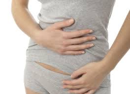
Special Considerations: Age, Pregnancy, and Immunosuppression
Certain populations present diagnostic challenges because their symptoms may be attenuated or atypical, leading to underestimation of severity.
Older adults often have blunted pain responses — both because of age-related changes in pain perception and because of comorbidities and medications that modify the inflammatory response. An older patient with acute cholecystitis or a perforated ulcer may present with only mild discomfort and low-grade fever, while underlying peritonitis is developing. The absence of dramatic pain in an elderly patient does not exclude a serious cause. A low threshold for investigation is warranted, particularly when there is any change in baseline function, appetite, or abdominal tenderness on examination.
Pregnant women experience anatomical shifts as the uterus enlarges, displacing the appendix upward and the bowel laterally. This means that conditions like appendicitis — which typically causes RLQ pain — may present with right upper abdominal pain in the second or third trimester. Gallstone disease is more common in pregnancy due to hormonal effects on bile composition and gallbladder motility. Upper abdominal pain in pregnancy may also represent HELLP syndrome — a combination of haemolysis, elevated liver enzymes, and low platelets — which is a life-threatening obstetric complication requiring emergency delivery.
Immunosuppressed patients — including those on chemotherapy, high-dose corticosteroids, or biological therapies, and those with HIV — may have dampened inflammatory responses that mask the usual signs of serious intra-abdominal infection. Fever may be absent even in the presence of established peritonitis. A normally borderline lipase level or white cell count may represent significant underlying disease in this population. Any new upper abdominal pain in an immunosuppressed patient warrants a lower threshold for hospital assessment, blood tests, and prompt imaging rather than expectant management at home.
Frequently Asked Questions
In primary care, functional dyspepsia, gastroesophageal reflux disease, and peptic ulcer disease are the leading causes. Among patients requiring hospitalisation, gallbladder disease — biliary colic and acute cholecystitis — accounts for the greatest proportion. Acute pancreatitis is a significant cause of emergency admissions and is associated with serious morbidity when severe.
Upper abdominal pain is an emergency when it is sudden and severe at onset, when it accompanies systemic illness (fever, shock, altered consciousness), when jaundice and fever are both present (cholangitis), when there is any sign of GI bleeding, or when the abdomen feels rigid. Any genuine uncertainty about severity should be resolved by seeking emergency assessment rather than waiting for confirmation.
Pancreatic pain is typically a deep, boring, or band-like pain in the central upper abdomen that radiates to the back. It is constant rather than intermittent, severe, and associated with nausea and vomiting. It characteristically improves slightly by sitting forward or adopting a foetal position, and worsens on lying flat or eating. These features together are highly suggestive of a pancreatic origin.
Yes. Inferior and posterior myocardial infarctions can present as epigastric pain, sometimes with nausea and vomiting but without classic chest pain. This pattern is more common in older adults and those with diabetes, whose pain perception may be blunted. Any upper abdominal pain with accompanying sweating, breathlessness, or radiation toward the chest or left arm warrants an ECG.
Charcot’s triad is the combination of RUQ pain, fever and rigors, and jaundice — the classic presentation of ascending cholangitis. It results from bacterial infection of the bile ducts, usually caused by an obstructing gallstone. The triad is present in approximately 50–70% of cholangitis cases; some patients present with only two components. When the full triad is present, the diagnosis is cholangitis until proven otherwise, and emergency biliary decompression is required.
No. While postprandial RUQ pain triggered by fatty food is suggestive of biliary disease, several other conditions also produce pain after eating. Peptic ulcer pain may be triggered or relieved by food depending on ulcer location. Functional dyspepsia and gastritis typically worsen with meals. Mesenteric ischaemia — arterial insufficiency to the bowel — causes fear of eating and postprandial pain in older patients with vascular disease. Location, character, and associated features are all needed for an accurate assessment.
Serum lipase (or amylase) is elevated in pancreatitis. Liver function tests — bilirubin, ALT, AST, alkaline phosphatase — indicate hepatic or biliary disease. A raised white cell count and CRP reflect infection or inflammation. Troponin identifies cardiac cause. Serum lactate is elevated in mesenteric ischaemia and systemic sepsis. No single test is diagnostic for all causes; the blood panel is interpreted alongside the clinical presentation and imaging findings.
- NICE CKS: Acute pancreatitis (2023)
- NICE CKS: Gallstones — management (2023)
- BMJ Best Practice: Approach to upper abdominal pain
- UpToDate: Evaluation of abdominal pain in the emergency department
- Tintinalli JE et al. Tintinalli’s Emergency Medicine: A Comprehensive Study Guide, 9th ed. McGraw-Hill, 2020.
- Liang TB et al. Ascending cholangitis: management and outcomes. World J Gastroenterol. 2021;27(14).
- NHS: Abdominal aortic aneurysm
- UK Small Aneurysm Trial Participants. Mortality results for randomised controlled trial of early elective surgery. Lancet. 1998;352(9141):1649–55.
The section on Charcot’s triad was something I genuinely did not know. My mother had exactly that combination last year — fever, jaundice, and pain on the right side — and the family thought it might be the flu or something she ate. Fortunately the GP sent her straight to hospital. She had a bile duct stone blocking drainage and needed ERCP urgently. Reading this makes me realise how much worse it could have been if she had waited another day. This is the kind of information more people should know.
Thank you for sharing your mother’s experience, Sandra. Charcot’s triad is one of those presentations that is well-known in emergency medicine but rarely reaches public awareness, and that gap costs lives. The mechanism is worth understanding: when a bile duct stone obstructs drainage, bile becomes static and bacteria colonise the biliary system, multiplying rapidly in a closed space under pressure. The resulting bacteraemia can progress to septic shock within hours. ERCP to decompress the duct is the definitive intervention because antibiotics alone cannot clear an infection while the obstruction remains in place. Your mother’s GP acted exactly right by sending her immediately.
As someone who had a heart attack with epigastric pain and no chest pain, I want to say how important the cardiac section is. I told the paramedics I thought I had eaten something bad. They ran an ECG immediately and found inferior ST elevation. I had no idea that heart attacks could feel like stomach pain. The distinction about inferior versus anterior MI and referred pain to the abdomen needs to be in every public health resource. Thank you for including it here.