
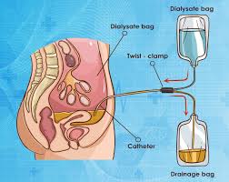
Peritoneal dialysis explained simply: it is a form of kidney replacement therapy that uses the body’s own peritoneal membrane — the thin tissue lining the inside of the abdomen — as a natural filter to remove waste products and excess fluid from the blood when the kidneys can no longer perform this function adequately. Unlike hemodialysis, which requires blood to be removed from the body and filtered by a machine, peritoneal dialysis works entirely inside the body: a cleansing fluid called dialysate is instilled into the abdominal cavity through a permanently implanted catheter, allowed to dwell for a set period while waste diffuses from the blood through the peritoneal membrane into the dialysate, and then drained out and replaced with fresh solution. This process — fill, dwell, drain — is repeated multiple times each day or night, and because it is performed continuously or very frequently rather than three times per week, peritoneal dialysis provides more stable removal of waste and fluid than conventional hemodialysis. Most peritoneal dialysis patients perform their own treatment at home, giving them substantially more independence than in-center hemodialysis. This guide covers how peritoneal dialysis works, the two main types (CAPD and APD), the catheter and access care, adequacy monitoring, complications, diet, and how to determine whether peritoneal dialysis is the right treatment choice. For a broader introduction to dialysis treatments, see the dialysis overview guide; for a detailed comparison with hemodialysis, see the hemodialysis explained guide.
How Peritoneal Dialysis Works: The Peritoneal Membrane as a Natural Filter
The peritoneal membrane is a thin, highly vascularized serous membrane that lines the inner wall of the abdominal cavity (the parietal peritoneum) and covers the abdominal organs (the visceral peritoneum). It has a total surface area of approximately 1–2 square meters in an adult, with an extensive network of capillaries running just beneath its surface — these capillaries are what make it effective as a dialysis membrane. In peritoneal dialysis, this membrane serves as the filter: waste products and excess fluid move from the blood circulating through the peritoneal capillaries, across the peritoneal membrane, and into the dialysate solution sitting in the peritoneal cavity. The two primary transport mechanisms are diffusion and osmotic ultrafiltration. Diffusion drives the movement of dissolved solutes — urea, creatinine, potassium, phosphorus, and other uremic toxins — from the area of higher concentration (the blood, where uremic toxins accumulate with kidney failure) to the area of lower concentration (the dialysate, which is formulated with zero or very low concentrations of these waste products). The greater the concentration gradient, the faster diffusion occurs; as solute levels in the dialysate rise over the dwell period, the gradient narrows and diffusion slows, which is why dwells are ended and fresh dialysate instilled rather than waiting indefinitely. Osmotic ultrafiltration is the mechanism by which excess fluid is removed from the body: standard peritoneal dialysis solutions contain glucose (dextrose) at concentrations of 1.5%, 2.5%, or 4.25%, creating a high osmotic pressure in the dialysate that draws water from the blood across the peritoneal membrane into the peritoneal cavity. The volume of fluid removed (ultrafiltration volume) depends on the glucose concentration used — higher glucose means more osmotic pull and greater fluid removal — and the duration of the dwell. Icodextrin, a glucose polymer solution, is used for long overnight or daytime dwells in patients who require sustained ultrafiltration without the rapid glucose absorption that limits standard glucose solutions during long dwell periods. The basic cycle of peritoneal dialysis consists of three phases: fill (typically 2 liters of fresh dialysate instilled into the peritoneal cavity via gravity over 10–15 minutes), dwell (dialysate remains in the cavity for a set period — 4–6 hours for standard CAPD exchanges, 60–90 minutes for APD cycles, 8–12 hours for an overnight dwell), and drain (spent dialysate drained out of the cavity via gravity into a drain bag over 20–30 minutes). Each complete fill-dwell-drain cycle removes a calculated volume of uremic waste and excess fluid. Over the course of 24 hours, multiple cycles together provide the total dialysis dose. The continuous or very frequent nature of peritoneal dialysis — as opposed to the three-per-week schedule of conventional hemodialysis — means that waste products and fluid are removed more gradually and consistently, resulting in lower peak uremic toxin levels, more stable blood pressure, and less dietary restriction. The NIDDK provides detailed patient information about how peritoneal dialysis works at the NIDDK peritoneal dialysis page.
Types of Peritoneal Dialysis: CAPD vs APD and How to Choose
Peritoneal dialysis is performed in two main modalities — Continuous Ambulatory Peritoneal Dialysis (CAPD) and Automated Peritoneal Dialysis (APD) — that differ primarily in whether exchanges are performed manually or by a machine. The choice between them depends on the patient’s peritoneal transport characteristics, lifestyle, work schedule, and residual kidney function, and most patients can succeed with either modality if properly trained. Continuous Ambulatory Peritoneal Dialysis (CAPD) involves three to five manual exchanges per day, typically one in the morning, one at midday, one in the evening, and an overnight long dwell. Each exchange takes approximately 30–40 minutes: the patient drains the spent dialysate, connects a fresh bag, fills the peritoneal cavity, and goes about their normal activities while the dialysate dwells. CAPD requires no machine, only gravity and a transfer set (the sterile connection device between the catheter and bags), which makes it portable and suitable for people who travel or who cannot or choose not to use a machine. The overnight dwell (8–12 hours) uses a long-dwell formulation, often icodextrin, to continue osmotic ultrafiltration throughout the night without glucose overexposure. CAPD is generally preferred for patients who are “low transporters” on peritoneal equilibration testing — patients whose peritoneal membranes transfer solutes more slowly benefit from the longer dwell times that CAPD provides, allowing more time for waste to diffuse across the membrane into each exchange. Automated Peritoneal Dialysis (APD) uses a cycler machine — an automated device about the size of a laptop bag — to perform multiple exchanges overnight while the patient sleeps, typically over 8–10 hours. The most common APD regimen is Continuous Cycling PD (CCPD): the cycler performs 4–8 short exchanges overnight (each dwell 60–90 minutes), and then the patient begins the day with fresh dialysate left in the abdomen as a daytime dwell (either a standard long dwell or an icodextrin dwell). Nocturnal Intermittent PD (NIPD) is similar but the abdomen is left dry (drained) during the day, which is sometimes preferred for patients with abdominal hernias or back pain worsened by intraperitoneal volume. APD is particularly well-suited for patients who are “high transporters” — whose peritoneal membranes transfer solutes quickly, which means long dwells lead to glucose reabsorption and loss of ultrafiltration drive before adequate waste removal is achieved; shorter APD dwell times take advantage of the fast solute transfer early in the dwell. APD is also preferred for patients who want to keep their daytime schedule free of dialysis exchanges, such as those who work full-time or have young children. Hybrid regimens combine overnight APD cycling with one additional manual daytime exchange to increase the weekly dialysis dose — often used when adequacy testing shows that the overnight APD session alone is not providing sufficient clearance. The Peritoneal Equilibration Test (PET) is the standardized test used to classify peritoneal transport type: the patient is instilled with a 2-liter dextrose 2.27% exchange for a 4-hour dwell, with blood and dialysate samples collected at intervals (0, 2, and 4 hours). The ratio of dialysate-to-plasma creatinine concentration at 4 hours (D/P creatinine) classifies the patient as high (>0.81), high-average, low-average, or low (<0.50) transporter, guiding optimal modality and dwell time prescription. The comprehensive KDIGO guidelines on dialysis modality selection are at the KDIGO CKD guidelines page.
Peritoneal Dialysis Access: The PD Catheter, Tunnel, and Exit-Site Care
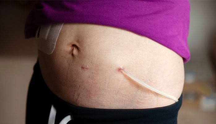
Peritoneal dialysis requires a permanently implanted catheter — a flexible silicone or polyurethane tube — that passes through the abdominal wall into the peritoneal cavity to allow dialysate to flow in and out. The most widely used type is the Tenckhoff catheter, which comes in straight and coiled configurations (the coiled tip sits in the pelvis and is less likely to migrate). The catheter has one or two Dacron cuffs — small rings of porous polyester material — at fixed positions along its length. The deep cuff is positioned just inside the abdominal wall at the peritoneal surface, and the superficial cuff is positioned in the subcutaneous tissue approximately 2 centimeters from the skin exit site. Over 2–4 weeks following insertion, fibrous tissue grows into the Dacron cuffs and anchors the catheter firmly in position, creating a physical barrier against the migration of bacteria from the skin surface along the catheter toward the peritoneal cavity — this fibrous ingrowth is also why the catheter is not used immediately after placement. PD catheter placement is performed surgically (open minilaparotomy or laparoscopic technique) or peritoneoscopically (Y-TEC placement), typically under local or general anesthesia as an outpatient or short-stay procedure. Most programs begin peritoneal dialysis training 2–6 weeks after catheter insertion to allow the tunnel and cuffs to mature; urgent start PD (beginning within days of catheter insertion, using supine position, low fill volumes, and careful technique) is increasingly offered for patients who need dialysis before the standard break-in period is complete. Exit-site care is one of the most critical daily practices for preventing PD complications: the exit site (where the catheter emerges from the skin) must be cleaned daily with soap and water or an antiseptic solution per the patient’s program protocol, covered with a dry sterile dressing, and inspected regularly for signs of infection (redness, swelling, crusting, discharge, pain, or odor). Nasal colonization with Staphylococcus aureus is the most important risk factor for exit-site and tunnel infections; many PD programs routinely screen patients for nasal S. aureus carriage and prescribe mupirocin ointment applied to the exit site (or nares) twice daily for carriers, which substantially reduces infection rates. A tunnel infection — infection in the subcutaneous tissue along the catheter’s path from the exit site to the peritoneum — is more serious than a simple exit-site infection and presents with tenderness, swelling, or erythema along the catheter tunnel, sometimes visible by ultrasound; it often requires catheter removal because antibiotics alone frequently fail to eradicate bacteria that colonize the catheter surface within the tunnel. Patients are taught to check the exit site daily and report any change promptly, because early treatment of exit-site infections with oral antibiotics (directed by culture) can prevent progression to tunnel infections and peritonitis. The catheter transfer set — the external segment of tubing between the catheter and the dialysate bags — is changed every 4–6 months or whenever contaminated to minimize the risk of bacterial colonization. Patients must avoid submerging the catheter (no swimming in pools, lakes, or hot tubs) and protect the exit site from trauma.
Peritoneal Dialysis Adequacy: Kt/V, Residual Kidney Function, and Monitoring
Peritoneal dialysis adequacy measures how much dialysis the patient is receiving and whether that amount is sufficient to remove enough uremic toxins to maintain health and minimize complications. The primary adequacy measure is weekly urea Kt/V — the same kinetic model used in hemodialysis but calculated on a weekly rather than per-session basis, because peritoneal dialysis clearance is continuous rather than intermittent. The current international standard (from KDIGO, ISPD, and KDOQI guidelines) is a total weekly Kt/V ≥ 1.7, where the total Kt/V is the sum of peritoneal Kt/V (the clearance provided by the dialysis exchanges themselves) and the residual Kt/V (the clearance contributed by any remaining native kidney function). This is why residual kidney function is especially important in peritoneal dialysis: a patient with residual urine output of 500–1,000 mL/day may have a residual Kt/V of 0.3–0.6, meaning the dialysis prescription need only provide peritoneal Kt/V of 1.1–1.4 to reach the total target of 1.7. Residual kidney function (RKF) is better preserved in peritoneal dialysis patients than hemodialysis patients, most likely because PD’s continuous, hemodynamically gentle clearance avoids the intermittent hypotension and ischemic episodes that hasten native kidney function decline in hemodialysis. Preserving RKF in PD patients not only contributes to dialysis adequacy but also provides additional benefits: better fluid management (residual urine output helps control fluid balance), reduced dietary restrictions, better preservation of middle molecule clearance, lower cardiovascular risk, and better patient survival. For this reason, preserving RKF by avoiding nephrotoxic medications and contrast agents is especially important in PD patients. Adequacy assessment is performed by collecting a 24-hour dialysate effluent sample and a 24-hour urine sample simultaneously, analyzing the urea and creatinine concentrations in both, and using patient blood urea nitrogen (BUN), weight (to calculate volume of distribution of urea), and total effluent and urine volumes to calculate total weekly Kt/V and total creatinine clearance. Adequacy testing is typically done at 1 month after starting PD, then every 6 months (or more frequently if clinical status changes, residual urine output declines, or symptoms suggest under-dialysis). Symptoms of inadequate dialysis — persistent uremia including fatigue, nausea, poor appetite, fluid retention, and difficulty controlling blood pressure — should prompt immediate adequacy assessment. When adequacy is insufficient, the prescription is intensified: adding exchanges, increasing fill volume, switching from CAPD to APD or hybrid, or adding icodextrin to improve ultrafiltration. The NKF has patient-accessible information about peritoneal dialysis adequacy at the NKF peritoneal dialysis page.
Peritoneal Dialysis Complications: Peritonitis, Infections, and Membrane Changes
Peritoneal dialysis is associated with several specific complications that differ from hemodialysis risks; understanding these complications and their warning signs allows patients to seek prompt treatment and avoid serious consequences. Peritonitis — infection of the peritoneal cavity — is the most serious and most common major complication of peritoneal dialysis, and the leading cause of PD technique failure (inability to continue PD, requiring transfer to hemodialysis). Peritonitis typically results from contamination during bag connections or disconnections (touch contamination) or from migration of bacteria from exit-site or tunnel infections into the peritoneum. The hallmark symptom is cloudy dialysate effluent — the spent dialysate bag appears opaque or milky rather than clear, due to an influx of white blood cells (predominantly neutrophils) responding to infection. Other symptoms include abdominal pain, tenderness, fever, nausea, and general malaise. Any episode of cloudy effluent should be treated as peritonitis until proven otherwise: the dialysate effluent is sent urgently for cell count (peritonitis is defined as >100 white blood cells/µL with >50% neutrophils), Gram stain, and culture, and empiric intraperitoneal (IP) antibiotic therapy covering both gram-positive and gram-negative organisms is started immediately — the most effective route of antibiotic delivery is directly into the peritoneal cavity (added to the dialysate bag), not systemic IV or oral, because this achieves much higher antibiotic concentrations at the site of infection. The most common causative organisms are gram-positive bacteria from the skin, particularly Staphylococcus aureus, Staphylococcus epidermidis, and Streptococcus species; gram-negative peritonitis (from enteric organisms) suggests bowel perforation or transmural migration and carries a worse prognosis. Fungal peritonitis, though rare, almost always requires catheter removal. Recurrent peritonitis episodes or refractory peritonitis that fails to respond to antibiotics within 5 days typically requires catheter removal and transfer to hemodialysis, because persistent infection damages the peritoneal membrane and because the catheter itself may be colonized by a biofilm that cannot be eradicated with antibiotics alone. Exit-site infections are treated with oral antibiotics guided by culture sensitivity; S. aureus exit-site infections require aggressive treatment because of their propensity to cause tunnel infections and peritonitis. Hernias — umbilical, inguinal, incisional, or epigastric — are common in PD patients because the intraperitoneal fill volume increases abdominal pressure; they are repaired surgically, usually with temporary reduction in fill volume or a switch to supine or nocturnal-only PD post-repair. Hydrothorax is an uncommon but significant complication in which dialysate leaks from the peritoneal cavity into the pleural space through a defect in the diaphragm, presenting as sudden shortness of breath; it typically requires temporary cessation of peritoneal dialysis while the defect heals spontaneously or is surgically repaired. Ultrafiltration failure — progressive loss of the peritoneal membrane’s ability to remove fluid across the osmotic gradient — develops over years of continuous PD exposure to glucose-based dialysate, due to neoangiogenesis (increased peritoneal vascularity that speeds glucose absorption and collapses the osmotic gradient before adequate ultrafiltration occurs) and peritoneal fibrosis. Management includes using lower dextrose concentrations when possible, icodextrin for long dwells, and biocompatible low-glucose-degradation-product (low-GDP) solutions to slow membrane changes. Encapsulating peritoneal sclerosis (EPS) is a rare but life-threatening complication of long-term PD (typically >8 years) in which the peritoneum develops progressive fibrous thickening that encases the bowel, eventually causing intestinal obstruction; it can present during PD or months to years after transition to hemodialysis or kidney transplant, and is treated with surgical enterolysis in severe cases. The StatPearls peritoneal dialysis chapter provides detailed clinical information at the StatPearls peritoneal dialysis reference.
Peritoneal Dialysis Diet: A More Flexible Approach Than Hemodialysis
One of the meaningful practical advantages of peritoneal dialysis over conventional hemodialysis is less restrictive dietary requirements — a direct consequence of the continuous or near-continuous nature of PD clearance compared to the three-per-week schedule of in-center hemodialysis. Because peritoneal dialysis removes waste products and potassium continuously around the clock, uremic solutes and electrolytes do not accumulate to the same peak levels between sessions that they do in hemodialysis patients. Potassium restriction in peritoneal dialysis is generally much less strict than in hemodialysis: most PD patients do not require formal potassium restriction at all, because continuous peritoneal clearance of potassium maintains serum levels within a safe range even with a relatively liberal diet. In fact, some PD patients — particularly those receiving icodextrin-based regimens or those with very low residual kidney function — can develop hypokalemia (low blood potassium) from excessive potassium removal by dialysis, and may actually need to increase potassium intake or take supplementation; serum potassium is monitored monthly to guide dietary potassium advice. Phosphorus restriction remains important in PD patients but is typically less stringent than in hemodialysis: phosphate binders (taken with meals to prevent phosphorus absorption from food) are still required, and high-phosphorus foods (dairy, processed foods, dark colas, nuts, organ meats) should be limited, but the target serum phosphorus is achievable with a less restrictive diet than in HD. Protein intake is a critical dietary consideration unique to peritoneal dialysis: the peritoneal membrane is permeable to proteins, and each liter of drained effluent contains approximately 0.5–0.8 grams of protein (primarily albumin, but also other plasma proteins and amino acids). Total daily protein losses into the dialysate in a typical CAPD patient are 5–15 grams per day — a significant ongoing loss that requires higher dietary protein intake to maintain nutritional status. Peritoneal dialysis dietary guidelines recommend a protein intake of 1.2–1.3 grams per kilogram of ideal body weight per day (compared with 1.0–1.2 g/kg/day for hemodialysis patients); inadequate protein intake in PD patients leads to hypoalbuminemia, which is a strong predictor of mortality. Glucose absorption from dialysate is a distinctive nutritional consideration in PD: glucose from the dialysate is absorbed through the peritoneal membrane into the bloodstream, contributing 200–600 kcal/day depending on the dextrose concentrations used. This absorbed glucose can cause hyperglycemia (especially in diabetic patients who may need increased insulin doses), weight gain, hypertriglyceridemia, and difficulty managing metabolic syndrome. Patients who develop significant glucose-related metabolic complications often benefit from icodextrin substitution for long dwells (icodextrin is not absorbed and does not cause systemic glucose elevation), low-dextrose regimens when clinically feasible, and dietary adjustments to account for the caloric load from absorbed dialysate glucose. Fluid restriction in peritoneal dialysis is generally less strict than in hemodialysis, particularly in patients with residual urine output: most PD patients produce some urine and the continuous ultrafiltration of PD removes fluid around the clock, so patients typically tolerate 1,500–2,500 mL of fluid per day depending on urine output and ultrafiltration volume. Patients should monitor their weight daily and report significant interdialytic weight gain to their care team. Sodium restriction to under 2,000 mg/day remains important to control thirst and fluid intake. Patients starting peritoneal dialysis should work with a renal dietitian experienced in PD dietary management to develop an individualized eating plan that accounts for their specific peritoneal transport characteristics, residual kidney function, diabetes status, and protein-calorie needs.
Who Is a Good Candidate for Peritoneal Dialysis?
Peritoneal dialysis is not appropriate for every patient with kidney failure, but it is suitable for a larger proportion of patients than is commonly appreciated — and many patients who might benefit from PD are never offered it because clinicians default to hemodialysis without a full discussion of both options. Understanding who is and is not a suitable candidate for peritoneal dialysis helps patients advocate for the choice that best fits their medical situation and life circumstances. Patients who are particularly well-suited for peritoneal dialysis include: those who want to perform dialysis at home and have a suitable living environment (clean space for storage of supplies, running water, access to power for APD machines); patients who work, study, or have caregiving responsibilities that make the fixed three-weekly hemodialysis schedule impractical; patients who have significant residual kidney function they wish to preserve (PD better preserves RKF than HD, which extends the period before full replacement therapy is required); patients with cardiovascular instability (PD’s gentle, continuous fluid removal avoids the hemodynamic stress of rapid volume shifts in hemodialysis, making it preferable for patients with heart failure, severe coronary artery disease, or significant hypotension during HD); pediatric patients (children tolerate PD well and home therapy avoids frequent travel to dialysis centers); patients in remote or rural areas where access to a hemodialysis center is difficult; and patients who prefer the autonomy and flexibility of a home therapy. Medical contraindications to peritoneal dialysis include: prior major abdominal surgery that left extensive intraperitoneal adhesions (which create pockets and loculations that prevent dialysate from circulating freely throughout the peritoneal cavity, dramatically reducing the effective surface area for dialysis); active inflammatory bowel disease with intestinal perforation risk; large unrepairable abdominal hernias (which are worsened by the intraperitoneal pressure of dialysate fills, though hernias can often be surgically repaired and PD resumed afterward); pleuroperitoneal communication (hydrothorax risk that makes dialysate instillation dangerous); patients with ostomies through the abdominal wall (increased infection risk at the ostomy site and technical difficulties); and profound obesity in some cases (very high fill volumes may be required to achieve adequate clearance, and BMI >40 is associated with significantly worse PD outcomes). Non-medical barriers to PD that must be assessed before starting: patients must be capable of learning and reliably performing the sterile exchange procedure — this requires adequate dexterity, vision, cognition, and motivation, as well as a suitable home environment. Patients who cannot perform exchanges independently may still be candidates for assisted PD, in which a trained healthcare aide or family member performs exchanges at the patient’s home — a model widely used in France, the UK, and increasingly globally, enabling elderly or physically limited patients to benefit from home therapy. The decision between PD and hemodialysis is ideally made jointly by the patient and their nephrology team after a thorough education session that covers both options. For patients proceeding toward dialysis, the preparing for dialysis guide covers the full preparation process, and the life on dialysis guide describes what daily life looks like on long-term dialysis treatment. The comprehensive clinical evidence base for peritoneal dialysis — including survival data comparing PD and HD — is reviewed in the KDIGO kidney disease management guidelines.
Sources: NIDDK Peritoneal Dialysis · KDIGO CKD Guidelines · National Kidney Foundation · StatPearls: Peritoneal Dialysis
I was diagnosed with stage 5 CKD last month and my nephrologist mentioned peritoneal dialysis as an option alongside hemodialysis. This article helped me understand what PD actually involves in a way the clinic pamphlets didn’t. The explanation of the fill-dwell-drain cycle and the difference between CAPD and APD was very clear. I wasn’t aware that CAPD requires no machine at all — knowing I could do it without being attached to equipment overnight is making me seriously consider PD over hemodialysis.
The section on peritonitis was something my PD nurse told me to read about but I hadn’t found a good explanation until this article. The detail about cloudy effluent being the key warning sign and why intraperitoneal antibiotics work better than oral ones makes a lot of sense now. I’ve been on CAPD for eight months and this is the complication I’m most worried about. The note about the PET test and high vs low transporters is also something my nephrologist mentioned but never fully explained — understanding that high transporters do better with shorter APD cycles helps me see why my team recommended the cycler machine over CAPD for me.
Thank you both for sharing your experiences. Sandra, the choice between PD and hemodialysis is worth taking your time on — the fact that CAPD works without a machine and can fit around a more normal daily schedule is one of the things that makes it appealing for people who want to maintain independence. The key is making sure the home environment is set up for proper technique and storage, which your PD team will go through in detail during training. Tom, you’re right to take peritonitis awareness seriously, and it sounds like you’re already thinking about it the right way: watch for cloudy effluent, report it the same day, and your clinic will guide you through treatment immediately. Eight months in with no peritonitis is a great sign. Understanding your transport type (and why the cycler was recommended for you) makes it much easier to follow the prescription confidently — well done for wanting to understand the reasoning.