
Pink water in the toilet. A rusty tinge to urine. A brief flicker of red that disappears and doesn’t return. These moments — startling, then apparently resolving — are among the most medically significant symptoms a person can experience and among the most commonly dismissed.
Blood in the urine, known medically as hematuria, is not a minor nuisance. In adults, it carries a meaningful risk of representing bladder cancer, kidney cancer, or upper tract urothelial carcinoma. The urologic and oncologic communities agree: a single episode of grossly visible blood in the urine in an adult requires a complete urologic evaluation. Not observation. Not a wait-and-see approach. A complete evaluation.
Two Types of Hematuria, One Clinical Rule
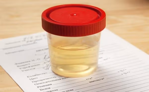
Gross hematuria is visible to the naked eye — the urine is pink, red, orange, or brown, sometimes just for a single void. One episode of gross hematuria in an adult is enough to initiate a full workup.
Microscopic hematuria is invisible to the naked eye but detected on urinalysis — defined as three or more red blood cells per high-power microscopic field. While it carries a lower cancer risk than gross hematuria, it is not benign by definition.
The critical clinical rule: There is no safe threshold of blood in urine below which investigation can be avoided in an adult. Even a single episode of gross hematuria — even if self-resolving, even if the patient is on blood thinners — warrants complete urologic evaluation. Anticoagulation does not cause hematuria; it may unmask bleeding from an underlying lesion that would have gone undetected at normal coagulation.
Bladder Cancer: Painless Hematuria Is the Red Flag
Bladder cancer is the cancer most consistently associated with blood in the urine, occurring in approximately 90 percent of cases. The defining clinical feature is painlessness. Urinary bleeding from infection is accompanied by burning and frequency. Bleeding from kidney stones is accompanied by flank pain. Bladder cancer bleeds without pain.
A middle-aged or older adult with pink or red urine and no other urinary symptoms is experiencing painless hematuria — and that description is bladder cancer until proven otherwise.
- Smoking: By far the most powerful modifiable risk factor — 2-3× increased risk. Carcinogens excreted in urine directly expose the bladder urothelium
- Occupational exposures: Aromatic amines (dye manufacturing, rubber, aluminum smelting)
- Cyclophosphamide (chemotherapy drug): Metabolized to acrolein, directly toxic to bladder urothelium
- Chronic indwelling catheter: Chronic bladder irritation; risk of squamous cell carcinoma
- Schistosoma haematobium (parasitic infection endemic in Middle East, sub-Saharan Africa): SCC of bladder
Cancer Causes of Blood in Urine
| Cancer | Hematuria Type | Key Associated Features | Diagnostic Test |
|---|---|---|---|
| Bladder cancer | Gross, painless (classic) | Smoking history; may have irritative symptoms | Cystoscopy (gold standard) |
| Renal cell carcinoma | Gross or microscopic; episodic | Flank pain; paraneoplastic syndromes | CT abdomen with contrast |
| Upper tract urothelial (renal pelvis/ureter) | Gross or microscopic | Lynch syndrome association; clot passage | CT urogram + ureteroscopy |
| Prostate cancer (locally advanced) | Gross (bladder neck invasion) | Obstructive LUTS; elevated PSA | PSA + mpMRI + biopsy |
Carcinoma In Situ of the Bladder: The Silent Threat
Carcinoma in situ (CIS) of the bladder is a flat, high-grade malignant lesion confined to the urothelial surface — it does not form a visible papillary mass. CIS is entirely invisible on CT and ultrasound. It cannot be found with high sensitivity on urine cytology. The only reliable way to diagnose it is cystoscopy with bladder biopsies.
CIS produces severe irritative symptoms — urgency, frequency, dysuria — sometimes without any hematuria. This presentation is easily misdiagnosed as a urinary tract infection, leading to months of antibiotic courses before the true diagnosis is made. CIS carries a high risk of progression to muscle-invasive bladder cancer if untreated.
Renal Cell Carcinoma: The Internist’s Tumor
Renal cell carcinoma is diagnosed in approximately 81,000 Americans annually. The classic presentation — hematuria + flank pain + palpable abdominal mass — is now uncommon. Most RCC is detected incidentally when patients undergo abdominal imaging for unrelated reasons.
RCC has earned the name “internist’s tumor” because of its remarkable range of paraneoplastic manifestations: unexplained fever, erythrocytosis (excess red blood cells from tumor-produced erythropoietin), hypercalcemia, hypertension, and abnormal liver function tests — all of which may appear before any urinary symptom. When hematuria does occur in RCC, it tends to be episodic — appearing and resolving over days. This intermittent pattern should not be reassuring.
- Any visible blood in urine — even a single episode, even if it self-resolves
- Painless visible blood in urine in anyone 45 or older
- Gross hematuria in a current or former smoker
- Hematuria with passage of tissue or clots
- Microscopic hematuria persisting after treated UTI is cleared (repeat UA in 6 weeks)
- Hematuria + weight loss or fatigue (systemic cancer concern)
The Complete Workup for Blood in Urine
Step 1 — Urinalysis with microscopy: Confirm RBCs; check for pyuria (infection), RBC casts (glomerulonephritis), and protein.
Step 2 — Urine culture: If UTI suspected, treat and repeat UA in 6 weeks. Persistent hematuria after infection is cleared = proceed to full evaluation.
Step 3 — CT urogram: The best single imaging study for hematuria evaluation. Performed in three phases (non-contrast, nephrographic, urographic), it evaluates kidneys for masses, collecting systems for UTUC, and bladder. Cannot replace cystoscopy for bladder cancer detection.
Step 4 — Cystoscopy: The gold standard for bladder evaluation. Cannot be replaced by CT — small flat lesions (CIS, small papillary tumors) are invisible on imaging but visible on cystoscopy. Flexible office cystoscopy is widely available and well tolerated.
Step 5 — Urine cytology: High specificity for high-grade urothelial cancer; low sensitivity for low-grade tumors. Used as an adjunct, not replacement for cystoscopy.
Frequently Asked Questions
References
- Barocas DA, et al. Microhematuria: AUA/SUFU Guideline. J Urol. 2020.
- NCCN Clinical Practice Guidelines: Bladder Cancer; Kidney Cancer. 2024.
- Siegel RL, et al. Cancer Statistics 2023. CA Cancer J Clin. 2023.
- Escudier B, et al. Renal cell carcinoma: ESMO Clinical Practice Guidelines. Ann Oncol. 2019.
- Antoni S, et al. Bladder cancer incidence and mortality: a global overview. Eur Urol. 2017.
- Cowan NC. CT urography for hematuria. Nat Rev Urol. 2012.