
Poor Circulation in the Legs: Causes and Symptoms
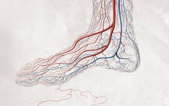
Poor circulation in the legs is a broad term describing inadequate blood flow to or from the lower extremities — a condition affecting an estimated 8 to 12 million Americans and tens of millions worldwide. It is not a single disease but a manifestation of several distinct underlying vascular conditions, each with different mechanisms, symptoms, diagnostic approaches, and treatments. The two most clinically important categories are peripheral artery disease (PAD), which reduces arterial inflow to the legs from atherosclerotic plaque, and chronic venous insufficiency (CVI), which impairs venous return from the legs due to damaged or incompetent venous valves.
Understanding which type of poor circulation is present — arterial or venous, or occasionally both simultaneously — is the essential first step in management, because the treatments are fundamentally different and applying venous treatments to arterial disease (such as compression stockings in severe PAD) can cause harm. A thorough clinical evaluation starting with the ankle-brachial index (ABI) rapidly and accurately identifies which patients have arterial insufficiency, directing them to appropriate vascular evaluation and treatment.
Arterial Poor Circulation — Peripheral Artery Disease
Peripheral artery disease (PAD) is the most medically serious cause of poor circulation in the legs, and it carries cardiovascular significance beyond the leg symptoms themselves: PAD is a manifestation of systemic atherosclerosis, and patients with PAD have dramatically elevated rates of myocardial infarction, ischemic stroke, and cardiovascular death — the same atherosclerotic process that blocks the leg arteries is active in the coronary and carotid arteries simultaneously. PAD affects approximately 8 to 10 million Americans aged 40 and above, with prevalence increasing steeply with age (affecting approximately 15 to 20 percent of adults aged 70 and above).
The hallmark symptom of PAD-related poor arterial circulation in the legs is intermittent claudication — a reproducible cramping, aching, or tightening pain in the calf (most commonly), thigh, or buttock that occurs with walking a predictable distance and is completely relieved by rest within 10 minutes without change of position. Claudication occurs because the narrowed arteries can supply adequate blood flow at rest but cannot increase flow sufficiently during exercise to meet the metabolic demand of the contracting muscles — the resulting ischemia produces pain that stops the patient from walking further. Unlike musculoskeletal leg pain, claudication is strictly correlated with walking distance (the same distance triggers symptoms reliably), involves specific muscle groups in the distribution of the obstructed artery (calf claudication for superficial femoral or popliteal disease; thigh and buttock claudication for iliac disease), and resolves completely with rest (not just slowing down).
As PAD progresses, symptoms escalate through a predictable pattern. Rest pain — severe burning or aching pain in the foot or toes that occurs at rest, typically worse at night when the legs are elevated (reducing gravitational assistance to perfusion) and relieved by hanging the legs over the bed edge — indicates critical ischemia from arterial supply insufficient even at rest. Critical limb ischemia (CLI) — the most severe stage, formerly called “critical limb-threatening ischemia” (CLTI) — produces non-healing ulcers (typically at pressure points: toes, heel, lateral foot) or dry gangrene, representing irreversible tissue loss from chronic or acute severe ischemia. CLI carries approximately 20 to 30 percent limb loss risk and 20 percent mortality risk at 6 months without revascularization.
Other symptoms of arterial poor circulation include: coldness of the affected foot or leg (asymmetric with the contralateral limb or with the hands); pallor on elevation and dependent rubor (foot turns pale when elevated above heart level, then becomes deep red/purple when lowered — reflecting the exhaustion of vasodilatory reserve in chronically ischemic vessels); absent or diminished pulses in the femoral, popliteal, posterior tibial, or dorsalis pedis arteries; loss of hair on the lower leg and foot (from chronic ischemia of hair follicles); and nail changes (thickened, slow-growing, opaque nails from ischemia).
Venous Poor Circulation — Chronic Venous Insufficiency
Chronic venous insufficiency (CVI) is far more prevalent than PAD — affecting approximately 25 to 40 percent of adults to some degree — and causes a different constellation of symptoms from arterial insufficiency. In CVI, the venous valves that normally prevent blood from flowing backward in the leg veins become incompetent (from prior DVT damage, prolonged standing, obesity, or primary valve incompetence), allowing venous reflux — backward flow of blood down the superficial or deep veins when standing. This produces venous hypertension in the lower leg, which over time causes tissue fluid accumulation (edema), skin changes (hyperpigmentation from hemosiderin deposition, lipodermatosclerosis — a fibrous thickening of the lower leg skin), varicose veins, and ultimately venous ulcers.
The classic symptoms of venous poor circulation are: leg swelling (pitting edema, typically worse by end of day and after prolonged standing, improved with elevation and overnight rest); aching heaviness in the lower legs with prolonged standing (from venous congestion) that is relieved by elevation (opposite of arterial rest pain, which worsens with elevation); varicose veins (dilated, tortuous, bulging superficial veins visible under the skin, most common in the greater saphenous territory on the medial calf and thigh); skin discoloration around the ankles (brown or reddish-brown hyperpigmentation from hemosiderin deposition as red blood cells extravasate through capillaries distended by venous hypertension); and venous ulcers (the most severe manifestation — shallow, irregular, exudative ulcers typically located above the medial malleolus, with surrounding hyperpigmentation and lipodermatosclerosis, and notoriously slow to heal without appropriate venous pressure management).
An important distinction: while venous insufficiency causes significant morbidity and quality of life impairment, it does not carry the same systemic cardiovascular significance as PAD. A patient with venous insufficiency has compromised leg circulation but is not at dramatically elevated risk of heart attack or stroke from the venous disease itself. However, venous insufficiency and PAD frequently coexist in the same patient (because they share some risk factors — obesity, hypertension, diabetes), and the presence of venous disease should not preclude evaluation for arterial disease when clinical features suggest both.

Other Causes of Poor Leg Circulation
Beyond PAD and CVI, several other conditions cause or contribute to poor circulation in the legs:
Deep vein thrombosis (DVT) — acute clot formation in the deep veins of the leg — initially impairs venous return from the affected limb, causing acute unilateral swelling, warmth, redness, and pain. After the acute phase, DVT damages venous valves through the inflammatory and fibrotic changes that occur as the clot organizes, leading to the post-thrombotic syndrome (PTS) — chronic venous insufficiency from DVT-damaged valves — in approximately 20 to 40 percent of DVT patients.
Raynaud’s phenomenon — episodic vasospasm of the small arteries and arterioles, most often affecting the fingers and toes — causes dramatic color changes (white pallor from vasospasm, then blue from deoxygenation, then red from reactive hyperemia) triggered by cold exposure or emotional stress. Primary Raynaud’s (without underlying disease) is common (affecting 3 to 5 percent of the population, predominantly young women) and benign. Secondary Raynaud’s (associated with autoimmune diseases — scleroderma, lupus, rheumatoid arthritis — or with occupational vibration exposure) can cause digital ischemia and tissue loss in severe cases.
Peripheral edema from systemic disease: Bilateral leg swelling without the specific skin changes of venous insufficiency suggests systemic causes — heart failure (most common; characterized by pitting edema worse in dependent areas, often with orthopnea and exertional dyspnea), nephrotic syndrome (massive proteinuria reducing plasma oncotic pressure), cirrhosis (portal hypertension and hypoalbuminemia), and hypoalbuminemia from malnutrition. These require treatment of the underlying condition rather than venous or arterial interventions.
Lymphedema: Impaired lymphatic drainage — from lymph node dissection (post-cancer surgery), radiation therapy, or congenital lymphatic dysplasia — causes progressive non-pitting edema that does not respond fully to elevation and is characterized by skin thickening, fibrosis, and susceptibility to recurrent cellulitis. Unlike venous edema, lymphedema has a characteristically hard, indurated feel to the swollen tissue (fibrosis) and characteristically involves the dorsum of the foot (creating the Stemmer sign — inability to pinch skin on the dorsum of the second toe into a tent fold).
Treatment of Poor Circulation — Arterial vs Venous Approaches
Treatment of poor leg circulation depends entirely on the underlying cause — arterial and venous insufficiency require fundamentally different management:
Arterial poor circulation (PAD): The treatment framework addresses both the local leg symptoms and the systemic cardiovascular risk that PAD represents. All PAD patients require cardiovascular risk factor optimization: high-intensity statin therapy (significantly reduces cardiovascular events and may improve walking distance through plaque stabilization effects on leg artery disease), blood pressure control below 130/80 mmHg, smoking cessation (the single most impactful intervention for PAD — smoking dramatically accelerates PAD progression, and cessation is associated with improved claudication distance and reduced limb loss risk), antiplatelet therapy (aspirin or clopidogrel reduces cardiovascular events in PAD), and diabetes management. For claudication symptoms specifically, supervised exercise therapy — structured walking programs that train patients to walk to pain, rest, then continue — improves claudication distance by 100 to 150 percent in clinical trials and is recommended as first-line therapy before revascularization for stable claudication. Revascularization (endovascular balloon angioplasty and stenting, or surgical bypass) is reserved for critical limb ischemia, lifestyle-limiting claudication refractory to medical and exercise therapy, and acute limb ischemia.
Venous poor circulation (CVI): Compression therapy is the cornerstone of CVI treatment — graduated elastic compression stockings (30 to 40 mmHg for moderate to severe CVI) reduce venous hypertension, improve edema, slow skin change progression, and significantly accelerate venous ulcer healing. Leg elevation above heart level (ideally 30 to 45 minutes three to four times daily) reduces venous pressure and edema. Endovenous ablation (laser or radiofrequency thermal ablation of the incompetent saphenous vein) has replaced surgical stripping as the preferred treatment for symptomatic great saphenous vein insufficiency — it eliminates the reflux source, reduces varicose vein recurrence, and significantly improves quality of life with minimal recovery time.
The Society for Vascular Surgery PAD patient resource explains the causes, diagnosis, and treatment options for peripheral arterial poor circulation. The CDC peripheral artery disease page covers risk factors, symptoms, and when to seek evaluation for poor leg circulation. The NHLBI PAD information guide reviews arterial circulation problems in the legs with guidance on treatment and secondary prevention.
Related reading: Peripheral Artery Disease Symptoms | Deep Vein Thrombosis | Blood Clots: Warning Signs | High Blood Pressure and Stroke | Stroke Prevention for Adults
Sources
- Gerhard-Herman MD, et al. 2016 AHA/ACC Guideline on the Management of Patients with Lower Extremity Peripheral Artery Disease. J Am Coll Cardiol. 2017;69(11):e71-e126.
- Fowkes FGR, et al. Comparison of Global Estimates of Prevalence and Risk Factors for PAD in 2000 and 2010. Lancet. 2013;382(9901):1329-1340.
- Conte MS, et al. Global Vascular Guidelines on the Management of Chronic Limb-Threatening Ischemia. J Vasc Surg. 2019;69(6S):3S-125S.
- Nicolaides AN, et al. Management of Chronic Venous Disorders of the Lower Limbs. Int Angiol. 2018;37(3):181-254.
- Murphy TP, et al. Supervised Exercise vs Stenting for Claudication (CLEVER). Circulation. 2012;125(1):130-139.
Risk Factors for Poor Leg Circulation
The major risk factors for poor circulation in the legs differ by type — arterial versus venous — though several risk factors promote both:
Risk factors predominantly for arterial PAD: Smoking is the strongest modifiable risk factor for PAD — smokers have 3 to 5-fold higher PAD prevalence than non-smokers, and smoking is present in approximately 70 to 80 percent of PAD patients. Diabetes substantially increases PAD risk (2 to 4-fold) and dramatically worsens outcomes — diabetic PAD patients have higher rates of multilevel disease, infrapopliteal disease distribution (more distal and harder to revascularize), and major amputation. Hypertension and dyslipidemia accelerate the underlying atherosclerosis driving arterial PAD. Advanced age is a powerful independent risk factor — prevalence rises sharply above age 65. Chronic kidney disease (CKD) is an underrecognized PAD risk factor, partly because CKD patients have high vascular calcification that impairs ABI interpretation and partly because they are often excluded from PAD trials.
Risk factors predominantly for venous insufficiency: Female sex (hormonal influences on vein wall collagen and valve competence; pregnancy-related venous hypertension); obesity (increased intra-abdominal pressure impairing venous return); prolonged standing or sitting occupations (varicose vein risk increased 2 to 3-fold in occupations requiring standing 4 hours or more per day); prior DVT (the most important cause of secondary venous insufficiency — DVT destroys venous valves through the recanalization and scarring process); family history of varicose veins (50 percent of patients with varicose veins have a first-degree family member with varicose veins — suggesting significant genetic contribution); and prior leg injury or surgery disrupting lymphatic and venous drainage.
Shared risk factors: Diabetes, hypertension, obesity, and advanced age increase risk for both arterial and venous poor circulation. This overlap explains why mixed arterio-venous disease (simultaneous PAD and CVI) is common in older adults with metabolic syndrome.
When to See a Doctor — Warning Signs That Need Urgent Evaluation
Most symptoms of poor leg circulation develop gradually and allow for elective medical evaluation, but several presentations require urgent or emergency assessment:
Acute limb ischemia — sudden severe worsening of leg pain with color change (pale or mottled), coldness, numbness, paralysis, or loss of pulses — is a vascular emergency requiring immediate hospital evaluation. Acute limb ischemia is caused by sudden arterial occlusion (embolism from the heart or from a proximal aneurysm, or acute in situ thrombosis of a stenosed segment) and threatens the limb within hours. The “6 P’s” of acute limb ischemia are: Pain, Pallor, Pulselessness, Paresthesia (numbness/tingling), Paralysis, and Poikilothermia (coldness). The presence of paresthesia or paralysis indicates advanced ischemia requiring emergency revascularization within 6 hours to prevent irreversible muscle and nerve damage. Call 911 or go to the emergency department immediately for any acute onset severe leg pain with pallor, coldness, or weakness.
Critical limb ischemia warning signs: Non-healing foot sore or ulcer despite appropriate wound care, black discoloration of a toe (early gangrene), severe rest pain preventing sleep, or any wound on the foot in a diabetic patient with PAD requires urgent vascular surgery evaluation within 24 to 72 hours. The window for limb salvage narrows rapidly as tissue ischemia progresses — delays of even a few days in referral can convert a revascularizable limb to one requiring major amputation.
Acute DVT: Sudden, significant unilateral leg swelling (especially if one leg is clearly larger than the other), warmth, redness, and pain — particularly in the calf, popliteal fossa, or thigh — requires same-day evaluation to exclude deep vein thrombosis. DVT carries risk of pulmonary embolism (when clot breaks off and travels to the lungs), which can be life-threatening. If a DVT is suspected, the evaluation should not be delayed overnight.
For gradually developing symptoms — aching legs with standing, varicose veins, mild ankle swelling, or stable claudication — elective appointment with a primary care physician or vascular specialist is appropriate. The evaluation will include history, physical examination (pulse assessment, skin inspection, ulcer characterization), and ABI testing — often with referral for vascular duplex ultrasound if PAD or venous disease is confirmed. Early evaluation and treatment of poor circulation prevents progression to the severe complications of critical limb ischemia or chronic venous ulceration.
Lifestyle Measures That Improve Leg Circulation
Regardless of the cause of poor leg circulation, several lifestyle measures support vascular health and symptom management:
Walking and structured exercise: For arterial poor circulation, supervised exercise therapy (structured treadmill walking programs) improves claudication distance by 100 to 150 percent over 3 to 6 months — more than most medications and comparable to angioplasty for claudication symptom relief in clinical trials. The mechanism involves both metabolic adaptations in muscle and improvements in collateral circulation. Even unsupervised home walking programs provide meaningful benefit and are appropriate for patients who cannot access formal supervised programs. For venous poor circulation, walking activates the calf muscle pump, improving venous return and reducing venous hypertension — making regular walking one of the most effective treatments for symptomatic venous insufficiency.
Leg elevation: Elevating the legs above heart level (lying with feet on pillows or a wedge, or using a recliner that elevates the feet) reduces venous pressure in the lower legs and improves edema from venous insufficiency or DVT. For arterial disease, elevation is generally neutral at moderate angles but can worsen symptoms at high elevation angles in severe PAD (reducing gravitational perfusion pressure).
Weight management: Obesity directly worsens venous insufficiency (increased intra-abdominal pressure compresses the inferior vena cava, elevating lower extremity venous pressure) and accelerates PAD through diabetes, hypertension, and dyslipidemia. Even modest weight loss of 5 to 10 percent in obese patients with venous insufficiency significantly reduces edema and venous pressure.
Foot care in PAD: Daily inspection of the feet and between the toes for any cuts, cracks, blisters, or discoloration is essential for PAD patients — particularly those with diabetes, where peripheral neuropathy eliminates the pain warning that would normally alert a patient to a foot injury. Any foot wound in a diabetic PAD patient requires professional evaluation within 24 hours, because the combination of ischemia and neuropathy can allow painless ulcers to progress to limb-threatening infections with remarkable speed.
PAD and Cardiovascular Risk — The Systemic Implications of Leg Artery Disease
Peripheral artery disease is not simply a disease of the legs — it is a systemic marker of advanced atherosclerosis affecting the entire arterial tree. Patients with diagnosed PAD have approximately 20 to 30 percent 5-year mortality from cardiovascular events (myocardial infarction, ischemic stroke, cardiovascular death) — a mortality rate comparable to having established coronary artery disease. This elevated systemic cardiovascular risk exists regardless of the severity of the leg symptoms: a patient with asymptomatic PAD (low ABI without claudication) has the same cardiovascular event risk as a patient with severe claudication, because the ABI reflects the burden of systemic atherosclerosis rather than the degree of leg ischemia specifically.
This systemic risk has an important implication: diagnosing PAD should trigger comprehensive cardiovascular risk evaluation and treatment — not just referral to a vascular surgeon for the leg symptoms. The REACH (Reduction of Atherothrombosis for Continued Health) registry, which followed over 60,000 patients with established atherosclerotic disease across three vascular beds (coronary, cerebrovascular, peripheral), demonstrated that patients with PAD had rates of cardiovascular events comparable to those with prior MI or stroke — confirming that PAD should be treated with the same intensity of secondary prevention as established coronary or cerebrovascular disease. High-intensity statin therapy, blood pressure control below 130/80 mmHg, and antiplatelet therapy are therefore standard of care for all PAD patients regardless of whether revascularization is planned.
The connection between PAD and stroke risk is particularly relevant: patients with PAD have a 2 to 3-fold increased risk of ischemic stroke compared to patients without PAD, reflecting the concurrent atherosclerosis in the carotid and intracranial arteries that typically accompanies peripheral arterial disease. Carotid artery stenosis — a common co-existing condition in PAD patients — should be evaluated in those with neurological symptoms or auscultatory carotid bruits. For PAD patients who are being evaluated for peripheral revascularization by surgical bypass, preoperative cardiac evaluation is standard because of the high prevalence of significant coronary artery disease (often clinically silent) that increases perioperative risk.
Medications Used for Poor Arterial Circulation
Several medications are used specifically for the leg symptoms of PAD, in addition to the cardiovascular risk reduction medications discussed above:
Cilostazol is an FDA-approved medication for claudication — a phosphodiesterase III inhibitor that reduces platelet aggregation and dilates peripheral arteries. Clinical trials demonstrate that cilostazol increases pain-free walking distance by 35 to 50 percent and maximum walking distance by 40 to 60 percent compared to placebo — making it the most effective pharmacological agent for claudication symptom management. Cilostazol should not be used in patients with heart failure (all phosphodiesterase III inhibitors are contraindicated in heart failure due to increased mortality in that setting). Common side effects include headache, diarrhea, and palpitations from its vasodilatory effects.
Vorapaxar is a PAR-1 (protease-activated receptor-1) antagonist that reduces platelet activation by blocking the thrombin receptor on platelets. The TRA 2P-TIMI 50 trial demonstrated that vorapaxar reduced major cardiovascular events including acute limb ischemia and peripheral revascularization in PAD patients when added to standard antiplatelet therapy. It is indicated for secondary prevention in PAD patients without prior stroke or TIA (where bleeding risk is too high).
Rivaroxaban plus aspirin (low-dose combination): The COMPASS trial demonstrated that rivaroxaban 2.5 mg twice daily plus aspirin 100 mg daily reduced major adverse cardiovascular events by 24 percent and major adverse limb events (including acute limb ischemia and major amputation) by 46 percent compared to aspirin alone in patients with PAD — a dramatic reduction in the limb-specific outcomes that are the most feared consequences of poor arterial circulation. This combination is now recommended by ACC/AHA guidelines for PAD patients at higher cardiovascular risk who are not at high bleeding risk.
Understanding the cause, extent, and systemic implications of poor leg circulation empowers patients to seek timely evaluation, engage effectively with treatment decisions, and maintain adherence to the lifestyle and medical interventions that can prevent disease progression to critical limb ischemia, major amputation, or systemic cardiovascular events. If you have symptoms consistent with poor leg circulation — especially claudication, cold feet, persistent leg swelling, or any foot wound — seeing a physician for ABI measurement and vascular evaluation is the essential first step.
Came across this while researching poor circulation in the legs: causes for a family member. The section on managing this condition day-to-day was especially useful for meal planning. This is going into my health binder that I bring to every doctor’s visit.
Thank you for covering poor circulation in the legs: causes so thoroughly without being overly technical. The section on managing this condition day-to-day was especially useful for meal planning. I wish I had found this article when I was first diagnosed — would have saved a lot of confusion.
I have been reading about poor circulation in the legs: causes for weeks and this is the most thorough guide I have found. I have tried following advice from several sources but this is the most consistent with what my specialist told me. Exactly the kind of evidence-based information that is hard to find in one place.
This is one of the clearest explanations of poor circulation in the legs: causes I have found. I appreciated how the article addressed both the clinical side and the practical adjustments. Keep up this kind of thorough health journalism — it genuinely helps patients like me.
I have been reading about poor circulation in the legs: causes for weeks and this is the most thorough guide I found. The article answered questions I didn’t even know I had until I started reading. This is exactly why I prefer this website over generic health platforms.
Finally a resource that explains poor circulation in the legs: causes in plain language. What I liked most was that the article didn’t just say what to avoid — it also gave alternatives. Shared this with three friends who are dealing with related issues. Very useful resource.