
Blood tests can reveal that a kidney is injured; urine tests can show what is leaking out; imaging can assess the kidney’s structure and size. But none of these tests can answer the most fundamental question in nephrology: what specific disease process is occurring inside the kidney’s filtering units? That question can only be answered by a kidney biopsy — a procedure in which a small sample of kidney tissue is obtained through a needle and examined under multiple microscopes.
A kidney biopsy is not ordered routinely and is not required for most kidney problems. It is reserved for clinical situations where knowing the precise diagnosis will meaningfully change treatment — situations where empiric treatment without a biopsy would either expose the patient to unnecessary immunosuppressive therapy or would miss a treatable condition that could be specifically targeted. Understanding why a biopsy may be recommended, what the procedure involves, and what results mean helps patients approach the decision with informed clarity rather than unfounded fear.
What Is a Kidney Biopsy
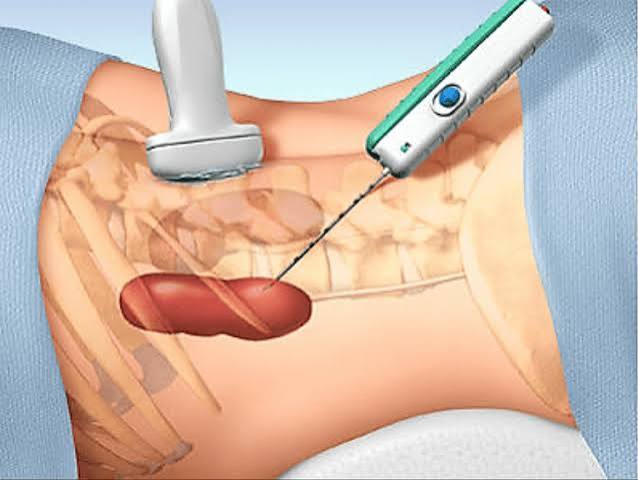
A kidney biopsy is a procedure in which a long, thin needle is passed through the skin of the back or flank, guided into the kidney using real-time ultrasound imaging, and used to remove a small cylinder of kidney tissue approximately 1 to 2 centimeters long and 1 to 2 millimeters in diameter. This core of tissue is then processed in the pathology laboratory and examined using three different microscopic techniques:
- Light microscopy (LM): The tissue is stained with multiple dyes (hematoxylin and eosin, PAS stain, silver methenamine, Masson’s trichrome) to reveal the structure of the glomeruli, tubules, interstitium, and blood vessels. Light microscopy shows abnormal cell proliferation, scarring, inflammation, deposits, and crescents (a sign of rapidly progressive disease).
- Immunofluorescence (IF): Antibody-tagged fluorescent probes are applied to the tissue to detect deposits of immunoglobulins (IgA, IgG, IgM) and complement components (C3, C1q). The pattern of deposits — where they are located and which proteins are present — is highly diagnostic for specific glomerular diseases.
- Electron microscopy (EM): The tissue is viewed at extremely high magnification to visualize structures invisible to light microscopy — the foot processes of podocytes, the basement membrane’s thickness and texture, and the internal structure of any deposits (granular for immune complex diseases; fibrillar for amyloid).
Transplant kidney biopsies target the transplanted kidney in the pelvis and are performed both on a scheduled protocol basis (to detect early rejection before symptoms appear) and for specific indications (rising creatinine, new proteinuria).
Why a Kidney Biopsy May Be Recommended
Nephrotic syndrome in adults: Defined by severe proteinuria (more than 3.5 grams per day), low blood albumin, and edema. In children age 1 to 8, nephrotic syndrome is presumed to be minimal change disease and treated empirically with steroids without biopsy. In adults, however, the differential is broad — minimal change disease, FSGS, membranous nephropathy, diabetic nephropathy, amyloidosis, lupus nephritis, and others — and each requires a different treatment. More on heavy proteinuria is in our article on protein in urine.
Nephritic syndrome: Combines blood in the urine (hematuria, often with red blood cell casts on urine microscopy), significant proteinuria, hypertension, and declining kidney function. The differential includes IgA nephropathy, lupus nephritis, ANCA-associated vasculitis, anti-GBM disease (Goodpasture syndrome), and others. A biopsy is essential because several of these conditions require aggressive immunosuppression and others do not. See our article on blood in urine for how hematuria is evaluated.
Rapidly progressive glomerulonephritis (RPGN): A nephrology emergency. RPGN presents as acute kidney injury with red blood cell casts on urine microscopy and a rapidly deteriorating GFR over days to weeks. On biopsy, RPGN typically shows crescents — proliferating cells that progressively destroy the glomerular tuft. Treatment with high-dose IV steroids and often cyclophosphamide or rituximab must begin within days to prevent irreversible kidney failure. Biopsy is performed on an urgent basis.
Unexplained AKI or CKD: When kidney function deteriorates acutely without an evident cause (not explained by dehydration, obstruction, or nephrotoxic medications), or when eGFR declines progressively with no identified etiology, a biopsy may reveal a treatable glomerular or interstitial disease. More on monitoring declining kidney function is in our guide to kidney function tests.
Systemic diseases with kidney involvement: In lupus nephritis, the ISN/RPS classification system (Classes I through VI) determines treatment intensity: Class III and IV require aggressive immunosuppression with mycophenolate mofetil and steroids, while Class V and VI are managed differently. Amyloidosis requires Congo red staining showing apple-green birefringence under polarized light — the pathognomonic finding — plus amyloid subtyping (AL from myeloma; AA from inflammation; ATTR transthyretin) because each type has completely different treatment. ANCA vasculitis requires histologic confirmation of pauci-immune crescentic glomerulonephritis before committing to high-dose immunosuppression.
What Happens Before the Procedure
- Blood pressure control: BP must be below 160/100 on the procedure day. Uncontrolled hypertension significantly increases bleeding risk.
- Coagulation testing: INR must be below 1.5 and platelets above 50,000 to 100,000 per microliter.
- Anticoagulant management: Warfarin held 5 days; DOACs held 2 to 3 days; aspirin and clopidogrel typically held 5 to 7 days.
- Blood type and screen: In case transfusion is needed.
- IV access and informed consent: Risks reviewed including bleeding (transfusion in ~1–3%), angiographic embolization if pseudoaneurysm develops, and very rarely nephrectomy.
What to Expect During the Kidney Biopsy
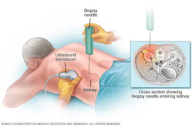
The patient lies face-down (prone) on a procedure table. The skin over the lower back is cleaned and draped. The nephrologist uses real-time ultrasound to identify the kidney’s lower pole — targeted because it has fewer large blood vessels. A local anesthetic (lidocaine) is injected into the skin and tissues. Most patients also receive a small dose of IV midazolam and fentanyl to reduce anxiety without full sedation.
A 16-gauge or 18-gauge spring-loaded core biopsy needle is advanced under ultrasound guidance into the kidney cortex. The patient holds their breath; the spring fires, collecting a core of tissue approximately 15 to 20 mm long. This is repeated 2 to 3 times. Most patients describe a brief sensation of pressure or dull impact — not sharp pain — at each needle firing. The procedure takes 30 to 60 minutes total.
After the Biopsy — Monitoring and Recovery
The patient rests flat for 4 to 6 hours. Vital signs are checked every 15 to 30 minutes; urine is checked at each void for blood. Some pink-tinged urine is normal. Most patients are discharged the same day after stable vital signs are confirmed.
Home instructions: Avoid heavy lifting, vigorous exercise, and contact sports for 1 to 2 weeks. Avoid NSAIDs — use acetaminophen for pain. Drink adequate fluids. Return to the emergency department immediately if gross hematuria with blood clots develops, urine output stops, or severe progressive flank pain occurs.
Risks and Complications
Common and expected: Brief gross hematuria in the first 24 to 48 hours occurs in the majority of patients and resolves spontaneously. A small perirenal hematoma is present on ultrasound in 60 to 90% of patients but is clinically silent in the vast majority.
Transfusion (~1 to 3%): Clinically significant bleeding requiring transfusion. Risk is higher with uncontrolled hypertension, thrombocytopenia, or coagulopathy.
Angiographic embolization (<1%): An AV fistula or pseudoaneurysm may develop, typically presenting as recurrent gross hematuria 1 to 2 weeks post-biopsy. Treated with minimally invasive catheter-based embolization.
Absolute contraindications: Uncorrectable bleeding disorder, uncontrolled hypertension, active urinary infection overlying the biopsy site, and an uncooperative patient. A solitary functioning kidney is a relative contraindication requiring careful individual risk-benefit discussion.
What Kidney Biopsy Results Can Show
IgA nephropathy: Dominant IgA deposits in the mesangium on immunofluorescence; mesangial hypercellularity on light microscopy. The most common glomerular disease worldwide. Treatments include angiotensin inhibition, dapagliflozin (FDA-approved for IgA nephropathy), and immunosuppression for rapidly progressive forms.
Membranous nephropathy: Thickened glomerular basement membrane with silver stain “spikes”; granular IgG deposits on the outer surface of the GBM on IF. Most primary membranous is caused by anti-PLA2R antibodies. First-line treatment is rituximab.
FSGS: Segmental scarring of some glomeruli. Primary FSGS is treated with high-dose steroids; only 30 to 40% achieve remission. Secondary FSGS is managed by treating the underlying cause.
Minimal change disease (MCD): Light microscopy and IF are normal; EM shows diffuse foot process effacement. The most steroid-responsive glomerular disease; most patients achieve full remission within 8 to 16 weeks of prednisone.
Amyloidosis: Congo red stain shows apple-green birefringence under polarized light — pathognomonic. Mass spectrometry on biopsy tissue identifies the amyloid type (AL = chemotherapy; ATTR = tafamidis or patisiran).
After Receiving the Biopsy Results
Preliminary results are typically available within 2 to 3 days; the full report including EM requires 7 to 10 days. A “non-diagnostic” biopsy (fewer than 5 to 10 glomeruli) occurs in approximately 5 to 10% of cases and may require repeat biopsy. Once a diagnosis is made, treatment is targeted specifically at the identified disease. A patient with membranous nephropathy and high PLA2R antibody titer receives rituximab; MCD receives steroids; IgA nephropathy receives angiotensin inhibition and dapagliflozin — radically different approaches that are impossible to choose correctly without the biopsy. For further context on what kidney tests measure, see our guide on urinalysis.
Frequently Asked Questions
Is a kidney biopsy painful? Most patients describe the procedure as significantly less painful than expected. The local anesthetic eliminates most discomfort; each needle firing produces a brief sensation of pressure rather than sharp pain. Post-procedure, mild to moderate flank aching for 24 to 48 hours responds to acetaminophen (not NSAIDs).
How long does it take to get kidney biopsy results? Preliminary results from light microscopy and immunofluorescence are typically available in 2 to 3 days. Full results including electron microscopy are usually available in 7 to 10 days. Specialized analyses such as mass spectrometry for amyloid typing may extend the timeline to 2 to 3 weeks. A follow-up appointment is typically scheduled to review the full results in person.
What happens if the biopsy shows a serious kidney disease? The most important consequence of a specific diagnosis is gaining access to targeted treatment. Many serious glomerular diseases — membranous nephropathy, lupus nephritis, ANCA vasculitis, MCD — are treatable and can achieve full or partial remission with appropriate therapy. Even when biopsy reveals advanced disease (extensive scarring with fibrosis), the diagnosis enables specific conversations about prognosis, timeline to kidney replacement therapy, and transplant planning — which are ultimately more useful than uncertainty.
Who Performs a Kidney Biopsy and Where Is It Done
Percutaneous kidney biopsies are performed by nephrologists (kidney specialists) or interventional radiologists, depending on institutional protocols. In most academic medical centers, nephrologists perform native kidney biopsies in a procedure suite or interventional radiology suite, while transplant biopsies may be performed bedside in a clinic or transplant unit. Interventional radiology is involved when CT guidance is required — typically in patients with obesity limiting ultrasound visualization depth, kidneys positioned unusually, or prior failed ultrasound-guided attempts.
Most native kidney biopsies are performed as outpatient procedures, with a 4- to 8-hour observation period and same-day discharge for patients who remain stable. Some institutions admit all patients overnight for 23-hour observation; others selectively admit higher-risk patients. Transplant biopsies, which access the easily reached pelvic transplant kidney, are frequently performed as outpatient office procedures at many transplant centers.
Special Situations: When Kidney Biopsy Is More Complex
Single functioning kidney: A patient with a solitary kidney — either congenitally single, previously removed, or with a failed contralateral kidney — is at higher risk from kidney biopsy because any complication affecting the remaining kidney could precipitate dialysis. The decision to biopsy a single kidney requires a careful individual risk-benefit discussion between the nephrologist and patient. In some cases, the clinical indication is strong enough (RPGN, suspected acute interstitial nephritis on potentially reversible AKI) that the benefit of the diagnosis clearly outweighs the risk. In others, the biopsy result would not change management (end-stage small kidneys, clinical diagnosis of diabetic nephropathy in a patient with longstanding diabetes and typical retinopathy), and the biopsy is appropriately deferred.
Severe obesity: In patients with BMI above 35 to 40, the depth from the skin surface to the kidney may exceed the effective range of standard biopsy needles, and ultrasound image quality degrades with increasing tissue depth. These patients may require CT guidance, prone positioning with additional padding, or a longer biopsy needle. The risk of post-biopsy bleeding may also be higher, as fat surrounding the kidney may not tamponade a bleeding vessel as effectively as in patients with less perirenal fat.
Multiple renal cysts (ADPKD): In autosomal dominant polycystic kidney disease, the kidneys are filled with cysts ranging from millimeters to centimeters in size. Biopsy of a polycystic kidney carries higher risk because the needle may inadvertently puncture a cyst (usually not clinically significant) or traverse multiple cysts before reaching cortical tissue with glomeruli. In most patients with ADPKD, biopsy is rarely needed because the diagnosis is established by imaging and genetic testing, and the mechanism of progression (cyst expansion and compression of normal parenchyma) does not require histologic confirmation. Biopsy may be considered in ADPKD patients who develop unusual clinical features suggesting a superimposed glomerular disease.
Anticoagulation that cannot be safely held: Patients on therapeutic anticoagulation for mechanical heart valves, prior arterial thromboembolism, or recent VTE may require careful bridging strategies before kidney biopsy. Patients on warfarin for mechanical mitral valves are at high thromboembolic risk during the days when warfarin is held; parenteral heparin bridging is standard in these patients. The specific strategy should be coordinated between the nephrologist, cardiologist, and hematologist. In some cases where biopsy cannot be safely performed percutaneously, a transjugular renal biopsy can be performed by interventional radiology — the biopsy needle is passed through the jugular vein, through the heart, and into the renal vein, allowing the kidney to be sampled from within the collecting system without traversing the kidney capsule; this approach has a lower bleeding risk because the access route avoids the kidney capsule, though it obtains smaller tissue cores and may be less diagnostic.
Questions to Ask Your Nephrologist Before a Kidney Biopsy
Coming to a pre-biopsy appointment with prepared questions helps patients make an informed decision and understand the clinical rationale for the procedure:
- Why exactly do you think I need a biopsy, and what diagnoses are you considering? Understanding the specific differential diagnosis — the list of conditions the biopsy is meant to distinguish between — helps patients understand how the biopsy result will change their care.
- What are the treatment options if the biopsy shows the most likely diagnosis? If the most likely diagnosis requires aggressive immunosuppression (cyclophosphamide, rituximab, high-dose steroids), knowing this in advance allows the patient to ask about side effects and monitoring requirements before starting therapy.
- What would we do if the biopsy is non-diagnostic? Understanding the plan for the approximately 5 to 10% of biopsies that return insufficient tissue (fewer than 5 to 10 glomeruli) — whether the approach would be to repeat the biopsy, accept a presumptive diagnosis, or consider open biopsy — prepares the patient for this outcome.
- Am I at higher than average risk for biopsy complications, and if so, what modifications will be made? Patients with diabetes (which may impair vascular healing), hypertension, chronic kidney disease (impaired platelet function in advanced CKD), or prior renal surgery should ask how these factors are being addressed in the pre-biopsy preparation.
- When will I hear the results, and who will call me? Knowing the expected timeline and who is responsible for communicating results prevents patients from being in limbo for days without knowing when to expect their call. In most centers, the ordering nephrologist reviews the pathology report and calls or messages the patient within 1 to 3 days of the preliminary result, then schedules a follow-up visit to review the full report including EM.
A kidney biopsy is one of the more significant diagnostic procedures a nephrology patient will undergo, and the information it provides can be life-changing — enabling treatment of diseases that would otherwise progress silently to kidney failure. Approaching it with accurate information about what it involves and what it can reveal transforms a potentially anxiety-provoking procedure into a meaningful step toward a specific diagnosis and targeted treatment.
When Kidney Biopsy Results Lead to Further Testing
A kidney biopsy result is often the beginning of a diagnostic process rather than the final step. Many biopsy findings generate additional testing that either confirms the biopsy diagnosis, identifies the underlying cause, or assesses the patient’s treatment options.
After a diagnosis of membranous nephropathy: The biopsy confirms the histologic pattern (subepithelial IgG deposits, GBM spikes). The next step is identifying whether the disease is primary (autoimmune, caused by anti-PLA2R or anti-THSD7A antibodies) or secondary (caused by an underlying condition). A serum anti-PLA2R antibody titer is drawn — positive in approximately 70 to 80% of primary membranous nephropathy. Secondary causes must be excluded: hepatitis B surface antigen, hepatitis C antibody, ANA and anti-dsDNA (for lupus), and cancer screening (chest imaging, colonoscopy if age-appropriate, PSA in older men) because membranous nephropathy can be a paraneoplastic manifestation of solid tumors in patients over 60. Once primary vs. secondary is established, the treatment approach differs dramatically: primary membranous nephropathy is treated with rituximab; secondary membranous requires treating the underlying disease.
After a diagnosis of ANCA-associated vasculitis: The biopsy shows pauci-immune crescentic glomerulonephritis (crescents without significant immune deposits on IF). This is immediately followed by ANCA serology if not already done — PR3-ANCA (associated with granulomatosis with polyangiitis / GPA) and MPO-ANCA (associated with microscopic polyangiitis / MPA) — and by assessment of other organ involvement: chest CT for lung involvement (pulmonary hemorrhage, nodules in GPA), ENT evaluation for sinusitis and saddle-nose deformity (GPA), and nerve conduction studies if peripheral neuropathy is suspected (MPA). ANCA vasculitis is a systemic disease; kidney biopsy confirms the kidney’s involvement but the full disease burden extends well beyond the kidney.
After a diagnosis of lupus nephritis: ISN/RPS Class III or IV on biopsy triggers serological monitoring (anti-dsDNA titers, complement C3/C4 levels) that will be used to assess treatment response alongside urine protein-to-creatinine ratio and eGFR during induction therapy. Repeat biopsy at 6 months to 1 year may be considered in patients not responding to induction therapy, to assess whether active nephritis has resolved or whether irreversible fibrosis has developed.
After finding chronic, irreversible changes: When biopsy shows predominantly global glomerulosclerosis (most or all glomeruli destroyed), severe tubular atrophy, and extensive interstitial fibrosis, the finding indicates end-stage or near-end-stage kidney disease unlikely to respond to immunosuppression. In this context, the focus shifts from disease-specific treatment to optimizing the rate of progression (aggressive BP control, renin-angiotensin system blockade, dietary protein restriction, SGLT2 inhibitor use), discussing kidney replacement therapy options (hemodialysis, peritoneal dialysis, transplant), and listing for transplant evaluation if appropriate. A patient who receives this biopsy result deserves a candid and compassionate conversation about prognosis and the timeline to kidney replacement therapy — information that the biopsy has now made concrete and that allows the patient to plan their life accordingly.
Sources: National Kidney Foundation — Kidney Biopsy | NIDDK — Kidney Biopsy | American Society of Nephrology | Urology Care Foundation | Related: Kidney Function Tests | Urinalysis: What It Can Show | Blood in Urine Test Results | Protein in Urine: Causes and Meaning
Really well-written article on kidney biopsy: why it may be. I especially valued the explanation of why these recommendations exist, not just what they are. I wish I had found this article earlier — would have saved a lot of confusion.
I shared this article on kidney biopsy: why it may be with my doctor and they appreciated the level of detail. The specific numbers and thresholds mentioned are exactly what I needed to understand my results. Will definitely be coming back to this site for more health information.
My doctor recommended I look into kidney biopsy: why it may be and this article covered it perfectly. The specific numbers and thresholds mentioned are exactly what I needed to understand my results. This gave me real confidence going into my next specialist appointment.