
Skin Changes and Diabetes: What to Look For
The skin is one of the most visible indicators of internal metabolic health, and skin changes and diabetes are linked in ways that reflect both the early warning signs of insulin resistance and the ongoing consequences of elevated blood glucose on tissue biology. Some skin changes appear before diabetes is diagnosed — acanthosis nigricans, for instance, is a visible marker of insulin resistance that may precede a formal diabetes diagnosis by years. Others emerge alongside or after diagnosis, driven by the microvascular damage, impaired immunity, autonomic neuropathy, and protein glycation that elevated blood glucose produces over time. Recognizing the full range of skin changes associated with diabetes — from those that signal risk to those that signal active complication — gives both people with diabetes and their clinicians a more complete picture of metabolic status and skin health. The context of how blood sugar elevation causes these changes is covered in our guide on why blood sugar matters for long-term health, and the full spectrum of early signs of high blood sugar provides the broader symptom context in which skin changes often occur.
Acanthosis Nigricans: The Skin Sign of Insulin Resistance
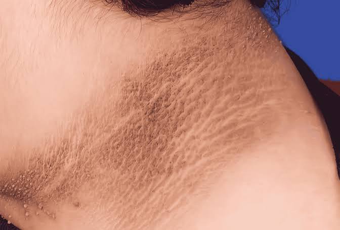
Acanthosis nigricans is the skin change most directly linked to insulin resistance — and therefore to the prediabetes and Type 2 diabetes that insulin resistance often precedes or accompanies. It presents as areas of darkened, thickened, velvety skin in skin folds and creases: most commonly the back of the neck, the armpits, the groin, and beneath the breasts. The affected areas look darker than the surrounding skin (ranging from tan to deep brown or gray-black, depending on baseline skin tone) and have a texture that is thick and raised rather than flat. When you run a finger across an area of acanthosis nigricans, it feels like soft velvet rather than normal smooth skin.
The mechanism behind acanthosis nigricans is the direct stimulation of skin cell receptors by elevated insulin levels. In insulin resistance — where peripheral tissues fail to respond normally to insulin, forcing the pancreas to overproduce insulin to compensate — the resulting hyperinsulinemia (chronically elevated insulin in the blood) stimulates receptors called insulin-like growth factor (IGF-1) receptors on keratinocytes (the primary cells of the outer skin layer) and fibroblasts. This overstimulation causes accelerated proliferation of these skin cells, producing the characteristic thickening, and triggers increased production of melanin-producing cells in the affected areas, producing the darkening. The result is a direct visible consequence of the hormonal dysregulation that drives insulin resistance — which is why acanthosis nigricans serves as such a useful clinical marker. Understanding what insulin resistance is and how it develops helps explain why this skin change is a metabolic marker, not merely a cosmetic issue.
Acanthosis nigricans is not painful and does not cause medical complications in itself — but its significance lies in what it indicates about metabolic health. Its presence, particularly in children or young adults, is a strong signal for blood glucose testing and evaluation of diabetes risk factors. In people already known to have insulin resistance or prediabetes, the severity of acanthosis nigricans can track with the degree of insulin resistance — improving when insulin sensitivity improves (through weight loss, exercise, and metformin or other medications) and worsening when it deteriorates. It is also worth noting that acanthosis nigricans can occur in conditions other than insulin resistance — particularly certain cancers and rare hormonal disorders — so evaluation by a healthcare provider is appropriate when it appears, especially if it develops rapidly or in unusual locations.
Diabetic Dermopathy: The Most Common Diabetes Skin Change
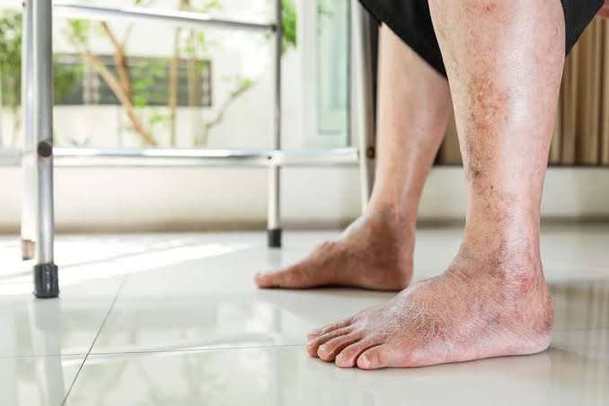
Diabetic dermopathy — sometimes called “shin spots” — is the most common skin change seen in people with established diabetes, occurring in approximately 55 percent of people with diabetes overall and more than 70 percent of those who have had diabetes for over fifteen years. It presents as small (0.5–2 cm), round or oval, reddish-brown or tan-colored spots on the shins (the front of the lower legs), typically multiple and scattered in a somewhat asymmetric pattern. Over time, the spots may fade to a lighter brown or near-skin-color and develop a slightly atrophic (thinned) appearance. They are flat or only slightly raised, painless, and not itchy.
The lesions of diabetic dermopathy reflect microvascular damage — the same small blood vessel damage that drives diabetic retinopathy, nephropathy, and peripheral neuropathy. Repeated minor trauma to the shin skin (which has less subcutaneous fat padding than other body areas and is more susceptible to minor bumps and impacts) combined with impaired microvascular blood flow and impaired tissue repair results in small deposits of hemosiderin (breakdown product of blood) and fibrin in the skin, producing the characteristic brown spots. While dermopathy itself is harmless, its presence is a reliable indicator that microvascular complications are present or at risk, and should prompt evaluation for diabetic retinopathy and nephropathy in people who have not had recent screening. For people newly noticing shin spots, reviewing whether other diabetes complications have been screened for recently is a valuable next step in comprehensive diabetes management.
- Acanthosis nigricans: Darkened, velvety skin in folds — marker of insulin resistance, may predate diabetes diagnosis
- Diabetic dermopathy: Shin spots — most common diabetes skin change; reflects microvascular disease
- Necrobiosis lipoidica: Yellowish, waxy patches on shins — rare but distinctive; associated with long-standing diabetes
- Diabetic skin infections: Bacterial (boils, cellulitis) and fungal (Candida) — elevated glucose feeds pathogens, impaired immunity reduces defense
- Eruptive xanthomas: Yellow papules from elevated triglycerides — signal very poorly controlled diabetes or lipid disorder
- Generalized dry skin (xerosis): Common in diabetes due to autonomic neuropathy reducing sweat and sebum production
Necrobiosis Lipoidica: A Rare but Distinctive Diabetes Skin Condition
Necrobiosis lipoidica diabeticorum (NLD) is a rare but clinically distinctive skin condition that occurs predominantly in people with diabetes, though it can rarely affect those without it. It presents as waxy, yellowish-orange or reddish-brown plaques, most commonly on the shins, that develop slowly over months to years. The plaques have a shiny surface with visible blood vessels running through them, often a violet or red-brown border, and a depressed (atrophic) center. As the condition progresses, the central area may ulcerate, particularly after minor trauma — and these ulcers can be chronic and difficult to heal, given the impaired wound healing already present in diabetes.
NLD affects women more than men and is most common in people with Type 1 diabetes, particularly those with long duration and imperfect glucose control, though it occurs in Type 2 as well. The mechanism involves granulomatous inflammation and degeneration of collagen in the dermis — a process driven by microvascular damage, altered collagen structure from glycation, and immune dysregulation. Treatment of NLD is challenging and often unsatisfying — topical corticosteroids, intralesional steroid injections, and laser therapy have been tried with variable results, and improving glucose control, while clearly beneficial for preventing new lesions, does not reliably cause existing lesions to regress. For lesions that have ulcerated, wound care principles similar to those described in our guide on slow wound healing and diabetes apply, with dermatology consultation appropriate for management of this specific condition. The rarity of NLD means it may initially be mistaken for other skin conditions — if a yellowy, waxy shin lesion is not diagnosed, dermatology evaluation is the appropriate next step.
Skin Infections in Diabetes
People with diabetes are significantly more susceptible to skin infections — both bacterial and fungal — than the general population, and these infections may be more severe, progress more rapidly, and be harder to treat when blood glucose is poorly controlled. Several overlapping mechanisms explain this susceptibility: elevated glucose in skin and body fluids provides a rich medium for bacterial and fungal growth; impaired neutrophil and macrophage function (driven by hyperglycemia) reduces the immune response to invading organisms; impaired blood flow to skin (from microvascular disease) reduces the delivery of immune cells and antibiotics to infected tissue; and neuropathy-related skin dryness and cracking creates entry points for bacteria.
Bacterial infections: Staphylococcus aureus is the most common cause of skin infections in diabetes — producing folliculitis (infection of hair follicles), boils (furuncles), carbuncles (clusters of boils), and cellulitis (spreading skin and tissue infection). Erythrasma — an infection caused by Corynebacterium minutissimum producing brownish patches in skin folds — is also more common in diabetes. Recurrent skin infections with Staphylococcus aureus, particularly boils, are in fact a recognized presentation that sometimes prompts blood glucose testing and leads to a first diabetes diagnosis in people who were unaware they had elevated blood sugar. The symptoms of type 2 diabetes guide covers the full range of presentations, including skin-related ones, that may precede formal diagnosis.
Fungal infections: Candida albicans infections in skin — particularly in skin folds (intertrigo), in the genital area, and beneath the breasts — are substantially more common in diabetes because elevated glucose feeds Candida growth and impaired immunity reduces the defense against it. Tinea (ringworm), another fungal infection, is also more prevalent and may be more extensive in people with poorly controlled glucose. Onychomycosis — fungal infection of the nails, producing thickened, discolored, brittle nails — affects more than one third of people with diabetes and is important to treat not only for cosmetic reasons but because thickened fungal nails can impair foot hygiene and become a source of repeated trauma that leads to nail-bed infections or ingrown nails. Proper nail trimming and treatment of onychomycosis with appropriate antifungal therapy is an underemphasized aspect of diabetes foot care that is reviewed in detail alongside the broader foot health considerations in our guide on tingling feet and diabetes.
Generalized Dry Skin and Its Consequences
Xerosis — generalized dry skin — is one of the most common skin complaints in diabetes, affecting a majority of people with the condition at some point. Several diabetes-related mechanisms reduce the skin’s natural moisturizing capacity: autonomic neuropathy affecting the small nerve fibers that control sweating (eccrine glands) and sebum production (sebaceous glands) reduces both sweat and oil output, leaving skin without its natural surface moisture and protective lipid layer; impaired blood flow to the skin’s microcirculation reduces the delivery of water and nutrients to skin cells; and glycation of skin structural proteins (collagen and elastin) changes the mechanical properties and water-binding capacity of the dermis and epidermis.
Dry skin in diabetes is more than a cosmetic issue. The skin’s barrier function depends on its surface lipid layer and intact surface cells — when skin is dry and the lipid layer is disrupted, the barrier is compromised, making skin more susceptible to bacteria and fungi entering through microscopic cracks, and making wounds more likely to form from friction or minor trauma. The hands and feet are particularly affected, and cracked skin on the heel — a common manifestation of severe xerosis in diabetes — can create entry points for bacterial cellulitis that are disproportionately serious given the impaired immune response and circulation that characterize diabetes. Moisturizing the feet daily with a good emollient (urea cream, shea butter, or other thick moisturizing lotion) is a standard component of diabetes foot care for exactly this reason. For people monitoring blood sugar levels at home, maintaining the skin condition of the fingertips used for glucose testing is also a practical consideration — using the sides of fingertips rather than the pads, rotating lancing sites, and keeping fingertips moisturized reduces the cumulative skin trauma of frequent lancing. For guidance on the broader context of testing and monitoring, our guide on home blood sugar monitoring covers the practical aspects of glucose testing. And for anyone experiencing multiple skin changes described above alongside other blood sugar symptoms, the diagnostic pathway covered in our guide on what diabetes is and the testing described in our guide on the A1C test provide the framework for confirming whether blood glucose is the underlying driver of these skin changes and how significantly it is elevated.
Eruptive Xanthomas: When Blood Fats Appear in Skin
Eruptive xanthomas are a striking and diagnostically important skin manifestation of severely poorly controlled diabetes — particularly when triglyceride levels are also very high. They present as sudden crops of small (2–5 mm), yellowish-orange papules surrounded by reddish halos, appearing most commonly on the buttocks, elbows, knees, and backs of the hands, and sometimes distributed more widely across the trunk and extremities. Unlike the other skin changes described in this guide, eruptive xanthomas are not subtle — they appear rapidly and in numbers that make them immediately noticeable.
The mechanism is triglyceride overflow into the skin when blood triglyceride levels are extreme (typically above 1000 mg/dL, though sometimes seen at lower levels). Poorly controlled diabetes significantly raises triglyceride levels through multiple mechanisms: insulin deficiency reduces the activity of lipoprotein lipase (the enzyme that clears triglyceride-rich lipoproteins from the blood), and elevated glucose prompts the liver to produce more VLDL (very low-density lipoprotein) particles laden with triglycerides. When triglycerides reach sufficiently high levels, triglyceride-laden macrophages migrate into the dermis and deposit lipids there, producing the yellow color (from carotenoids in the triglycerides) and the raised papule appearance of eruptive xanthomas. The sudden appearance of eruptive xanthomas should prompt immediate evaluation of triglyceride levels and blood glucose control — severely elevated triglycerides carry their own risk of acute pancreatitis, which is a life-threatening emergency. Treatment with insulin to lower blood glucose typically produces rapid improvement in triglyceride levels and resolution of the eruptive xanthomas within weeks. The presence of eruptive xanthomas in someone not known to have diabetes warrants immediate blood glucose and lipid testing, as it often signals the first recognition of severely decompensated diabetes or diabetic hyperlipidemia.
Scleredema Diabeticorum: Skin Thickening in Long-Standing Diabetes
Scleredema diabeticorum is a skin change associated with long-standing poorly controlled diabetes — particularly Type 2 diabetes — that is often missed or attributed to other causes. It presents as a symmetrical thickening and hardening of the skin on the upper back and neck, sometimes extending to the shoulders, arms, and face. The affected skin feels indurated (woody or board-like) when pressed, does not pit with pressure (unlike edema), and is typically not itchy or painful. The overlying skin color is usually normal or only slightly reddened.
The mechanism is accumulation of glycosaminoglycans (large sugar-protein complexes) in the dermis, driven by the activation of fibroblasts through AGE-mediated and growth factor pathways that chronically elevated blood glucose promotes. The excess glycosaminoglycans between collagen fibers expand the dermis and restrict its normal mobility. Unlike the scleredema that occurs after streptococcal infection in children (which resolves spontaneously), diabetic scleredema is typically persistent and may progress over years without meaningful spontaneous improvement. It is more cosmetically concerning and functionally limiting than medically dangerous in most cases — though severe cases affecting the neck and shoulders can restrict range of motion. No treatment has been consistently effective, and the condition does not reliably respond to glucose improvement alone, though aggressive glucose management is the most reasonable foundation for limiting progression. When skin thickening on the upper back develops in a person with long-standing diabetes and is brought to medical attention, it is often initially mistaken for lymphedema, obesity-related skin changes, or other conditions — awareness of scleredema diabeticorum as a diabetes complication improves the chance of correct attribution and appropriate expectations for management.
Pruritus: Itching as a Diabetes Skin Symptom
Generalized itching — pruritus — is more common in people with diabetes than in the general population, and it can occur through multiple mechanisms. Dry skin (xerosis) is the most common cause of itching in diabetes, as dry, compromised skin is more easily irritated by friction, clothing, and minor environmental changes. Candida skin infections in moist skin folds produce localized itching that can be intense and distressing. Peripheral neuropathy — the nerve damage described in our guide on tingling feet and diabetes — can produce a form of neuropathic itch that is distinct from inflammatory itch: it is driven by abnormal sensory nerve firing rather than skin inflammation, and may not respond to antihistamines or topical treatments that work for conventional itch.
Localized genital itching is particularly common in diabetes and strongly associated with Candida vaginitis (in women) or Candida balanitis (in men) — both driven by elevated glucose in genital secretions that promotes fungal overgrowth. In women, recurrent vulvovaginal candidiasis — yeast infections that keep returning despite standard antifungal treatment — is a recognized early presentation that prompts blood glucose evaluation, and it is one of the symptoms of type 2 diabetes that specifically affects women and may be the first symptom severe enough to bring someone to medical attention. Treatment of the fungal infection provides temporary relief, but without addressing the elevated glucose that fuels the overgrowth, recurrence is nearly inevitable. Sustained glucose improvement — tracked through the A1C test and daily monitoring with home blood sugar monitoring — is the definitive intervention for glucose-driven recurrent candidiasis, alongside appropriate antifungal treatment of each episode.
Practical Skin Care for People With Diabetes
Given the multiple ways diabetes compromises skin health, a proactive skin care routine is an important and underemphasized component of comprehensive diabetes management. The following principles are particularly relevant:
Daily moisturizing: Apply a thick emollient (urea-based cream, petroleum jelly, shea butter lotion) to the skin of the legs, feet, and any particularly dry areas immediately after bathing, when the skin is still slightly moist, to lock in surface hydration. Avoid applying moisturizer between the toes, where retained moisture can promote fungal growth — keep the toe webspaces dry while moisturizing the rest of the foot. For the hands, reapplying hand cream after each handwashing reduces the cumulative drying effect of frequent soap exposure.
Gentle skin care products: Use mild, fragrance-free soaps and cleansers that do not strip natural skin oils. Avoid hot water, which further dries skin and can cause capillary dilation that is more pronounced in people with impaired autonomic regulation. Pat skin dry rather than rubbing, particularly over areas of thin or fragile skin.
Daily foot inspection: Because neuropathy reduces pain sensation in the feet, minor injuries, cuts, and pressure areas may go unnoticed. A daily inspection of the entire foot — including the soles and between the toes — using a mirror or assistance if needed, allows early detection of skin changes before they progress to wounds or infections. Any change in skin color, new callus formation, blister, cracked skin, or wound warrants prompt attention.
Sun protection: People with diabetes may have more difficulty mounting effective inflammatory and immune responses to UV damage, and some diabetes-related skin conditions are worsened by sun exposure. Using sunscreen and protective clothing reduces additional skin stress that compounds the existing metabolic skin vulnerabilities.
Managing skin health in diabetes is ultimately inseparable from managing blood glucose — most diabetes-related skin changes are driven by or worsened by elevated glucose, and improving glucose control is the most effective long-term intervention for all of them. Understanding what prediabetes looks like and catching blood sugar elevation early — before years of elevated glucose have produced microvascular damage, neuropathy, and the immune impairment that drives most diabetes skin complications — is the most powerful way to prevent the skin consequences of diabetes from developing at all. For those already managing diabetes, consistent attention to glucose control alongside the practical skin care steps above provides the best foundation for maintaining skin health throughout the course of the condition.
Sources: American Diabetes Association. Standards of Medical Care in Diabetes — 2024. Diabetes Care. 2024;47(Suppl 1):S20–S42. • Duff M, et al. Skin Manifestations of Diabetes Mellitus. Clinical Diabetes. 2015;33(1):40–48. • National Institute of Diabetes and Digestive and Kidney Diseases. Diabetes and Skin Complications. NIDDK; 2023.
As someone dealing with this personally, the skin changes and diabetes: what to section was very helpful. This is the kind of evidence-based writing that actually changes how people approach their health. Thank you for making complex medical information accessible without dumbing it down.
I never fully understood skin changes and diabetes: what to until I read this. The practical tips made this immediately actionable, not just theoretical. This is going into my health folder that I bring to every doctor’s visit.
I have been reading about skin changes and diabetes: what to for weeks and this is the most thorough guide I found. I especially valued the explanation of why these recommendations exist, not just what they are. I wish I had found this article earlier — would have saved a lot of confusion.