

Lower back pain affects approximately 80 percent of adults at some point in their lives. The vast majority is musculoskeletal — a strained muscle, a herniated disc, or prolonged poor posture — and resolves on its own within a few weeks. But some lower back pain comes from the kidneys. Kidney stones, pyelonephritis, kidney cancer, and polycystic kidney disease can all produce pain in the back or flank that is clinically urgent and easily mistaken for the more common musculoskeletal variety. The difference matters because the appropriate response is entirely different. This article covers how to tell kidney pain from back pain, which kidney conditions cause it, and when back pain requires immediate kidney evaluation.
Kidney Anatomy — Why Kidney Problems Cause Back Pain
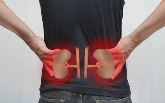
The kidneys sit in the retroperitoneal space — behind the abdominal organs, on either side of the spine — at approximately the level of the T12 to L3 vertebrae. This is precisely the region most people describe as the lower back or flank. Each kidney is enclosed in a fibrous capsule containing pain-sensitive nerve fibers; when the kidney swells from obstruction, infection, or tumor growth, the stretching capsule generates pain felt in the flank and back.
The costovertebral angle (CVA) is the anatomical landmark for kidney pain assessment — formed by the junction of the 12th rib and the lateral border of the erector spinae muscle on each side of the spine. Tenderness with fist percussion of the CVA is the classic physical examination finding for kidney pathology.
When a kidney stone passes from the kidney into the ureter, ureteral spasm produces colicky pain — waves of intense pain radiating from the flank downward toward the groin, inner thigh, and genitalia. This radiation pattern is characteristic of ureteral rather than musculoskeletal pain and is one of the clearest clinical distinguishing features.
Kidney Pain vs. Musculoskeletal Back Pain — Key Differences
| Feature | Kidney / Flank Pain | Musculoskeletal Pain |
|---|---|---|
| Location | Unilateral flank or CVA | Lower lumbar, bilateral, midline |
| Quality | Deep ache or colicky waves | Aching stiffness, sharp with movement |
| Movement effect | Not clearly worsened by position | Clearly worse with bending/lifting |
| Radiation | Flank → groin, inner thigh, genitalia | Local or down the leg (sciatica) |
| Urinary symptoms | Often present (blood, dysuria, frequency) | Absent |
| Fever / chills | Present in infection | Absent |
| CVA tenderness | Often positive | Negative |
The most reliable differentiating features: (1) unilateral flank location with CVA tenderness, (2) urinary symptoms such as blood in urine or burning with urination, and (3) fever with chills — none of which occur with simple musculoskeletal back pain.
Kidney Conditions That Cause Lower Back or Flank Pain
Kidney Stones — The Most Common Kidney Cause
Kidney stones are the most common kidney-related cause of acute flank and back pain. When a stone passes into the ureter, ureteral spasm produces the classic presentation: sudden onset severe colicky flank pain radiating to the groin, with nausea and vomiting. Blood in the urine (hematuria) is present in approximately 90 percent of cases. The article on blood in urine covers hematuria evaluation in detail.
Approximately 1 in 11 Americans will develop a kidney stone in their lifetime. Common risk factors include low fluid intake (the most important modifiable factor), high-oxalate diet, high sodium intake, obesity, diabetes, and family history. (NIDDK, 2024)
Small stones (less than 5 mm) pass spontaneously in about 68 percent of cases with adequate hydration and pain management. Larger stones require urological intervention — extracorporeal shockwave lithotripsy, ureteroscopy, or percutaneous nephrolithotomy. Alpha-blockers (tamsulosin) improve spontaneous stone passage rates by relaxing ureteral smooth muscle.
Pyelonephritis — Kidney Infection
Pyelonephritis is a bacterial infection of the kidney parenchyma, typically caused by bacteria ascending from the bladder. The classic triad: flank pain, fever (often above 38.5°C / 101.3°F), and dysuria. CVA tenderness on examination is often marked. Rigors — uncontrolled shaking chills — indicate bacteremia and signal a need for hospitalization and IV antibiotics.
E. coli is responsible for approximately 80 percent of cases. Urinalysis shows WBCs, bacteria, nitrites, and often RBCs. Urine culture guides antibiotic selection. Uncomplicated cases in healthy adults are treated with oral fluoroquinolones or trimethoprim-sulfamethoxazole for 7 to 14 days; complicated cases (pregnancy, diabetes, immunosuppression) require hospitalization. (NIDDK, 2024)
Renal Cell Carcinoma — Kidney Cancer
Renal cell carcinoma (RCC) is the most common kidney cancer, representing approximately 85 percent of renal malignancies, with about 80,000 new U.S. cases annually. (ACS, 2024) The classic symptomatic triad — flank pain, gross hematuria, and a palpable flank mass — is present in only about 10 percent of cases and usually indicates advanced disease. Most RCC is now found incidentally on imaging.
When RCC causes back or flank pain, it is typically a dull, persistent, unilateral ache from tumor compressing or distending the renal capsule, possibly accompanied by hematuria, unexplained weight loss, fatigue, and night sweats. Any persistent dull unilateral back or flank ache with these features, or with hematuria, in an adult over 50 should trigger imaging. Risk factors include smoking (doubles RCC risk), obesity, and hypertension. Monitoring kidney health numbers including urinalysis for hematuria is the most accessible screening tool.
Polycystic Kidney Disease (PKD)
Autosomal dominant PKD (ADPKD) — affecting approximately 1 in 400 to 1 in 1,000 adults — causes progressive bilateral cyst growth that enlarges the kidneys and produces chronic back and flank pain. PKD-related pain has several mechanisms: gradual capsule stretching from kidney enlargement (chronic bilateral aching), acute cyst hemorrhage (sudden severe flank pain + cola-colored or dark urine — see the article on dark urine), kidney stones, and UTI or cyst infections.
ADPKD typically becomes symptomatic in the third to fifth decade. A family history of PKD — autosomal dominant with 50% inheritance risk per affected parent — is the critical diagnostic clue. Approximately 50% of ADPKD patients progress to end-stage renal disease by age 60.
Renal Infarction
Renal infarction — arterial occlusion of the renal artery or a branch — is rare but frequently misdiagnosed as kidney stone or musculoskeletal pain. Most common cause: cardiac embolism in atrial fibrillation. The presentation is sudden severe flank or back pain, nausea, and hematuria without the classic colicky radiation of a stone. Markedly elevated LDH (disproportionate to other liver enzymes) is the characteristic laboratory finding. CT angiography is required for diagnosis.
Red Flags — When Back Pain May Be a Kidney Emergency
Go to the emergency department immediately:
- Severe colicky flank pain radiating to groin + nausea/vomiting — renal colic from kidney stone
- Flank pain + high fever (≥38.5°C / 101°F) + rigors — pyelonephritis with bacteremia risk
- Sudden severe back pain in a patient with atrial fibrillation or aortic disease — renal infarction or aortic dissection
- Back pain + markedly reduced urine output + leg edema — possible acute kidney injury from obstruction
See a doctor the same day:
- Flank or back pain + visible blood in urine (pink, red, brown, or cola-colored)
- Dull persistent unilateral flank or back pain lasting more than 2 weeks with unexplained weight loss
- Flank pain + dysuria in a pregnant woman
- Any back pain + cloudy urine + low-grade fever
Schedule within 1–2 weeks:
- Recurrent dull unilateral flank ache without urinary symptoms or fever, with family history of PKD or kidney stones
How Kidney-Related Back Pain Is Evaluated
Urinalysis with microscopy is the first test. Blood, WBCs, nitrites, and protein each point toward different causes. Urine culture identifies the organism when infection is suspected. CT (non-contrast) is the gold standard for kidney stones; CT with contrast evaluates masses, infection complications, and infarction. Ultrasound detects hydronephrosis, PKD cysts, and masses without radiation — preferred in pregnancy. Blood tests include CBC (WBC count for infection), BMP (creatinine/BUN for kidney function), and LDH (markedly elevated in renal infarction). CT angiography when renal infarction is clinically suspected.
Preventing Kidney-Related Back Pain
Kidney stone prevention: The single most effective measure is adequate fluid intake — targeting pale yellow urine and urine output above 2 liters per day. Reducing sodium, limiting high-oxalate foods (spinach, nuts, chocolate, rhubarb), and maintaining a healthy weight all reduce risk. Recurrent stone formers benefit from 24-hour urine analysis to identify specific metabolic targets.
Pyelonephritis prevention: Reducing lower UTI frequency prevents ascending kidney infection. Strategies include adequate hydration, front-to-back wiping, voiding after intercourse, and — in postmenopausal women — vaginal estrogen therapy to restore the protective vaginal microbiome.
CKD and PKD progression: Blood pressure control (target below 130/80 mmHg), glycemic control in diabetics, and — for ADPKD specifically — tolvaptan to slow cyst growth reduce kidney enlargement and pain frequency. Long-term monitoring of chronic kidney disease progression through eGFR and proteinuria provides the framework for intervention before kidney failure occurs.
Frequently Asked Questions
Can kidney problems cause lower back pain?
Yes. Kidney stones, pyelonephritis, renal cell carcinoma, polycystic kidney disease, and renal infarction can all produce back or flank pain. The kidneys sit at the level of the lower thoracic and upper lumbar vertebrae — directly in the region most people call the lower back. When the kidney swells, becomes infected, or develops a stone or tumor, pain is felt in this region.
How do I know if my back pain is kidney related?
The most reliable clues: unilateral location in the flank or costovertebral angle, blood in the urine, fever or chills, burning during urination, colicky pain radiating to the groin, or pain not clearly worsened by body position. Musculoskeletal back pain is bilateral or midline, clearly worse with bending or lifting, improved with rest, and without urinary symptoms or fever.
Where is kidney pain felt in the back?
In the flank — between the lower ribcage and the hip, on one side of the back. Specifically at the costovertebral angle (CVA), where the 12th rib meets the spine on the posterior back. From there it may radiate downward toward the groin with ureteral involvement. (Mayo Clinic, 2024)
What does kidney pain feel like vs. back pain?
Kidney pain is typically a dull, deep ache in the flank unaffected by body position — or severe colicky waves radiating from flank to groin (with a stone). Musculoskeletal back pain is worse with bending, twisting, or lifting, better with rest, and localizes to the lumbar spine rather than the flank. Kidney pain is often accompanied by nausea, vomiting, or urinary symptoms absent in musculoskeletal pain.
When should I go to the ER for back pain?
Immediately for: severe flank pain radiating to groin with nausea (kidney stone), flank pain with high fever and shaking chills (pyelonephritis), or sudden severe back pain in someone with atrial fibrillation (renal infarction). Same day for: visible blood in urine with back or flank pain, or flank pain with burning urination in pregnancy.
Lower back pain is almost always musculoskeletal. But kidney conditions — kidney stones, pyelonephritis, renal cell carcinoma, and polycystic kidney disease — produce back and flank pain requiring a very different and often urgent response. The distinguishing features are not subtle: fever with chills, blood in the urine, colicky pain radiating to the groin, and pain unrelated to body position each point clearly toward the kidney. When any of these features are present, the evaluation begins with a urine dipstick and imaging — not rest and ibuprofen.
Kidney-Related Back Pain in Special Groups
Adults Over 50
In adults over 50, both the probability of kidney stones and the probability of renal malignancy increase substantially compared to younger adults. Kidney stones peak in incidence between 30 and 60 years of age, and older adults who have had one stone have a 50 percent lifetime recurrence risk without preventive measures. More critically: hematuria in this age group should be treated as a malignancy indicator until proven otherwise. Any older adult presenting with persistent dull unilateral flank or back pain — even without visible blood in urine — warrants CT imaging and urinalysis rather than a trial of physical therapy alone.
Older adults with longstanding hypertension or diabetes are also at elevated risk for renovascular disease — renal artery stenosis can cause progressive kidney dysfunction and occasionally produces a chronic, dull flank-level ache, though more often it is asymptomatic. Routine monitoring of kidney function with eGFR and creatinine is the appropriate management rather than symptom-based detection.
Women
Women face specific scenarios where kidney-related back pain takes on added clinical urgency. During pregnancy, the growing uterus can partially obstruct the ureters (typically the right ureter, which lies across the iliac vessels), increasing the risk of hydronephrosis, urinary stasis, and ascending UTI. Pyelonephritis is the most common serious bacterial infection of pregnancy and occurs in approximately 1 to 2 percent of pregnancies — often presenting with right-sided flank pain, fever, and nausea.
Any pregnant woman with flank pain and fever should be evaluated and likely hospitalized rather than managed at home. The same flank-fever combination that might prompt outpatient oral antibiotics in a healthy non-pregnant adult requires intravenous antibiotics and monitoring in pregnancy because of the risk to both the mother (sepsis, preterm labor) and the fetus.
Postmenopausal women with recurrent UTIs — from the decline in estrogen altering the urinary tract microbiome — are at increased risk for ascending infection and pyelonephritis compared to premenopausal women. Awareness of this elevated baseline risk means that back pain plus urinary symptoms in this group should prompt earlier evaluation rather than a wait-and-see approach.
Men Over 50
Men over 50 face two specific kidney-related back pain scenarios. First, benign prostatic hyperplasia (BPH) can cause incomplete bladder emptying and urinary stasis, increasing the risk of UTI and ascending pyelonephritis. A man in this age group with new flank pain, fever, and a history of urinary hesitancy or incomplete emptying should have pyelonephritis rather than musculoskeletal pain as the primary consideration until urinalysis and culture are completed.
Second, renal cell carcinoma peaks in incidence between 60 and 74 years in men, who are affected approximately twice as often as women. Persistent unilateral flank or back ache in a male smoker over 60 — even without hematuria, since early RCC often causes no hematuria — warrants imaging evaluation. Hematuria in a male over 50 should never be attributed to BPH without imaging to exclude a kidney or bladder mass.
Living With PKD-Related Back Pain
For the approximately 1 in 400 to 1 in 1,000 adults living with autosomal dominant PKD, chronic back and flank pain is a major quality-of-life concern. Cyst enlargement is progressive and continuous, meaning the chronic aching pain that begins in the third to fourth decade typically increases over time as the kidneys enlarge further. Managing this pain is a substantive clinical challenge.
Acute cyst hemorrhage: The most painful PKD events are cyst hemorrhages — sudden, severe, localized flank pain with visible blood in the urine (hematuria) from blood draining from the cyst into the collecting system. These episodes typically resolve over 2 to 7 days with bed rest and adequate hydration. While frightening, cyst hemorrhage is usually self-limited and does not require surgical intervention. During the episode, NSAIDs should be avoided (they can impair kidney function in PKD) — acetaminophen is the safer analgesic.
Cyst infection vs. cyst hemorrhage: The clinical distinction matters. Both present with flank pain and possibly fever, but cyst infection additionally produces persistent fever (often above 38°C) that does not resolve with hydration alone. Infected cysts require prolonged antibiotic courses with agents that penetrate cyst fluid (fluoroquinolones or trimethoprim-sulfamethoxazole — not cephalosporins, which have poor cyst penetration). CT and FDG-PET are used to localize infected cysts when standard imaging is inconclusive.
Chronic pain management in PKD: Tolvaptan (a vasopressin V2 receptor antagonist) reduces the rate of cyst growth and kidney enlargement in ADPKD, which over time reduces the burden of chronic pain. Tolvaptan requires monitoring for hepatotoxicity and is not appropriate for all patients. Physical modalities — stretching, heat application, posture modification — can provide adjunctive relief. Surgical intervention (cyst decompression or nephrectomy) is reserved for patients with severe, refractory pain unresponsive to conservative measures. Monitoring progression of chronic kidney disease with regular eGFR measurement is essential in PKD, as the rate of function decline directly predicts the timeline to dialysis or transplantation.
Back Pain After a Kidney Diagnosis
For people already diagnosed with a kidney condition — CKD, PKD, a history of kidney stones, or recurrent UTIs — back and flank pain takes on a different clinical meaning than it does for the general population. The baseline of “normal” is different, and the threshold for evaluation should be lower.
History of kidney stones: Any new episode of unilateral flank pain in someone with a known stone history should be treated as a new stone event until proven otherwise. CT without contrast is appropriate even for a presentation that seems identical to prior stone episodes — because stones change in size and location, and a stone that would have passed previously may require intervention this time. Persistent flank pain between stone episodes should prompt evaluation for urinary tract obstruction, which can be silent and progressive.
History of recurrent UTI or pyelonephritis: Back or flank pain combined with any urinary symptom (frequency, dysuria, urgency, malodorous urine) should trigger immediate urinalysis rather than a wait-and-see approach. Recurrent pyelonephritis can cause progressive renal scarring, and each episode adds cumulative risk of kidney function decline. Early treatment of ascending UTI before it reaches the kidney is the most effective prevention — meaning lower UTI symptoms should be evaluated and treated promptly in people with a prior pyelonephritis history, without waiting for flank pain to develop.
Known CKD: Back pain in a patient with established CKD should prompt evaluation for new complications — particularly obstruction from stones, which is more common in some CKD subtypes, and for renal osteodystrophy (bone disease from secondary hyperparathyroidism) that can cause vertebral fractures mimicking back pain. Routine eGFR and proteinuria monitoring remains the cornerstone of CKD management. The article on kidney health numbers provides a practical framework for understanding eGFR trends and what they mean for disease progression. Staying informed about chronic kidney disease stages and what changes in those numbers mean for daily management is an important part of living well with kidney disease.
Back and flank pain accounts for a significant proportion of urgent care and emergency department visits each year in the United States, and kidney causes — stones, infection, and tumor — represent a meaningful and frequently underestimated fraction of those presentations. Understanding the distinguishing features of kidney pain allows both patients and clinicians to move faster toward the correct diagnosis and the appropriate treatment, rather than defaulting to musculoskeletal explanations that delay the imaging and urinalysis that actually make the diagnosis. When the back hurts and the kidney is involved, the evidence is almost always in the urine.
Sources: NIDDK (niddk.nih.gov), Mayo Clinic (mayoclinic.org), American Cancer Society (cancer.org), NIDDK Urinary Tract Infections (niddk.nih.gov)
Finally a resource that explains lower back pain and kidney health in plain language. It is refreshing to see an article that acknowledges individual variation rather than one-size-fits-all advice. I wish I had found this article earlier — would have saved a lot of confusion.
Really well-written article on lower back pain and kidney health. I especially valued the explanation of why these recommendations exist, not just what they are. This is exactly why I prefer this website over generic health platforms.
Thank you for covering lower back pain and kidney health so thoroughly without being overly technical. The practical tips made this immediately actionable, not just theoretical. I wish I had found this article earlier — would have saved a lot of confusion.