
Capsule endoscopy is a non-invasive imaging technique in which a patient swallows a pill-sized wireless camera capsule that travels through the digestive tract by natural peristalsis, capturing thousands of images of the small intestine as it passes. Unlike conventional upper GI endoscopy and colonoscopy — which visualise the oesophagus, stomach, and large bowel but cannot access the majority of the small intestine — capsule endoscopy provides direct visualisation of the entire small bowel (jejunum and ileum), a region of the GI tract that spans approximately six to seven metres and was historically difficult to image without surgery. For patients with unexplained gastrointestinal bleeding, suspected Crohn’s disease of the small bowel, coeliac disease monitoring, or investigation of small bowel tumours, capsule endoscopy has transformed the diagnostic pathway by making direct small bowel mucosal imaging accessible in an outpatient setting without sedation, needles, or invasive procedures.
The capsule endoscopy system — most commonly the PillCam SB (Given Imaging/Medtronic) in clinical practice, though other systems including the Olympus Endocapsule and the Jinshan OMOM capsule are in use globally — consists of the capsule itself, a sensor array worn by the patient on a belt or vest, a data recorder that stores the images captured during the transit, and dedicated review software used by the gastroenterologist or radiologist to analyse the images. The capsule is approximately 11 × 26 mm, contains a CMOS camera, an LED illumination system, a battery lasting approximately eight to twelve hours, and a radio-frequency transmitter. It captures two to six frames per second and transmits images to the recorder worn by the patient in real time. The capsule is disposable — patients simply pass it naturally in the stool without noticing it.
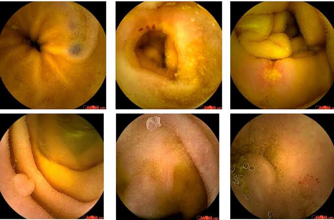
What Capsule Endoscopy Is Used For
Capsule endoscopy is used primarily when a clinical question specifically concerns the small bowel — the region of the GI tract that upper GI endoscopy and colonoscopy cannot adequately visualise. The three most important clinical indications are unexplained GI bleeding, suspected Crohn’s disease of the small bowel, and coeliac disease surveillance or investigation of non-responsive coeliac disease.
Obscure gastrointestinal bleeding: This is the most common indication for small bowel capsule endoscopy. “Obscure GI bleeding” refers to gastrointestinal bleeding that persists or recurs after a negative upper GI endoscopy and colonoscopy — investigations that together cover approximately ninety-five percent of GI bleeding sources, with the remaining five percent originating in the small bowel. Small bowel sources of bleeding include: angioectasias (vascular malformations, also called angiodysplasia — the most common cause of small bowel bleeding in adults over sixty); Dieulafoy lesions (large, aberrant arterioles erupting through the mucosa); small bowel tumours (gastrointestinal stromal tumours [GISTs], lymphomas, carcinoid tumours, adenocarcinomas); Meckel’s diverticulum (a congenital remnant of the omphalomesenteric duct found in approximately two percent of the population, containing ectopic gastric mucosa that can ulcerate and bleed); NSAIDs-associated small bowel ulceration and erosions; and Crohn’s disease ulceration. Capsule endoscopy has a diagnostic yield of fifty to seventy percent in obscure GI bleeding — significantly higher than alternative small bowel imaging modalities for mucosal lesions. The stool tests that may indicate GI bleeding are covered in the stool tests for digestive health article, and the abdominal imaging context is discussed in the abdominal ultrasound and CT scan articles.
Suspected Crohn’s disease: When conventional endoscopy (ileocolonoscopy) and cross-sectional imaging (CT or MRI enterography) have not confirmed small bowel Crohn’s disease despite clinical suspicion, capsule endoscopy provides direct mucosal assessment of the proximal and mid small bowel beyond the reach of the colonoscope. Capsule endoscopy detects small bowel Crohn’s lesions (aphthous ulcers, linear ulcers, cobblestoning, strictures) with high sensitivity — though in patients with known or suspected small bowel strictures, capsule endoscopy carries a risk of capsule retention (described below). A patency capsule or cross-sectional imaging to assess for structuring disease is performed before small bowel capsule endoscopy in patients at risk of obstruction.
Coeliac disease: Capsule endoscopy can visualise the classic endoscopic features of villous atrophy (scalloping of mucosal folds, reduction in fold height, mosaic pattern) across the entire small bowel and may identify additional complications of coeliac disease — refractory coeliac disease, ulcerative jejunitis, small bowel adenocarcinoma, and enteropathy-associated T-cell lymphoma (EATL). The serology and duodenal biopsy-based diagnosis of coeliac disease is covered in the coeliac disease testing article; capsule endoscopy plays a complementary role in monitoring and complication detection rather than primary diagnosis. Small bowel polyps and tumours: In patients with polyposis syndromes — Peutz-Jeghers syndrome, familial adenomatous polyposis (FAP), and MUTYH-associated polyposis — capsule endoscopy is used for surveillance of small bowel polyps, guiding when and where balloon-assisted enteroscopy or surgical intervention is required. Other indications include investigation of protein-losing enteropathy, suspected small bowel Behçet’s disease, and portal hypertensive enteropathy.
Preparing for Capsule Endoscopy
Preparation for capsule endoscopy combines fasting with bowel preparation, though the preparation requirements are less stringent than for colonoscopy because transit through the small bowel (rather than the colon) is the primary focus. Standard preparation protocols vary between centres, but typically include: fasting from midnight before the procedure; a low-residue or clear liquid diet for the twenty-four hours before swallowing the capsule; polyethylene glycol (PEG) or sodium phosphate-based bowel preparation to clear the small bowel of food residue and improve image quality; simethicone (an anti-foaming agent) to reduce the small bubbles in luminal fluid that can obscure mucosal detail. Some centres use pro-kinetic agents (metoclopramide, domperidone) to ensure complete small bowel transit within the battery life of the capsule, particularly in patients with known or suspected delayed gastric emptying.
Medications: iron supplements and medications known to slow small bowel motility should be discussed with the referring clinician. Patients with cardiac pacemakers or implantable cardiac devices were historically advised against capsule endoscopy due to potential radiofrequency interference, though this restriction has been relaxed for most modern devices following safety studies — device-specific review should be performed before proceeding. Patients with known swallowing difficulty should alert the endoscopy team as the capsule can be endoscopically delivered into the stomach or duodenum if oral swallowing is not reliable.
What Happens on the Day
On arrival, the sensor array (a set of sensors attached to the patient’s abdomen with adhesive pads, or incorporated into a recording belt) is attached and connected to the data recorder worn on the waist. The patient swallows the capsule with a small glass of water. No sedation is required. After swallowing, patients are typically asked to fast for a further two to four hours (to allow the capsule to clear the stomach and enter the small bowel without food interference), after which a light snack is permitted. Activity restriction during the procedure is minimal — patients can walk and carry out gentle daily activities but are advised to avoid strenuous exercise (which can displace the sensor array) and MRI scanning during the recording period. Patients return to the endoscopy unit after eight hours to return the recorder; some units allow the recorder to be returned by post.
The gastroenterologist or radiologist reviews the recording using dedicated software — this typically takes thirty to sixty minutes for an experienced reader. The software displays the images at variable speed (typically four to twenty frames per second), allows navigation through the recording, and in modern systems incorporates artificial intelligence (AI) assistance that flags potential lesions for review. AI-assisted reading of capsule endoscopy images has demonstrated improved adenoma detection rates and reduced reading time in clinical trials and is increasingly integrated into routine capsule endoscopy practice. The final report is usually available within one to two weeks of the procedure.
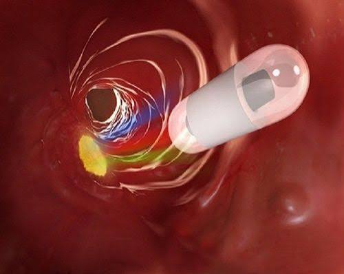
What Capsule Endoscopy Can and Cannot Show
Capsule endoscopy is highly sensitive for mucosal lesions — small ulcers, erosions, angioectasias, and polyps in the small bowel that cross-sectional imaging (CT or MRI) will miss. Its key limitation is that it cannot take biopsies, cannot perform therapeutic interventions, and provides only luminal mucosal images without information about the bowel wall thickness or extra-intestinal structures. When capsule endoscopy identifies a lesion requiring biopsy or treatment, balloon-assisted enteroscopy (single or double-balloon enteroscopy, or spiral enteroscopy) is used — a technique that allows deep intubation of the small bowel using an overtube system, enabling biopsy forceps and therapeutic instruments to reach the target lesion under direct vision.
The capsule endoscopy report characterises the location of findings by the percentage of small bowel transit time rather than by anatomical distance (since actual small bowel length varies between patients and cannot be reliably measured during transit). A lesion at twenty-five percent transit time is in the proximal small bowel (duodenojejunal junction area); at fifty percent, mid-jejunum; at seventy-five percent, mid-to-distal ileum. This transit time percentage is used by the enteroscopist to plan the depth of insertion required when balloon-assisted enteroscopy is subsequently performed — deciding whether an oral (antegrade) or anal (retrograde via ileocaecal valve) approach will more efficiently reach the lesion.
Capsule Retention: The Key Risk
The principal risk of capsule endoscopy is capsule retention — the capsule becoming lodged at a small bowel stricture (narrowing) and failing to pass. Capsule retention occurs in approximately one to two percent of unselected small bowel capsule endoscopy procedures. In patients with known or suspected small bowel strictures — Crohn’s disease with fibrotic stricturing, NSAID enteropathy, radiation enteritis, ischaemic strictures — the risk is significantly higher (up to thirteen percent in some series). A retained capsule typically causes no immediate symptoms but can ultimately cause small bowel obstruction requiring surgical or enteroscopic retrieval.
To reduce the risk of capsule retention in patients with potential small bowel strictures, a patency capsule is used first: this is a dissolvable dummy capsule of the same dimensions as the endoscopy capsule that dissolves within thirty to sixty hours if retained in the bowel, avoiding the complication of mechanical obstruction. Patients swallow the patency capsule; if it passes normally (confirmed by plain X-ray or handheld scanner at forty-eight hours), the real capsule endoscopy can proceed safely. If the patency capsule is retained, small bowel stricturing is confirmed — balloon-assisted enteroscopy or surgical investigation becomes the appropriate next step rather than capsule endoscopy. In patients without risk factors for stricturing disease, routine patency capsule testing before capsule endoscopy is not required.
Capsule Endoscopy vs Other Small Bowel Investigations
The choice of small bowel investigation depends on the clinical question, the likelihood of therapeutic intervention being required, the patient’s comorbidities, and local availability. CT enterography (CT with oral contrast and enterography technique) and MR enterography (preferred over CT for younger patients and those requiring repeated monitoring due to the absence of radiation) provide cross-sectional imaging of the bowel wall and mesentery — superior to capsule endoscopy for identifying transmural disease, strictures, fistulae, abscesses, and extra-intestinal complications of Crohn’s disease, but inferior to capsule endoscopy for mucosal lesions. For patients with suspected Crohn’s disease, MR enterography and capsule endoscopy are often performed as complementary investigations, with MR providing transmural and structural assessment while capsule endoscopy provides mucosal detail. The MRI article in this series covers the principles of MRI in gastrointestinal assessment in detail. Push enteroscopy — a longer version of a standard gastroscope advanced beyond the duodenojejunal junction — visualises only the proximal one to two metres of the small bowel but enables biopsy and therapy and may be sufficient for lesions localised to the proximal jejunum. Balloon-assisted enteroscopy can reach the majority of the small bowel via oral or anal approach and provides full therapeutic capability — biopsy, haemostasis, polypectomy, dilation — but is a more prolonged procedure requiring sedation and a longer recovery period than capsule endoscopy.
Frequently Asked Questions About Capsule Endoscopy
Will I feel the capsule as it passes through?
No — the capsule is very small and smooth, and the vast majority of patients do not feel it at any point during transit. The gastrointestinal tract has no tactile sensation for a smooth intraluminal object of this size. Patients occasionally notice mild bloating or wind during the recording period, but this is related to the bowel preparation rather than the capsule itself. Some patients experience mild nausea when swallowing the capsule, though this is uncommon. The capsule passes in the stool, typically within twenty-four to seventy-two hours — most patients do not notice it in the toilet. If the capsule has not been confirmed as passed within two weeks, an abdominal X-ray may be taken to confirm its passage, though in practice this is rarely required in patients without symptoms of obstruction.
I’ve been told I need a capsule endoscopy for recurrent anaemia — what findings are they looking for?
Iron-deficiency anaemia that recurs after upper GI endoscopy and colonoscopy have excluded the most common sources (gastric ulcer, coeliac disease, colonic cancer, haemorrhoids) points toward a small bowel source as the explanation. The most common small bowel findings in this context are angioectasias — small, spider-like vascular malformations in the mucosa that bleed intermittently and are below the threshold of detection for CT or MRI in most cases. Capsule endoscopy detects angioectasias with high sensitivity and characterises their distribution and number, informing decisions about treatment with balloon-assisted enteroscopy (argon plasma coagulation of angioectasias) or medical management. Other findings in this context include small bowel ulceration from NSAID use, Dieulafoy lesions, and, in younger patients, a Meckel’s diverticulum. The liver function tests and full blood count context for anaemia investigation — including the serum ferritin, B12, folate, and coeliac serology that accompany a typical investigation — are covered across the relevant articles in this series. For patients with a confirmed finding on capsule endoscopy that requires treatment, balloon-assisted enteroscopy provides the therapeutic access that capsule endoscopy cannot, completing the diagnostic-to-therapeutic pathway initiated by the capsule study.
Sources: BSG — Capsule Endoscopy Guidelines · ASGE — Capsule Endoscopy Technology Review · NIDDK — Capsule Endoscopy
Oesophageal and Colon Capsule Endoscopy
Although small bowel capsule endoscopy is the dominant clinical application, capsule technology has been extended to other regions of the GI tract. Oesophageal capsule endoscopy (PillCam ESO) uses a bi-directional camera capsule that captures images at a higher frame rate (eighteen frames per second) during oesophageal transit. It has been studied as an alternative to conventional upper GI endoscopy for the screening of Barrett’s oesophagus in primary care, offering a less invasive and potentially more acceptable screening pathway for patients who are averse to conventional endoscopy with sedation. Studies have demonstrated adequate sensitivity for identifying long-segment Barrett’s, though conventional endoscopy with biopsy remains the gold standard for Barrett’s confirmation and dysplasia grading. Colon capsule endoscopy (PillCam COLON) uses a bi-directional wide-angle capsule optimised for colonic transit and mucosal visualisation. After the same bowel preparation as conventional colonoscopy, the capsule is swallowed and images the entire colon during transit. Colon capsule endoscopy has demonstrated sensitivity for polyp detection comparable to CT colonography for lesions above 6 mm, and superior to CT colonography for small lesions. It is used primarily as an alternative for patients who are unable or unwilling to undergo conventional colonoscopy — for example, patients with anticoagulant requirements that make polypectomy unsafe, patients who declined colonoscopy after a positive FIT test, and incomplete colonoscopies requiring colonic assessment. Like small bowel capsule endoscopy, it cannot take biopsies or perform polypectomy — positive findings require conventional colonoscopy for management.
After Capsule Endoscopy: What Happens Next
After the recording session, patients detach the sensor array and return it to the endoscopy unit. They can eat and drink normally from approximately four hours after swallowing the capsule (or when the small bowel phase is likely complete). Activity is unrestricted after returning the recorder. The endoscopist’s review of the images — supported by AI detection algorithms in modern systems — produces a report characterising any findings by location (percentage of transit time), appearance, and clinical significance. If a significant lesion is identified — an angioectasia causing ongoing bleeding, a suspicious polyp, a deep ulcer, or a stricture — the managing gastroenterologist will discuss the findings and plan the next investigative or therapeutic step. For patients with negative capsule endoscopy (no significant findings) following obscure GI bleeding, the negative result has important clinical value: it reduces the likelihood of a significant structural small bowel pathology and directs investigation toward non-structural causes (haematological disorders, coagulopathies, drug effects) or intermittent vascular lesions too small to have been detected during the recording window. A negative capsule endoscopy does not definitively exclude all small bowel pathology — small lesions can be missed, particularly in areas of rapid transit or poor bowel preparation — and should be interpreted in the context of the overall clinical picture. Patients whose symptoms persist after a negative capsule study may require repeat capsule endoscopy, device-assisted enteroscopy, or consultation at a tertiary small bowel centre. The full digestive investigation framework — integrating blood tests, stool tests, conventional endoscopy, capsule endoscopy, and cross-sectional imaging — is covered in the article on CT for digestive and liver problems and the series overview on how doctors diagnose digestive and liver problems, providing patients with the integrated picture of how each investigation contributes to the diagnostic pathway.
Patients with Crohn’s disease of the small bowel who are undergoing capsule endoscopy as part of their initial assessment or disease monitoring should be aware that the Lewis score and the Capsule Endoscopy Crohn’s Disease Activity Index (CECDAI) — validated scoring systems applied during image review — allow the endoscopist to grade disease activity quantitatively and comparably across serial studies. This structured scoring provides a more reproducible and objective assessment of small bowel mucosal inflammation than a purely descriptive report, and is increasingly used in clinical trials and specialist Crohn’s management programmes to assess response to biological therapies (anti-TNF agents, vedolizumab, ustekinumab). For patients receiving treatment for small bowel Crohn’s, serial capsule endoscopy demonstrating mucosal healing — the absence of deep ulceration and a fall in Lewis or CECDAI score — represents the endoscopic equivalent of radiological response on MR enterography and provides a direct mucosal endpoint for treatment optimisation decisions. The integration of capsule endoscopy findings with blood test markers (CRP, faecal calprotectin), imaging findings (MR enterography), and clinical symptoms builds the complete picture used to manage complex small bowel Crohn’s disease — a context in which the abdominal ultrasound, CT, and MRI articles in this series together with capsule endoscopy provide complementary perspectives on the same disease process.
As capsule technology continues to advance — with AI-assisted lesion detection, magnetically guided capsules allowing active navigation (the NaviCam system), and multi-spectral imaging under investigation — the clinical scope of capsule endoscopy will expand beyond its current primary indications. For patients today, the key is understanding how capsule endoscopy fits within the existing investigative framework: an excellent non-invasive first-line small bowel mucosal investigation for appropriate clinical questions, complemented by conventional endoscopy, balloon-assisted enteroscopy, and cross-sectional imaging to complete the diagnostic and therapeutic picture. The full context of how digestive investigations are selected and sequenced is covered in the overview article on how doctors diagnose digestive and liver problems, available in this series alongside the dedicated colonoscopy and upper GI endoscopy articles.
I was referred for capsule endoscopy after two negative colonoscopies and an upper GI endoscopy — all trying to find the source of my recurring iron deficiency anaemia. The procedure was far simpler than I expected. The capsule section on angioectasias explains exactly what they found on mine — two in the mid-small bowel. I’ve since had balloon enteroscopy to treat them. This article would have been invaluable to read beforehand.
Thank you Liam — your experience is a very typical capsule endoscopy success story: conventional endoscopy exhausted, capsule endoscopy identifying the culprit, balloon enteroscopy providing targeted treatment at the same anatomical site identified on the capsule. Angioectasias are notoriously difficult to detect on cross-sectional imaging and represent the majority of obscure small bowel bleeding cases — which is exactly why capsule endoscopy occupies such an important position in the investigation pathway when upper and lower endoscopy are negative. Glad to hear the treatment has been completed, and thank you for sharing your experience to help others in a similar situation.
Really informative — I wasn’t aware of the patency capsule concept at all before reading this. My gastroenterologist mentioned it when I was referred for small bowel Crohn’s investigation, but I didn’t understand why I needed to swallow a dissolving capsule first. The explanation of capsule retention risk and why the patency test is necessary for patients with suspected stricturing makes complete sense now.