
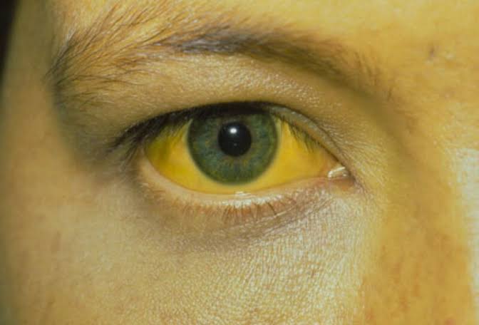
Yellowing of the skin or the whites of the eyes — medically called jaundice — is one of the most recognizable signs of liver and biliary disease. The yellow pigmentation comes from bilirubin, a breakdown product of hemoglobin that the liver normally processes and excretes. When bilirubin accumulates in the bloodstream above a threshold of approximately two to three milligrams per deciliter, it deposits in tissues and becomes visible, first in the sclera (whites of the eyes) — which are particularly sensitive to bilirubin accumulation — and then in the skin, mucous membranes, and, in severe cases, body fluids including urine.
Jaundice is not a diagnosis in itself but a physical sign pointing toward an underlying condition. The list of conditions that cause jaundice is broad — spanning liver diseases from viral hepatitis to cirrhosis, bile duct obstruction from gallstones to cancer, hemolytic anemias that overwhelm the liver’s bilirubin processing capacity, and benign metabolic variants that require no treatment. The clinical significance of jaundice depends almost entirely on its underlying cause, the speed of its development, and the associated symptoms that accompany it. Understanding what jaundice means — and which presentations require urgent evaluation — is essential for both patients who notice it in themselves and clinicians who encounter it in practice.
How Bilirubin Metabolism Produces or Prevents Jaundice
Bilirubin metabolism is a tightly regulated process that begins with the destruction of aging red blood cells and ends with bilirubin excretion in bile and urine. As red blood cells complete their approximately 120-day lifespan, the spleen and liver remove them from circulation and break down the hemoglobin they contain. The heme portion of hemoglobin is converted through a series of enzymatic steps to biliverdin and then to unconjugated (indirect) bilirubin, which is poorly water-soluble and circulates in the blood bound to albumin. The liver takes up this albumin-bound bilirubin, conjugates it with glucuronic acid to produce water-soluble conjugated (direct) bilirubin, and excretes it into bile through the bile ducts.
In the intestine, conjugated bilirubin is converted by bacteria to urobilinogen and stercobilin — the compound that gives stool its characteristic brown color. A small fraction of urobilinogen is reabsorbed and excreted in the urine, giving it its pale yellow color. When any step in this pathway is disrupted — excessive bilirubin production from hemolysis, impaired hepatocyte uptake or conjugation, blocked bile excretion — bilirubin accumulates in the blood. The distinction between unconjugated and conjugated hyperbilirubinemia is clinically important because it localizes the defect: elevated unconjugated bilirubin points toward hemolysis, impaired uptake, or conjugation defects; elevated conjugated (direct) bilirubin points toward hepatocellular disease or biliary obstruction.
The sclera of the eye becomes yellow before the skin in most patients because the scleral connective tissue has a high affinity for bilirubin. Icterus (yellow sclera) is detectable at serum bilirubin levels of approximately 2–3 mg/dL, while visible skin jaundice typically requires levels above 3–4 mg/dL. In patients with dark skin tones, scleral icterus is often the most reliable early sign of jaundice, as subtle skin yellowing may be difficult to detect visually. The best place to look for early jaundice in any patient is the conjunctiva of the lower eyelid in natural light — a pale yellow or golden tint there, even subtle, is a reliable indicator of elevated bilirubin.
Prehepatic Jaundice — Hemolysis and Ineffective Erythropoiesis
Prehepatic jaundice occurs when the rate of bilirubin production exceeds the liver’s processing capacity — not because the liver is diseased, but because so many red blood cells are being destroyed that even a normally functioning liver cannot conjugate and excrete all the resulting bilirubin. Hemolytic anemias are the paradigm: autoimmune hemolytic anemia, hereditary spherocytosis, sickle cell disease, G6PD deficiency, and malaria all destroy red blood cells at accelerated rates, flooding the liver with unconjugated bilirubin. The resulting jaundice is usually mild to moderate — serum bilirubin rarely exceeds 5–6 mg/dL in pure hemolysis because the liver can upregulate its processing capacity to some degree — and is accompanied by anemia and reticulocytosis (elevated immature red blood cell count reflecting increased bone marrow production).
Gilbert’s syndrome deserves special mention as a common cause of mild unconjugated hyperbilirubinemia that is entirely benign. Affecting approximately five to ten percent of the population, Gilbert’s syndrome results from a genetic variant in the UGT1A1 gene that reduces the efficiency of bilirubin conjugation in hepatocytes. Bilirubin levels are mildly elevated at baseline (typically 1–3 mg/dL) and rise further during fasting, illness, stress, or strenuous exercise. The jaundice is usually subtle — most often noticed as mild scleral icterus during a period of illness or fasting — and requires no treatment. The diagnosis is made by demonstrating isolated unconjugated hyperbilirubinemia with otherwise normal liver tests and normal complete blood count (excluding hemolysis). Patients with Gilbert’s syndrome benefit from reassurance that their intermittent jaundice is not a sign of liver disease and needs no further investigation once the diagnosis is established.
Hepatocellular Jaundice — Liver Disease as the Cause
Hepatocellular jaundice results from damage to hepatocytes — the liver cells responsible for bilirubin uptake, conjugation, and excretion — that impairs the liver’s ability to process bilirubin normally. Viral hepatitis is among the most common causes worldwide. Hepatitis A causes an acute, self-limiting jaundice that typically resolves within four to six weeks as the infection clears. Hepatitis B and E can produce acute icteric hepatitis, and hepatitis E is particularly important in pregnant women, in whom it can cause fulminant hepatic failure. Hepatitis C rarely causes acute jaundice at initial infection but produces chronic hepatitis that may eventually progress to cirrhosis with secondary jaundice from impaired hepatic function.
Alcoholic hepatitis — an acute inflammatory condition triggered by heavy alcohol intake — causes jaundice through direct hepatocyte damage and cholestasis. Severe alcoholic hepatitis is a medical emergency with a short-term mortality risk of 20–30%, and the combination of severe jaundice (bilirubin above 5 mg/dL), coagulopathy, and encephalopathy is a particularly ominous pattern. Drug-induced liver injury (DILI) from medications, herbal supplements, or dietary supplements can produce both hepatocellular jaundice (from hepatocyte necrosis) and cholestatic jaundice (from bile duct injury) depending on the offending agent. Acetaminophen toxicity is the leading cause of acute liver failure in most Western countries and produces rapid-onset jaundice alongside coagulopathy and encephalopathy that can progress to death within days without liver transplantation.
In cirrhosis, jaundice develops as decompensation progresses and the residual functional liver mass becomes insufficient to process the daily bilirubin load. The development of jaundice in a patient with previously compensated cirrhosis is a significant clinical event — it marks the transition to decompensated cirrhosis and substantially worsens prognosis. Jaundice in decompensated cirrhosis is often accompanied by ascites, hepatic encephalopathy, and coagulopathy as part of the syndrome of acute-on-chronic liver failure. Associated symptoms of liver disease including persistent fatigue and pale stool frequently accompany jaundice in advanced liver disease.
Obstructive Jaundice — Bile Duct Blockage
Obstructive (posthepatic) jaundice results from mechanical blockage of bile flow from the liver to the duodenum. The most common cause is choledocholithiasis — gallstones in the common bile duct — which may cause acute-onset jaundice with right upper quadrant pain and fever (Charcot’s triad of biliary obstruction). Gallstone-related obstructive jaundice is typically reversible with stone removal, and ERCP is highly effective at clearing common bile duct stones in the majority of patients. However, when a stone causes complete obstruction alongside infection (cholangitis), the condition becomes a medical emergency requiring antibiotics and urgent biliary decompression within hours to prevent sepsis and death.
Malignant biliary obstruction — from pancreatic cancer, cholangiocarcinoma, or ampullary cancer — typically presents as painless, progressive jaundice in an older adult. The absence of pain is a classically important feature that distinguishes malignant from gallstone obstruction, though this distinction is not absolute. Progressive jaundice with weight loss, loss of appetite, and pale stool in a patient over fifty without fever or acute pain should be treated as a potential malignancy until imaging proves otherwise. Biliary stenting via ERCP or percutaneous transhepatic cholangiography (PTC) can palliate malignant obstruction and relieve jaundice even when curative resection is not possible, substantially improving quality of life and enabling patients to tolerate systemic therapy.

When Yellow Skin or Eyes Requires Emergency Evaluation
Not all jaundice is an emergency, but some presentations are. Seek emergency evaluation immediately for jaundice accompanied by: high fever with chills and right upper quadrant pain (Charcot’s triad — possible ascending cholangitis requiring urgent biliary drainage); altered mental status or confusion (possible acute liver failure or hepatic encephalopathy); severe vomiting with inability to maintain hydration; signs of bleeding (hematemesis, black stool, or unusual bruising suggesting coagulopathy from liver failure); or rapid development of jaundice over hours to days in the context of medication changes, alcohol binge, or possible toxin exposure.
Prompt evaluation within days is appropriate for: new-onset jaundice in an otherwise stable adult; jaundice with dark urine and pale stool suggesting cholestasis; jaundice with progressive weight loss; a newborn with jaundice persisting beyond two weeks; or known liver disease with a new episode of jaundice. A serum bilirubin level with fractionation into conjugated and unconjugated components, along with liver function tests, provides the first layer of diagnostic information. Abdominal ultrasound is the initial imaging choice for evaluating biliary dilation. MRCP follows when bile duct pathology is suspected but not confirmed by ultrasound.
Frequently Asked Questions About Yellow Skin or Eyes
Can eating too many carrots cause yellow skin?
Yes. Carotenemia — yellow-orange discoloration from excessive dietary intake of carotenoids (found in carrots, sweet potatoes, squash, and supplements) — can cause skin yellowing that is sometimes confused with jaundice. The key distinction is that carotenemia does not affect the sclera (whites of the eyes) — the eyes remain white — while true jaundice yellows both the skin and sclera. Serum bilirubin is normal in carotenemia. The discoloration resolves over weeks after reducing carotenoid intake and requires no treatment.
Is jaundice in newborns the same as jaundice in adults?
Neonatal jaundice is distinct from adult-onset jaundice in both mechanism and significance. Most newborns develop mild physiological jaundice in the first days of life because the neonatal liver’s bilirubin conjugation capacity lags behind the rate of red blood cell breakdown during the transition from fetal to adult hemoglobin. Physiological neonatal jaundice peaks at days three to five, then resolves spontaneously. Pathological neonatal jaundice — from ABO or Rh incompatibility, G6PD deficiency, biliary atresia, or serious infection — is more severe, appears earlier, or persists longer and requires specific evaluation and treatment. Severe unconjugated hyperbilirubinemia in newborns, if untreated, can cause kernicterus — bilirubin deposition in the brain causing permanent neurological damage.
How quickly does jaundice develop?
The speed of onset provides diagnostic clues. Rapid development over hours to days is typical of acute hepatitis (viral, alcoholic, drug-induced), acute biliary obstruction from a gallstone, or acute hemolysis. Gradual development over weeks to months is more consistent with progressive hepatocellular disease (chronic hepatitis progressing to cirrhosis) or slowly growing malignant biliary obstruction. Gilbert’s syndrome produces fluctuating jaundice that waxes and wanes with physiological stressors over years without progressive worsening.
Can jaundice resolve without treatment?
It depends entirely on the cause. Jaundice from hepatitis A or mild alcoholic hepatitis typically resolves as the liver heals — the timeline ranges from weeks (hepatitis A) to months (alcoholic hepatitis if alcohol is stopped). Gilbert’s syndrome jaundice resolves when the triggering stressor (illness, fasting) resolves. Jaundice from bile duct obstruction by a gallstone requires stone removal to resolve. Malignant obstructive jaundice requires biliary drainage (stenting) or tumor resection to improve. Jaundice in advanced cirrhosis reflects irreversible hepatocellular loss and does not resolve without transplantation. The cause determines both the trajectory and the appropriate response.
What is the difference between hepatitis A, B, and C in terms of jaundice?
Hepatitis A is an acute, self-limiting infection transmitted by the fecal-oral route that commonly causes icteric hepatitis with jaundice, dark urine, and pale stool; it resolves completely in the vast majority of cases. Hepatitis B can cause acute icteric hepatitis but more commonly causes chronic hepatitis that progresses silently to cirrhosis over decades, with jaundice appearing only in advanced disease or acute reactivation. Hepatitis C rarely causes jaundice at initial infection and is most commonly identified incidentally on blood testing; chronic HCV progresses slowly to cirrhosis over fifteen to thirty years in many untreated patients. Understanding which type of hepatitis is causing symptoms has critical implications for treatment and transmission prevention, making viral serology testing essential in any patient with unexplained jaundice.
Sources: NIDDK — Viral Hepatitis · ACG — Jaundice · Mayo Clinic — Jaundice
Primary Biliary Cholangitis, Primary Sclerosing Cholangitis, and Intrahepatic Cholestasis
Primary biliary cholangitis (PBC) and primary sclerosing cholangitis (PSC) are autoimmune conditions that destroy the intrahepatic and extrahepatic bile ducts respectively, producing a syndrome of progressive cholestasis — impaired bile excretion — that causes jaundice, pruritus (itching), profound fatigue, and eventually biliary cirrhosis. PBC affects predominantly middle-aged women and is characterized by the presence of anti-mitochondrial antibodies (AMA), which are highly specific for the condition and detectable decades before clinical disease develops in some patients. Jaundice in PBC typically appears in the later stages of disease progression, after years of asymptomatic cholestasis; the earliest abnormality is usually an isolated elevation of alkaline phosphatase (ALP) with a disproportionately small rise in transaminases. Ursodeoxycholic acid (UDCA) is the first-line treatment for PBC and slows disease progression in most patients; obeticholic acid is used for those with inadequate UDCA response.
PSC is more common in men and is strongly associated with inflammatory bowel disease — particularly ulcerative colitis — in approximately seventy percent of cases. It is characterized by multifocal stricturing and dilation of the bile ducts producing a “beaded string” appearance on MRCP. Unlike PBC, PSC lacks an effective medical therapy that alters disease progression; management focuses on endoscopic treatment of dominant strictures, surveillance for cholangiocarcinoma (which complicates PSC in ten to fifteen percent of patients over a lifetime), and consideration of liver transplantation for end-stage disease. Jaundice in PSC may reflect progressive biliary obstruction, cholangitis from a dominant stricture, or the development of cholangiocarcinoma — any acceleration of jaundice in a PSC patient warrants urgent evaluation.
Intrahepatic cholestasis of pregnancy (ICP) is a condition specific to the second and third trimester in which elevated serum bile acids produce pruritus and jaundice from impaired bile secretion. It resolves promptly after delivery but carries significant risks for the fetus — including preterm birth, meconium-stained amniotic fluid, and stillbirth — making fetal surveillance and, in severe cases, early delivery important management considerations. Total bile acid levels above forty micromoles per liter are associated with substantially increased fetal risk. Ursodeoxycholic acid is the standard treatment for symptom relief in ICP and may reduce fetal risk, though evidence for the latter remains incomplete.
Autoimmune Hepatitis and Drug-Induced Liver Injury as Causes of Jaundice
Autoimmune hepatitis (AIH) is a chronic inflammatory liver disease in which the immune system attacks hepatocytes, producing an aminotransferase-dominant pattern with elevations of ALT and AST often in the hundreds to thousands. Jaundice in AIH reflects active hepatocellular injury and is most common at presentation of a previously undiagnosed case or during disease flares in known patients. The presence of autoantibodies — anti-nuclear antibody (ANA), anti-smooth muscle antibody (SMA), or anti-liver-kidney microsomal antibody (LKM-1) — combined with elevated IgG and compatible histology on liver biopsy confirms the diagnosis. Corticosteroids and azathioprine produce remission in the majority of patients within weeks to months, and jaundice typically resolves as liver inflammation subsides. AIH should be excluded in any patient with unexplained hepatocellular jaundice, particularly women of any age with a personal or family history of autoimmune disease.
Drug-induced liver injury (DILI) is among the most common causes of new-onset jaundice in hospitalized patients and a frequently underrecognized cause in outpatient practice. The spectrum of DILI-related jaundice spans a hepatocellular pattern (elevated transaminases with secondary bilirubin rise, as seen with isoniazid, nitrofurantoin, or statins), a cholestatic pattern (elevated ALP and bilirubin with modest transaminase elevation, as seen with amoxicillin-clavulanate, sulfonamides, and some antipsychotics), and a mixed pattern. Hy’s Law — a biochemical criterion combining significant hepatocellular injury (ALT greater than three times the upper limit of normal) with jaundice (bilirubin above two times the upper limit of normal) in the absence of biliary obstruction — identifies patients at high risk of fatal DILI and triggers urgent medication discontinuation and monitoring. The LiverTox database maintained by the National Institutes of Health is a comprehensive reference for evaluating causality between a specific medication and liver injury; clinicians and patients suspecting DILI should consult it and report suspected cases to the FDA MedWatch program. Cessation of the offending agent is the cornerstone of treatment; most cases of DILI-related jaundice resolve within weeks to months after the causative drug is stopped.
The broader clinical picture of jaundice must always include an assessment of the patient’s overall trajectory — not just the current bilirubin level. Isolated, stable mild hyperbilirubinemia in a young, otherwise well adult with a consistent history (Gilbert’s syndrome), or a single episode of transient jaundice with a clear precipitant (a viral illness, a brief alcohol excess, a recently started medication) carries a very different prognosis than progressive jaundice accumulating over weeks in an older patient without a clear precipitant. The pattern of associated liver test abnormalities — whether transaminases, alkaline phosphatase, or total bilirubin is disproportionately elevated — provides the single most important diagnostic clue to the type of liver injury before imaging results return. An ALT elevation ten to fifty times the upper limit of normal with only modestly elevated ALP points toward hepatocellular disease; an ALP elevation three to five times normal with modest transaminase elevation points toward cholestatic or biliary disease. Combining this pattern recognition with age, sex, alcohol history, medication review, and the presence or absence of systemic symptoms such as dark urine allows a confident differential diagnosis to be formed before a single imaging study is obtained. This structured approach reduces unnecessary testing, speeds time to diagnosis, and ensures that the small but important subset of patients with jaundice who need urgent intervention — such as ascending cholangitis requiring same-day ERCP, or acetaminophen-induced acute liver failure requiring N-acetylcysteine and transplant evaluation — are identified without delay.
For patients who are diagnosed with Gilbert’s syndrome, it is worth emphasizing that the condition is entirely benign and has no implications for long-term liver health. The gene variant responsible is extremely common in the general population, and the intermittent mild scleral icterus it produces during fasting or illness does not indicate liver damage, does not progress to liver disease, and does not require any dietary restriction beyond avoiding prolonged fasting. The main practical consideration for patients with Gilbert’s syndrome is medication dosing: several drugs metabolized by the UGT1A1 enzyme — including irinotecan, used in colorectal cancer chemotherapy — have reduced clearance in patients with Gilbert’s syndrome, and oncologists prescribing these agents should be made aware of the diagnosis to allow dose adjustment and closer toxicity monitoring. For most people, however, Gilbert’s syndrome is a finding to file away rather than worry about, and reassurance from a clinician who understands the mechanism is typically all that is needed.