

Of all the complications that develop in adults with diabetes, diabetic neuropathy symptoms are among the earliest to appear and the most directly linked to the life-altering consequences of long-standing nerve damage. Diabetic neuropathy symptoms — the burning, tingling, numbness, and pain that affect the feet, legs, and hands in distal symmetric polyneuropathy — often begin years before a person is diagnosed with Type 2 diabetes, reflecting the widespread nerve damage that has been accumulating during the years of undiagnosed elevated blood glucose. By the time a diagnosis of diabetes is confirmed, an estimated 10–20% of adults already have measurable neuropathy. Over a lifetime with diabetes, the cumulative prevalence approaches 50% — making neuropathy the most common of all diabetes complications and one that every adult with diabetes needs to understand, monitor for, and actively work to prevent or slow.
According to the National Institute of Diabetes and Digestive and Kidney Diseases (NIDDK), approximately 60–70% of adults with diabetes have some form of neuropathy — though mild cases are often undiagnosed. Distal symmetric peripheral neuropathy is the most common form, affecting approximately 50% of adults with long-standing Type 2 diabetes. Lower extremity amputation rates in adults with diabetes are 10 times higher than in the general population, and the vast majority of these amputations are preceded by a diabetic foot ulcer — which itself results from the loss of protective sensation caused by peripheral neuropathy. Painful diabetic neuropathy — the burning, electric shock, and hypersensitivity form — affects 15–25% of adults with diabetic peripheral neuropathy, significantly impairing sleep, mood, and quality of life.
What Causes Diabetic Neuropathy: The Mechanisms of Nerve Damage
Diabetic nerve damage is not a single uniform process — it results from multiple simultaneous mechanisms acting on different components of the peripheral nervous system, which is why neuropathy affects multiple nerve types (sensory, motor, and autonomic) in overlapping patterns and why its prevention requires addressing the root causes of hyperglycemia-driven vascular and metabolic damage rather than any single pathway:
- Endoneurial ischemia from microvascular disease: Peripheral nerves depend on a rich network of tiny vessels — the vasa nervorum — for their oxygen and nutrient supply. Hyperglycemia damages these microvasculature vessels through the same mechanisms that cause diabetic retinopathy and nephropathy: endothelial dysfunction, basement membrane thickening, pericyte loss, and reduced blood flow. The resulting “endoneurial ischemia” — oxygen deprivation of the nerve fibers themselves — is particularly damaging to the longest nerve fibers (those serving the feet) because they have the greatest dependence on intact microvascular supply along their entire length. This explains the “stocking-and-glove” distribution of peripheral neuropathy: the longest nerves fail first, beginning at the tips of the toes and ascending symmetrically upward. Studies using nerve biopsy have confirmed intraepidermal nerve fiber density reduction (a direct measure of small fiber loss) that correlates with duration and severity of hyperglycemia and with A1C levels.
- Oxidative stress and mitochondrial dysfunction in nerve axons: Hyperglycemia dramatically increases mitochondrial reactive oxygen species (ROS) production in peripheral nerve cells — specifically in Schwann cells (which produce the myelin that insulates nerve axons) and in the axons themselves. ROS damage mitochondrial DNA and membranes within nerve axons, impairing the axon’s ability to produce the ATP needed for ion pump function and signal conduction. In sensory neurons, mitochondrial dysfunction reduces the threshold for spontaneous firing — contributing to the painful symptoms of burning and electric shock that characterize painful diabetic neuropathy — while simultaneously impairing the ion channel function needed for normal tactile and proprioceptive signal transmission. The UKPDS demonstrated that each 1% reduction in A1C reduces the risk of developing neuropathy (measured by vibration perception threshold) by approximately 25%, directly confirming that glucose-driven oxidative stress is a primary driver.
- Polyol pathway activation and sorbitol accumulation in Schwann cells: In Schwann cells — the cells that produce the myelin sheath around peripheral nerve axons — chronic hyperglycemia activates the polyol pathway: excess glucose is converted to sorbitol (by aldose reductase) and then to fructose (by sorbitol dehydrogenase). Sorbitol accumulation causes osmotic stress that impairs Schwann cell function, while the NADPH consumed by this pathway reduces the cell’s capacity to regenerate glutathione (its primary antioxidant). Myoinositol depletion — another consequence of polyol pathway activation — reduces the Schwann cell’s ability to maintain normal sodium-potassium ATPase activity, which is essential for axon repolarization and conduction. This mechanism underlies the slowed nerve conduction velocities that can be detected on electromyography (EMG) even in early, asymptomatic neuropathy.
- Advanced glycation end-products (AGEs) and axon structural damage: Prolonged hyperglycemia leads to the glycation of nerve structural proteins — including tubulins, neurofilaments, and myelin basic protein — forming advanced glycation end-products that cross-link with and structurally deform the normal axon cytoskeleton. AGE-modified myelin is recognized as foreign by macrophages and triggers an inflammatory response that further damages the myelin sheath. The RAGE (receptor for AGEs) on endothelial cells of the vasa nervorum activates NF-κB signaling, promoting further inflammatory damage to the nerve’s blood supply. This AGE-mediated structural damage accumulates over years and is not rapidly reversed when glucose is improved — explaining why neuropathy, once established, progresses even with improved glycemic control and why the “legacy effect” of early good glucose control (from the DCCT/EDIC study) so powerfully reduces lifetime neuropathy risk. Our A1C testing schedule guide covers the monitoring frequency and targets that directly affect neuropathy risk.
Diabetic Neuropathy Symptoms: A Complete Guide by Type
Not all diabetic neuropathy symptoms look the same — they vary significantly depending on which nerve types are predominantly affected (sensory, motor, or autonomic) and which regions of the body are involved. Understanding the different presentations helps adults with diabetes and their healthcare providers identify neuropathy at the earliest detectable stage, before irreversible damage has accumulated:
- Distal symmetric peripheral neuropathy — sensory symptoms (most common): The sensory symptoms of DSPN begin insidiously in the toes and the balls of the feet and ascend symmetrically over months and years. Positive sensory symptoms — those that the person actively feels — include burning pain (often worse at night, when there is no competing sensory input from walking), electric shock sensations, stabbing or lancinating pain, deep aching, and allodynia (pain from normally non-painful stimuli, such as light bedsheet contact with the feet). Negative sensory symptoms — those representing loss of sensation — include numbness, tingling without pain, reduced ability to feel sharp objects, impaired temperature discrimination (inability to feel a hot or cold surface), and reduced vibration sense (detectable with a 128 Hz tuning fork pressed against the bony prominences of the foot). The most clinically dangerous symptom is the complete loss of protective sensation — the inability to feel a 10g monofilament pressed against the plantar foot surface — because this is the threshold below which foot injuries accumulate without causing pain. Adults can walk on a developing foot ulcer, a nail through a shoe, or a severely infected wound for days without discomfort.
- Motor neuropathy symptoms — muscle weakness and deformity: Motor fiber involvement in diabetic peripheral neuropathy is less prominent than sensory symptoms but clinically important because it contributes to the foot deformities that increase ulceration risk. Loss of intrinsic foot muscle function — the small muscles that flex and extend the toes — leads to imbalanced pull by the long flexors and extensors, producing characteristic toe deformities (hammer toes, claw toes) and abnormal weight distribution across the plantar foot. These deformities create high-pressure areas over bony prominences — particularly the metatarsal heads and the tips of deformed toes — where calluses form and ulcers develop. Proximal motor neuropathy (diabetic amyotrophy) presents differently: sudden-onset severe asymmetric proximal leg pain, weakness in hip flexors and knee extensors, and profound muscle wasting of the thigh — often misdiagnosed as a lumbar herniated disc before the characteristic bilateral but asymmetric involvement pattern and the setting of diabetes establish the correct diagnosis. Spontaneous gradual improvement over 12–18 months is typical, but recovery is incomplete in many adults.
- Small fiber neuropathy — pain without numbness, detectable by skin punch biopsy: Small fiber neuropathy (SFN) is a form of diabetic peripheral neuropathy in which the small unmyelinated C-fibers (pain and temperature) and thinly myelinated Aδ fibers are predominantly damaged while the large myelinated Aβ and Aα fibers (vibration, proprioception, motor) are relatively preserved. SFN causes burning pain, allodynia, and temperature dysregulation without significant numbness — and crucially, standard EMG and nerve conduction studies (which measure large fiber function) are normal. SFN can only be confirmed by measuring intraepidermal nerve fiber density on a skin punch biopsy — a small, outpatient procedure — or by quantitative sensory testing. Adults with SFN may be dismissed or misdiagnosed for years because their EMG is normal and their examination shows no large fiber signs, yet they experience debilitating neuropathic pain. This pattern is particularly common in prediabetes and early Type 2 diabetes, where it may be the first manifestation of metabolic nerve injury before fasting glucose or A1C meet diagnostic thresholds.
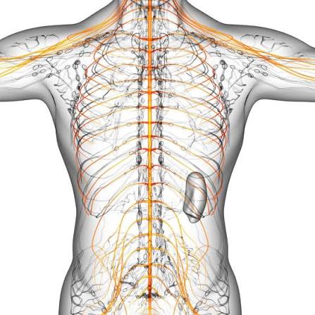
Autonomic Neuropathy Symptoms: When Automatic Body Functions Are Affected
Autonomic neuropathy — damage to the nerves of the autonomic nervous system that regulate automatic body functions — is estimated to be present in 20–40% of adults with long-standing diabetes, and it carries the most serious consequences of any neuropathy subtype because it affects cardiovascular regulation, digestion, bladder control, and sexual function. Unlike peripheral neuropathy, autonomic neuropathy symptoms are often not recognized as neuropathy-related, leading to years of symptomatic treatment without addressing the underlying cause:
- Cardiovascular autonomic neuropathy — resting tachycardia, orthostatic hypotension, and sudden death risk: Cardiovascular autonomic neuropathy (CAN) is diagnosed when the cardiac autonomic reflexes — as measured by heart rate variability during deep breathing, the Valsalva maneuver, and postural change — are blunted or absent. Early CAN manifests as a persistently elevated resting heart rate (over 90 bpm) without physiological cause, because the parasympathetic (vagal) input that normally slows the resting heart rate is damaged before the sympathetic input. Advanced CAN causes orthostatic hypotension — a drop in systolic blood pressure of ≥20 mmHg or diastolic ≥10 mmHg within 3 minutes of standing — which presents as dizziness, lightheadedness, pre-syncope, or falls on standing from a chair or bed. Most seriously, CAN is associated with a two-to-three-fold increase in mortality — primarily from ventricular arrhythmias and sudden cardiac death — because the autonomic nervous system normally protects the heart from fatal rhythms, and its dysfunction removes this protection. Adults with established CAN are at significantly elevated risk during anesthesia and surgery, requiring careful cardiac monitoring.
- Gastroparesis — delayed gastric emptying and its impact on glucose control: Gastroparesis — impaired gastric motility caused by vagal nerve damage — causes nausea, vomiting, early satiety, abdominal bloating, and erratic food absorption that profoundly destabilizes blood glucose control. Normally, the stomach empties its contents into the small intestine at a regulated rate; in gastroparesis, this emptying is delayed or intermittent — so a meal’s carbohydrate load may not be absorbed until hours after the insulin was administered (in insulin-using adults), causing post-injection hypoglycemia followed by delayed post-meal hyperglycemia. Gastroparesis is diagnosed with a 4-hour solid-meal gastric emptying scintigraphy study (the gold standard) and is treated with dietary modification (small frequent meals, liquid-form nutrition), prokinetic medications (metoclopramide, domperidone where available, erythromycin for refractory cases), and in severe cases, gastric electrical stimulation or jejunal feeding. Blood glucose management in adults with gastroparesis often requires switching to basal insulin-dominant regimens, continuous glucose monitoring with extended post-meal observation, or GLP-1 receptor agonists (with caution, as they also delay gastric emptying).
- Bladder and sexual dysfunction from autonomic nerve damage: Diabetic cystopathy — autonomic neuropathy of the bladder’s parasympathetic innervation — causes impaired bladder sensation (the person does not feel the urge to urinate until the bladder is very full), reduced detrusor muscle contractility (the bladder doesn’t squeeze effectively), and incomplete bladder emptying with elevated post-void residual volumes. The resulting urinary stasis increases urinary tract infection risk significantly — and adults with diabetes already have impaired immune function that makes UTIs more likely to ascend to the kidneys (pyelonephritis). Diagnosis is confirmed by measuring post-void residual urine volume by ultrasound. Treatment includes timed voiding schedules, alpha-blockers, and in severe cases, clean intermittent self-catheterization. Erectile dysfunction — affecting 35–75% of men with diabetes — and female sexual dysfunction (reduced arousal, lubrication, and orgasm in women with diabetes) result from combined autonomic neuropathy (impaired vascular regulation of genital blood flow) and vascular disease (atherosclerosis of pudendal arteries). Our diabetes complications: what adults should know guide provides a comprehensive overview of all complications including autonomic neuropathy sequelae. The NIDDK’s diabetic neuropathy information and the ADA’s neuropathy resources provide authoritative clinical guidance on diagnosis and management.
Diagnosing Diabetic Neuropathy: What to Expect
Diabetic neuropathy diagnosis combines clinical examination, symptom assessment, and electrophysiological or morphological testing. The ADA recommends that all adults with Type 2 diabetes undergo neuropathy screening at diagnosis and annually thereafter — and all adults with Type 1 diabetes should be screened beginning 5 years after diagnosis:
- Clinical examination — the foundation of neuropathy screening: The comprehensive foot examination that should be performed at every annual diabetes visit assesses multiple nerve fiber types: vibration sensation (128 Hz tuning fork applied to the first metatarsal head or great toe), protective sensation (10g Semmes-Weinstein monofilament applied at 10 standard plantar sites — the most clinically validated screening test for amputation risk), ankle reflexes (reduced or absent reflexes indicate large fiber damage), and pin-prick temperature sensation at the dorsum of the foot. The Michigan Neuropathy Screening Instrument (MNSI) combines a symptom questionnaire with a brief foot examination score and has been validated as a sensitive screening tool. Importantly, a “normal” comprehensive foot examination does not rule out small fiber neuropathy — the intraepidermal nerve fiber density punch biopsy is needed for definitive small fiber assessment.
- Nerve conduction studies and electromyography: Nerve conduction studies (NCS) and electromyography (EMG) measure the speed and amplitude of electrical signals in large myelinated sensory and motor nerve fibers. In diabetic peripheral neuropathy, NCS typically shows slowed conduction velocities (reflecting myelin damage) and reduced amplitudes (reflecting axon loss) in the sural nerve (sensory), peroneal nerve (motor), and tibial nerve — all of which serve the lower extremities. NCS findings confirm and quantify the severity of large fiber neuropathy and can detect subclinical (asymptomatic) neuropathy before clinical examination findings appear. EMG assesses denervation of muscle tissue and can help distinguish diabetic polyneuropathy from radiculopathy (nerve root compression) when the clinical picture is ambiguous.
- Quantitative sensory testing and skin punch biopsy: Quantitative sensory testing (QST) measures thresholds for detecting vibration (large fiber) and temperature change (small fiber) using computer-controlled stimuli — providing a quantitative measure of sensory function that is more sensitive than tuning fork or pinprick testing. Skin punch biopsy — in which a 3 mm sample of skin is taken from the distal leg and distal thigh — allows direct counting of intraepidermal nerve fibers under immunofluorescence microscopy. Reduced intraepidermal nerve fiber density (IENFD) compared with age-matched normal values is the diagnostic criterion for small fiber neuropathy and correlates directly with the severity of small fiber symptoms. IENFD measurement is available at academic medical centers and specialized neuropathy clinics. Our annual diabetes care checklist covers which neuropathy assessments should be performed at each annual diabetes visit.
Preventing and Slowing Diabetic Neuropathy Progression
The evidence base for neuropathy prevention is substantial: the DCCT/EDIC study demonstrated that intensive glucose control in Type 1 diabetes reduced clinical neuropathy by 60% over 6.5 years, and the legacy effect continued to show neuropathy risk reduction 13–14 years after the intensive intervention ended. The UKPDS demonstrated a 25% reduction in neuropathy (measured by vibration perception threshold) per 1% reduction in A1C in Type 2 diabetes. Beyond glucose control, addressing the full spectrum of neuropathy risk factors and managing established neuropathic pain are both important components of comprehensive care:
- Intensive glucose control — the most evidence-based neuropathy prevention strategy: Every percentage point of A1C reduction meaningfully reduces neuropathy risk. For adults with Type 2 diabetes, maintaining A1C below 7% (or as close to normal as can be safely achieved) is the single most important neuropathy prevention strategy. The ADA recommends A1C targets individualized by age, hypoglycemia risk, and comorbidities — with less stringent targets (7.5–8%) for older adults or those with frequent hypoglycemia. The complete A1C monitoring framework, including how often to measure and what results mean for complication risk, is in our A1C testing schedule guide.
- Blood pressure and lipid management contribute to neuropathy prevention: Hypertension independently damages the vasa nervorum — the microvasculature supplying peripheral nerves — and its control reduces neuropathy progression in addition to its cardiovascular and kidney-protective effects. The UKPDS demonstrated that tight blood pressure control reduced the need for retinal photocoagulation (a surrogate for microvascular damage) and the same microvascular protection extends to peripheral nerves. Dyslipidemia — particularly elevated triglycerides — has been associated with an increased risk of neuropathy independent of glucose control in large epidemiological studies, and statin therapy has shown peripheral nerve protective effects in some observational studies. Our blood pressure monitoring in diabetes guide and cholesterol monitoring in diabetes guide cover the full context of managing these neuropathy risk factors. The CDC’s diabetic neuropathy resources provide population-level data on neuropathy prevention impact.
- Treating painful diabetic neuropathy — medications that reduce neuropathic pain: For adults with established painful peripheral neuropathy, several medication classes have FDA approval or strong evidence for pain reduction. Pregabalin (Lyrica) and gabapentin (Neurontin) — calcium channel alpha-2-delta ligands — reduce neuronal hyperexcitability and are first-line options for painful diabetic neuropathy; pregabalin has Level A evidence from multiple randomized controlled trials. Duloxetine (Cymbalta) — a serotonin-norepinephrine reuptake inhibitor — is FDA-approved for painful diabetic neuropathy and also provides benefits for depression and anxiety (common comorbidities in adults with chronic neuropathic pain). Tricyclic antidepressants (amitriptyline, nortriptyline) provide effective neuropathic pain relief at doses below those needed for antidepressant effects, but their anticholinergic side effects and cardiac arrhythmia risk limit their use in elderly adults. Topical capsaicin (8% patch, applied in a clinic setting) and topical lidocaine (5% patch) provide localized relief for adults who cannot tolerate or do not respond to systemic medications. Opioids are not recommended for diabetic neuropathic pain due to their limited long-term efficacy and significant addiction and side-effect risks.
- Foot care and injury prevention — the most direct neuropathy-related action: For adults with established loss of protective sensation, consistent foot care practices directly prevent the injuries that lead to ulcers and amputations. Daily foot inspection — examining the entire plantar surface, between the toes, and the heels with a mirror or a family member’s assistance — identifies injuries before they progress to deep wounds. Appropriate footwear — properly fitted shoes with no internal seams that could create pressure points, cushioned soles, and rocker-bottom modifications for adults with metatarsal head pressure — significantly reduces plantar pressure peaks and ulceration risk. Never walking barefoot — indoors or outdoors — removes the most common source of undetected plantar foot trauma. Annual podiatric evaluation and more frequent podiatry visits for adults with prior ulceration or active foot pathology are standard of care. Our diabetic foot problems: prevention and care guide covers the complete foot inspection, footwear, and wound care framework in detail.
Sources: American Diabetes Association — Standards of Medical Care in Diabetes, diabetic neuropathy screening and management; NIDDK — diabetic neuropathies overview and classification; CDC — diabetic neuropathy and lower extremity amputation statistics; DCCT/EDIC — intensive glucose control reducing peripheral neuropathy by 60% in Type 1 diabetes; UKPDS — A1C reduction and vibration perception threshold improvement in Type 2 diabetes; distal symmetric peripheral neuropathy prevalence (50% over lifetime with diabetes); painful diabetic neuropathy prevalence (15–25% of adults with DSPN); small fiber neuropathy in prediabetes and early Type 2 diabetes; intraepidermal nerve fiber density measurement by skin punch biopsy; cardiovascular autonomic neuropathy — resting tachycardia, orthostatic hypotension, sudden death risk (2–3x increase); gastroparesis epidemiology and management; diabetic cystopathy and urinary tract infection risk; Michigan Neuropathy Screening Instrument (MNSI) validation; pregabalin, duloxetine, and gabapentin — evidence for painful diabetic neuropathy; tricyclic antidepressants for neuropathic pain; polyol pathway, AGE accumulation, and oxidative stress in peripheral nerve damage mechanisms; endoneurial ischemia and vasa nervorum damage in diabetic neuropathy; 10g monofilament screening for protective sensation loss and amputation risk.
Thank you for covering diabetic neuropathy: symptoms and prevention so thoroughly without being overly technical. The article answered questions I didn’t even know I had until I started reading. Looking forward to reading more articles from this website.
This breakdown of diabetic neuropathy: symptoms and prevention is exactly what patients need before a specialist appointment. The article answered questions I didn’t even know I had until I started reading. Forwarding this to others in my support group who are dealing with similar issues.
I never fully understood diabetic neuropathy: symptoms and prevention until I read this. The section on managing this condition day-to-day was especially useful for planning. Thank you for making complex medical information accessible without dumbing it down.