
A change in urine output — making less urine than usual, or stopping urination altogether — is one of the most significant signals the body can send about the state of kidney function. The kidneys produce urine as a byproduct of filtering the blood: they remove waste products, excess fluid, and electrolytes from circulation and package them for excretion. When urine output falls substantially, it almost always means that something has disrupted this filtration process — either by reducing blood flow to the kidneys, by directly damaging the kidney tissue, or by blocking the drainage pathway through which urine exits. Each of these mechanisms has a distinct cause, a different prognosis, and a different treatment.
Decreased urine output ranges from the mild and reversible — dehydration that resolves with hydration — to the severe and life-threatening, such as bilateral ureteral obstruction from cancer or acute kidney injury from a nephrotoxic medication or prolonged shock. Understanding which category applies to a given situation requires knowing the definitions, the patterns, and the warning signs that distinguish a manageable situation from an emergency.

Normal vs. Decreased Urine Output
Healthy adult kidneys produce approximately 1,000 to 2,000 mL of urine per day under normal conditions. The medical term for substantially reduced urine output is oliguria, defined as less than 400 mL per day, or less than 0.5 mL per kilogram of body weight per hour in clinical monitoring settings. Anuria is the near-complete cessation of urine production — less than 100 mL per day — and represents a critical state of kidney failure or complete obstruction that is nearly always a medical emergency.
A separately important early sign of kidney dysfunction is nocturia — waking at night to urinate more than once. In healthy kidneys, the body concentrates urine effectively during sleep. In early kidney disease, this concentrating ability is lost and urine is produced at the same rate around the clock, producing increased overnight urination. Nocturia is therefore an early warning sign of impaired kidney function. According to the National Institute of Diabetes and Digestive and Kidney Diseases, changes in urine frequency and quantity are among the earliest detectable signs of kidney disease, often preceding other symptoms by years.
Pre-Renal Causes: When Blood Flow to the Kidneys Drops
The most common cause of acutely decreased urine output is pre-renal — meaning the kidneys themselves are structurally intact but are producing less urine because they are receiving less blood flow. Without adequate blood flow, the pressure available to drive filtration across the glomerular capillaries falls below the threshold needed to produce urine at normal rates.
Dehydration is the most common pre-renal cause. When fluid losses from vomiting, diarrhea, sweating, or insufficient intake exceed intake, blood volume falls, blood pressure drops, and the kidneys conserve water — reducing urine output and concentrating the remaining urine to a dark, amber color. This reverses rapidly with rehydration unless the dehydration has been severe or prolonged enough to cause ischemic tubular injury. Heart failure causes decreased kidney perfusion when the heart’s pumping function is reduced; the kidneys respond as if the body is volume-depleted even though the underlying problem is poor cardiac output — the cardiorenal interaction detailed in the guide on high blood pressure as a kidney warning sign.
NSAIDs and ACE inhibitors/ARBs in specific settings can acutely reduce kidney filtration. NSAIDs block prostaglandins that dilate the afferent arteriole; removing this dilation in dehydrated or low-perfusion patients causes acute oliguric injury. ACE inhibitors and ARBs reduce efferent constriction; in patients with bilateral renal artery stenosis, this dramatically reduces glomerular filtration pressure. The key laboratory clue to pre-renal cause is a BUN-to-creatinine ratio greater than 20:1 and urine sodium below 20 mEq/L, indicating the kidneys are avidly conserving sodium in response to perceived volume depletion.
Intrinsic Kidney Causes
Acute tubular necrosis (ATN) is the most common form of intrinsic acute kidney injury leading to oliguria. Tubular cells — sensitive to ischemia and toxins — are killed by prolonged pre-renal injury, aminoglycoside antibiotics, contrast dye, myoglobin from rhabdomyolysis, or certain chemotherapy drugs. The classic finding is muddy-brown granular casts in the urine. ATN is treated supportively: the offending cause is removed, fluid balance is managed, and dialysis is started if needed while awaiting spontaneous tubular regeneration. The guide on kidney health numbers every adult should know explains how creatinine and BUN track AKI severity and recovery.
Acute glomerulonephritis and rapidly progressive glomerulonephritis (RPGN) cause oliguria through immune complex deposition and inflammation that damage the glomerular filtration barrier. RPGN is a medical emergency — it can progress from normal kidney function to dialysis dependence within days to weeks without immunosuppressive treatment. Red blood cell casts in the urine are the classic finding.
Post-Renal Causes: Obstruction
Benign prostatic hyperplasia (BPH) is the most common cause of urinary outflow obstruction in older men. An enlarged prostate compresses the urethra; in severe BPH, complete obstruction and urinary retention occur. The patient presents with suprapubic pain, inability to void despite a strong urge, and a distended bladder. Bladder catheterization immediately relieves the obstruction. According to the Mayo Clinic, urinary retention from BPH is one of the most common causes of decreased urine output in men over 60. Kidney stones can obstruct the ureter; bilateral obstruction — from stones or cancer compressing both ureters — can cause complete anuria and is a urological emergency. Neurogenic bladder from diabetes, multiple sclerosis, or spinal cord injury produces functional obstruction without anatomical blockage.
Decreased Urine Output in Advanced CKD and Dialysis
As CKD progresses through stages 4 and 5, the progressive loss of functioning nephrons gradually reduces urine output. One of the significant transitions in ESRD management is the progressive loss of residual kidney function (RKF) after dialysis begins — the natural decline of whatever urine-producing capacity remained at dialysis initiation. Residual kidney function, even at just 100 to 300 mL per day, provides meaningful benefits: it removes toxins, fluid, and phosphorus continuously between sessions, reducing the burden on the dialysis machine. The National Kidney Foundation emphasizes that preserving residual kidney function should be a clinical priority from the start of dialysis, as loss of RKF is associated with worse fluid control, more hospitalizations, and higher mortality. Strategies include avoiding nephrotoxic medications (NSAIDs, aminoglycosides, IV contrast when alternatives exist) and preventing excessive dehydration between sessions. The swelling that accumulates as residual function declines is covered in the guide on swollen feet and kidney problems.
When Decreased Urine Output Is an Emergency
Sudden onset of significantly reduced or absent urine output in a person who was previously urinating normally is an emergency. Anuria is always an emergency. Urinary retention — inability to void with suprapubic pain and a distended bladder — requires emergency catheterization. Oliguria accompanied by a rapidly rising creatinine, confusion, severe fluid overload, or dangerous potassium elevation requires emergent evaluation for dialysis initiation. The NIDDK resource on acute kidney failure explains the emergency nature of AKI and its treatment priorities. The foundational context for understanding CKD progression is at what is chronic kidney disease. The nausea that commonly accompanies severe AKI and advanced CKD is addressed in the companion guide on nausea and kidney problems.
Frequently Asked Questions
What is a normal amount of urine per day?
Healthy adult kidneys produce approximately 1,000 to 2,000 mL of urine per day. Less than 400 mL per day is oliguria and warrants evaluation. More than 3,000 mL per day (polyuria) can be an early sign of kidney disease when not explained by high fluid intake, because failing kidneys often lose concentrating ability before losing overall filtration capacity.
Can decreased urine output mean kidney failure?
Yes. Oliguria or anuria is a cardinal sign of both acute kidney injury and advanced chronic kidney disease. Not all decreased urine output is kidney failure — dehydration, obstruction, and certain medications can cause reversible oliguria. However, any new, unexplained reduction in urine output warrants evaluation of kidney function through blood tests (creatinine, BUN) and urine analysis.
What is the difference between oliguria and anuria?
Oliguria is significantly reduced output — less than 400 mL per day — indicating impaired but not completely absent kidney function. Anuria is near-complete cessation — less than 100 mL per day — and represents a critical state of kidney failure or complete obstruction. Both require evaluation, but anuria is more immediately dangerous because even minimal excretion of waste products is absent.
Why do dialysis patients stop making urine?
As CKD progresses to end-stage and dialysis begins, the remaining functional nephrons continue to decline — a process that continues even after dialysis starts. Residual kidney function is lost progressively over months to years on dialysis until urine production stops entirely. This is the expected natural history of ESRD. Strategies to slow this process include avoiding nephrotoxic drugs, preventing severe dehydration between sessions, and using biocompatible dialysis membranes.
How do doctors determine the cause of decreased urine output?
The evaluation starts with blood tests (creatinine, BUN, electrolytes), urinalysis (looking for casts, protein, blood), and assessment of volume status. A urine sodium measurement (low in pre-renal, high in ATN) helps distinguish the mechanism. Kidney ultrasound rapidly identifies hydronephrosis (obstruction) or small, shrunken kidneys (chronic CKD). If glomerulonephritis is suspected, additional blood tests and potentially kidney biopsy are needed. This systematic approach directs treatment accurately rather than requiring empirical management.
Acute Kidney Injury: When Urine Output Falls Suddenly
Acute kidney injury (AKI) — formerly called acute renal failure — is a sudden episode of kidney dysfunction in which GFR drops rapidly over hours to days, creatinine rises, and urine output may fall to oliguric or anuric levels. AKI is classified by the KDIGO (Kidney Disease: Improving Global Outcomes) criteria based on the magnitude of creatinine rise and the duration or degree of urine output reduction: Stage 1 is a creatinine rise of 0.3 mg/dL within 48 hours or a 50 percent rise within 7 days; Stage 2 is a twofold creatinine increase; Stage 3 is a threefold increase or a fall below 0.3 mL/kg/hour for 12 hours or more, or the initiation of dialysis. Oliguria — less than 0.5 mL/kg/hour for 6 or more hours — is a criterion for Stage 1 AKI at its simplest, reinforcing that urine output measurement is a clinically meaningful monitoring parameter in high-risk inpatients.
AKI is extremely common in hospitalized patients — affecting approximately 10 to 15 percent of all hospital admissions and up to 50 to 60 percent of ICU patients — and represents one of the highest-risk situations for progression to chronic kidney disease. Patients who survive an episode of AKI have significantly higher rates of CKD, ESRD, and cardiovascular events in the years following the AKI episode, even if creatinine appears to return to baseline. This post-AKI vulnerability makes prevention and early treatment of AKI episodes critically important — particularly for patients who already have CKD, in whom each AKI episode may permanently eliminate additional nephrons from the already-depleted pool. Common precipitants to watch for and avoid include nephrotoxic antibiotics, NSAIDs, iodinated contrast dye, inadequate hydration before procedures, and prolonged hypotension from any cause.
The Role of Urine Output Monitoring
In hospitalized patients, particularly those in the ICU, the operating room, or the post-operative setting, hourly urine output monitoring through an indwelling urinary catheter is a standard practice because urine output is one of the earliest sensitive indicators that kidney perfusion is failing. A fall in urine output to below 0.5 mL/kg/hour in the post-operative or ICU setting is a trigger for immediate clinical assessment: is the patient adequately hydrated? Is the blood pressure sufficient to perfuse the kidneys? Is there an obstruction (catheter kink)? Is there a new nephrotoxin exposure? Responding to early oliguria in this setting — restoring blood pressure and perfusion, correcting volume deficits, removing nephrotoxins — can prevent progression to more severe AKI that requires dialysis. For outpatients and CKD patients managed in the clinic, monitoring urine output through daily weight (which reflects fluid balance), monitoring for swelling, and noting changes in urine color and frequency is the practical equivalent of inpatient urine output monitoring.
Drug-Induced Causes of Decreased Urine Output
Medications are among the most common and most preventable causes of reduced urine output and AKI, and patients with CKD are disproportionately vulnerable because their kidneys have less functional reserve to absorb insults. Understanding which medications carry kidney risk — and communicating your kidney disease history to every prescribing clinician — is one of the most important patient-level actions for protecting kidney function.
NSAIDs (ibuprofen, naproxen, diclofenac, ketorolac) are the most commonly used over-the-counter drugs and among the most important contributors to drug-induced AKI. NSAIDs are nephrotoxic through two mechanisms: they reduce renal blood flow by blocking prostaglandins that dilate the afferent arteriole (causing pre-renal injury), and with prolonged use, they can cause tubulointerstitial nephritis (direct kidney inflammation). Even a short course of NSAIDs in a dehydrated CKD patient, or in a patient on ACE inhibitors or ARBs (a combination sometimes called “triple whammy” when a diuretic is added), can produce significant oliguric AKI. Acetaminophen is the recommended alternative for pain management in CKD.
Aminoglycoside antibiotics (gentamicin, tobramycin, amikacin) are highly effective but accumulate in kidney tubular cells and cause direct tubular toxicity. The risk is dose-related and cumulative, with the highest toxicity seen in patients receiving multiple courses or extended courses. Once-daily dosing strategies and careful monitoring of drug levels reduce but do not eliminate this risk. Alternative antibiotics are preferred when effective options exist for the organism being treated.
Iodinated contrast dye used in CT scans, cardiac catheterizations, and angiography can cause contrast-induced nephropathy (CIN) — an AKI that typically peaks 48 to 72 hours after exposure and resolves within a week in most patients, but can be severe and persistent in those with advanced CKD, diabetes, and volume depletion. Pre-procedure hydration with intravenous saline is the most effective preventive strategy. When contrast is necessary in a CKD patient, using the lowest effective volume of a low-osmolality or iso-osmolality contrast agent, combined with adequate pre-hydration, minimizes risk.
Proton pump inhibitors (PPIs) — among the most commonly prescribed medications worldwide (omeprazole, pantoprazole, lansoprazole) — have been linked to acute interstitial nephritis, an immune-mediated kidney inflammation that can reduce urine output and cause AKI. The association is recognized in pharmacovigilance databases and reinforced by multiple epidemiological studies, though the absolute risk per patient is low. In a CKD patient who develops unexplained AKI while on a PPI, discontinuing it as a diagnostic and therapeutic step is appropriate.
Communicating a complete medication list — including over-the-counter drugs, supplements, and herbals — to the nephrology team is the practical implementation of kidney protection in CKD. The guide on kidney health numbers every adult should know includes creatinine and GFR among the values that should be monitored more frequently in the weeks following any new medication start or dose change in CKD.
Urine Color and Appearance as Diagnostic Clues
In addition to the volume of urine produced, its color, clarity, and odor can provide meaningful diagnostic information about why urine output has changed. These observations are accessible to any patient and can be the first step in communicating a concerning change to a medical provider.
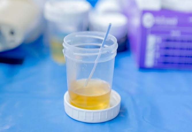
Dark amber or tea-colored urine with reduced volume most commonly indicates dehydration — the kidneys are concentrating urine maximally to conserve fluid. When this occurs after vomiting, diarrhea, or intense exercise in hot weather, it is usually reversible with oral rehydration. When it persists despite adequate fluid intake, it suggests the kidneys may not be excreting enough fluid — raising the possibility of worsening kidney function. Reddish-brown or cola-colored urine can indicate blood in the urine (hematuria) from glomerulonephritis, kidney stones, urinary tract infection, or injury. The presence of blood in the urine combined with reduced urine output should prompt urgent evaluation — particularly when accompanied by pain (suggesting stones) or when painless (more concerning for glomerulonephritis or bladder cancer).
Foamy urine — persistent foam that does not dissipate after flushing — suggests proteinuria. While some foam is normal with high urine flow rate, persistent foam specifically indicates the presence of protein in amounts that create surface tension when the urine hits the water. This is a visual sign of glomerular damage and an early marker of CKD in people who may not yet have other symptoms. Any new, persistent foaminess in urine should be mentioned to a healthcare provider and evaluated with a urine dipstick and urine albumin-to-creatinine ratio. Cloudy urine, particularly with an odor or burning sensation, suggests a urinary tract infection. Entirely clear urine — very pale, almost colorless — in a CKD patient may indicate impaired concentrating ability (isosthenuria) or excessive fluid intake relative to output.
Tracking Urine Output and Fluid Balance at Home
For patients with advanced CKD, on dialysis, or recovering from an AKI episode, tracking fluid intake and urine output at home is a practical and meaningful strategy for monitoring kidney function and fluid balance on a day-to-day basis. In dialysis patients who retain some residual function, knowing approximately how much urine they produce each day provides useful information for the dialysis team in calibrating ultrafiltration targets and assessing whether residual function is declining.
Measuring urine output at home does not require specialized equipment — a graduated plastic container is sufficient. Output is recorded over a 24-hour period by collecting all urine passed during that time. This 24-hour urine collection is also a standard clinical tool used to measure urine protein (for CKD staging and treatment monitoring) and creatinine clearance (as a GFR estimate). Patients who have been instructed by their nephrologist to collect a 24-hour urine sample should follow the provided instructions carefully — typically starting the collection after the first morning void and ending it after the first void the following morning. Keeping a fluid intake diary alongside the urine output measurement provides a fluid balance calculation that directly informs whether fluid is being retained or excreted as expected.
For non-dialysis CKD patients, the most practical daily monitoring is weight. Body weight measured every morning at the same time — before eating or drinking, after voiding — on the same scale reflects fluid balance accurately. A weight gain of more than 1 to 2 kg over 24 to 48 hours in a patient with known CKD and reduced urine output strongly suggests fluid retention and should prompt a call to the nephrology team. The guides on swollen feet and kidney problems and what is chronic kidney disease provide additional context on fluid management throughout CKD progression.
Sources: National Institute of Diabetes and Digestive and Kidney Diseases (NIDDK) — CKD and AKI; National Kidney Foundation — Residual Kidney Function; Mayo Clinic — Decreased Urine Output; published nephrology guidelines on AKI classification (KDIGO) and residual function preservation
I never fully understood decreased urine output: what it may until I read this. This is the kind of evidence-based writing that actually changes how people approach their health. This gave me real confidence going into my next specialist appointment.
As someone dealing with this personally, the decreased urine output: what it may section was very helpful. I have tried following advice from several sources but this is most consistent with what my specialist told me. Thank you for making complex medical information accessible without dumbing it down.
Really well-written article on decreased urine output: what it may. What I liked most was that the article didn’t just say what to avoid — it also gave alternatives. This is going into my health folder that I bring to every doctor’s visit.