

Blood in the stool is a symptom that should never be dismissed, yet many adults either ignore it, assume it is from hemorrhoids, or delay evaluation out of concern or embarrassment. While hemorrhoids are indeed the most common cause of rectal bleeding, they are not the only cause — and treating every instance of blood in the stool as benign without evaluation carries a real risk of missing a more serious diagnosis. Understanding the possible causes of blood in the stool and recognizing which patterns constitute warning signs is essential for knowing when to seek medical attention promptly.
Blood in the stool can appear in several distinct ways depending on where in the GI tract the bleeding originates. Bright red blood on the surface of stool or toilet paper reflects lower GI bleeding — rectum or anus. Dark red or maroon blood mixed into the stool suggests bleeding from the left colon. Black, tarry stool (melena) indicates upper GI bleeding — from the stomach, esophagus, or upper small bowel — where blood has been partially digested during transit. And occult blood — not visible to the naked eye but detected by testing — can be the only sign of slow, chronic bleeding from polyps or early colorectal cancer.
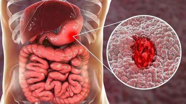
How to Interpret Blood in the Stool by Location and Color
The color and location of blood in or around the stool provides the most immediate clue to where the bleeding is occurring in the GI tract — an anatomical road map that helps prioritize urgency and guide evaluation.
Bright red blood on the toilet paper or surface of stool — without being mixed into the stool — most often indicates a source very close to the anal opening: hemorrhoids or an anal fissure. This pattern suggests the blood has not traveled far enough through the colon to darken. However, this same pattern can also result from rectal polyps, rectal cancer, proctitis, or a rapid arteriovenous malformation (AVM) in the rectum or left colon. The distinction matters because while hemorrhoidal bleeding is rarely dangerous, rectal cancer bleeding requires prompt colonoscopy.
Bright red blood mixed into the stool (rather than just on the surface or paper) is a more serious pattern suggesting the bleeding source is higher in the rectum or sigmoid colon, and that blood is being incorporated into the stool rather than coating it. This pattern increases suspicion for colorectal cancer, diverticular bleeding, ischemic colitis, or IBD and warrants timely colonoscopy.
Dark red or maroon blood throughout the stool indicates a mid-colonic source — typically the right colon or transverse colon — where blood darkens during transit but has not fully digested to melena. This pattern can also reflect a brisk upper GI bleed with rapid transit. Dark maroon stool with significant volume warrants urgent evaluation.
Black, tarry, sticky stool (melena) with a distinctively unpleasant odor indicates blood that has been digested by stomach acid and bacteria during its transit from the upper GI tract. The hemoglobin is converted to hematin, producing the black color. Common upper GI sources include peptic ulcers (gastric and duodenal), esophageal varices, and Mallory-Weiss tears. Melena is an urgent finding requiring immediate evaluation — it can precede hemodynamic instability in cases of significant upper GI hemorrhage. Our companion article on black stool: what it may mean covers the evaluation of melena in greater detail.
Occult blood — detected only on stool-based testing (fecal immunochemical test, FIT, or fecal occult blood test, FOBT) — means the amount of bleeding is too small to see visually. This is the pattern most commonly associated with early colorectal polyps and cancers, which bleed slowly and intermittently into the stool without producing visible discoloration. Annual stool-based screening is recommended precisely to catch this type of bleeding before it becomes visible or symptomatic.
Hemorrhoids — The Most Common Cause
Internal hemorrhoids are dilated vascular cushions in the submucosa of the anal canal. They are present to some degree in most adults — problems arise when they become enlarged, prolapse, or bleed. Hemorrhoidal bleeding is typically bright red, painless (internal hemorrhoids lack somatic nerve supply), associated with straining or hard stools, and appears on the surface of the stool or on the toilet paper — not mixed throughout the stool. Bleeding after each bowel movement that is small in volume and resolves on its own is the classic presentation.
External hemorrhoids — located below the dentate line and covered by squamous epithelium — cause pain when thrombosed (a clot forms within the external hemorrhoid, causing sudden, severe anal pain with a visible or palpable lump). External hemorrhoids rarely bleed unless irritated or traumatized.
Hemorrhoids are graded I through IV based on degree of prolapse: grade I (no prolapse, only bleeding), grade II (prolapse with straining but spontaneous reduction), grade III (prolapse requiring manual reduction), and grade IV (cannot be manually reduced). Lower-grade hemorrhoids typically respond to dietary modification (fiber, fluids, stool softeners, sitz baths), while grade III and IV hemorrhoids often require procedural management (rubber band ligation, sclerotherapy, or hemorrhoidectomy).
The critical clinical point is that hemorrhoidal bleeding should be a diagnosis of exclusion in older adults — particularly those over 45 — not an assumption made without examination. Colorectal cancer can coexist with hemorrhoids, and the presence of hemorrhoids does not exclude another bleeding source higher in the GI tract. Adults over 45 with rectal bleeding — even classic hemorrhoidal-pattern bleeding — who have not had a recent colonoscopy should have one to exclude a coincidental upper source.
Anal Fissures and Anorectal Causes
An anal fissure is a linear tear in the anoderm (the skin lining of the anal canal distal to the dentate line). Fissures cause severe anal pain during and after defecation — classically described as passing broken glass — followed by a dull throbbing ache lasting minutes to hours. Bright red blood on the paper or in the toilet bowl typically accompanies the pain. The pain distinguishes fissures from hemorrhoids: hemorrhoidal bleeding is usually painless.
Fissures most commonly result from passage of a hard, large stool that tears the delicate anoderm. They can also result from diarrhea (repeated trauma from frequent stools), anal intercourse, or, rarely, from Crohn’s disease or sexually transmitted infections (syphilis, HSV). Acute fissures (less than six weeks) often heal with conservative management: high-fiber diet, sitz baths, topical anesthetics, and stool softeners. Chronic fissures (more than six weeks) often develop a sentinel pile (a skin tag below the fissure) and internal hypertrophied papilla, and respond to topical nitroglycerin, calcium channel blocker cream, or botulinum toxin injection; surgical lateral internal sphincterotomy is reserved for refractory cases.
Other anorectal causes of blood in the stool include rectal prolapse (the rectal mucosa protrudes through the anus and bleeds from friction), solitary rectal ulcer syndrome (caused by paradoxical pelvic floor contraction and intussusception), and proctitis (inflammation of the rectal mucosa from radiation, infection, or IBD).
Diverticular Bleeding
Colonic diverticula are small pouches that develop in weak points of the colon wall, particularly in the sigmoid colon, though right-sided diverticula are more common in Asian populations. Most diverticula are asymptomatic. Diverticular bleeding occurs when a blood vessel running alongside a diverticulum erodes and bleeds into the bowel lumen. It is the most common cause of significant lower GI bleeding (as opposed to small-volume rectal bleeding from hemorrhoids) in adults over 50.
Diverticular bleeding typically presents as sudden, painless passage of large amounts of bright red or maroon blood — often described by patients as passing “blood clots” in the toilet. Despite the dramatic presentation, most episodes (approximately 70 to 80 percent) stop on their own. However, significant diverticular bleeding can cause hemodynamic instability requiring resuscitation, colonoscopy to identify and treat the bleeding diverticulum, or rarely surgery. Adults who experience a significant diverticular bleed have approximately a 25 percent risk of rebleeding over the following years.
Colorectal Cancer and Polyps
Colorectal cancer is the bleeding source that is most important not to miss. It can cause rectal bleeding in several ways: direct surface ulceration of the tumor produces bleeding that may be intermittent or continuous; partial luminal obstruction causes straining that traumatizes the tumor; and tumors in the right colon may bleed so slowly that no visible blood appears, manifesting only as iron-deficiency anemia.
The bleeding pattern associated with colorectal cancer is variable: it may be bright red (sigmoid and rectal cancers), dark red or occult (right-sided cancers), or intermittent (causing a positive fecal occult blood test on some but not all tests). Other associated features that increase suspicion for colorectal cancer include a change in bowel habits, narrowing stool caliber, unexplained weight loss, iron deficiency anemia, and tenesmus.
Colorectal polyps — the precursor to most colorectal cancers — can also bleed, though they rarely produce significant visible bleeding. A positive annual fecal immunochemical test (FIT) in an asymptomatic adult represents polyp or cancer bleeding until proven otherwise and should be followed by colonoscopy within four to six weeks. Adults who dismiss a positive FIT result or defer follow-up colonoscopy significantly increase their risk of delayed cancer detection.
Any adult over 45 with rectal bleeding — even bleeding that appears entirely characteristic of hemorrhoids — should have the source confirmed by colonoscopy if a colonoscopy has not been performed within the recommended surveillance interval. Our article on changes in bowel habits: what adults should know discusses colorectal cancer screening in broader context.
Inflammatory Bowel Disease and Infectious Colitis
Ulcerative colitis (UC) characteristically produces bloody mucoid diarrhea — blood mixed with mucus, passed with urgency and frequency. In mild to moderate UC, bleeding may be low volume; in severe UC, bloody diarrhea can occur 10 or more times daily, causing significant anemia. The blood in UC is typically mixed throughout the stool (not just on the surface) and accompanied by urgency and tenesmus. Crohn’s colitis can produce a similar picture when the colon is involved; small bowel Crohn’s rarely causes visible rectal bleeding.
Infectious colitis from bacteria such as Campylobacter, Shigella, and Salmonella can cause bloody diarrhea (dysentery pattern) indistinguishable from IBD on clinical grounds alone. The distinguishing factors are the timeline (acute onset with fever in infectious colitis; subacute progressive onset over weeks to months in IBD), travel or exposure history, and stool cultures. Entamoeba histolytica causes amoebic dysentery with bloody stool and deserves consideration in patients with travel to endemic regions.
Peptic Ulcer Disease and Upper GI Bleeding
Peptic ulcer disease — erosion of the gastric or duodenal mucosa from Helicobacter pylori infection or NSAID use — is the most common cause of significant upper GI bleeding. When an ulcer erodes into a blood vessel, bleeding can range from slow (producing melena) to rapid and life-threatening (producing hematemesis — vomiting blood — and a precipitous drop in blood pressure). Upper GI bleeding from any cause should be considered when black tarry stool is present, when the patient is hemodynamically unstable, or when hematemesis accompanies GI symptoms.
Other upper GI causes of blood in the stool include esophageal varices (dilated veins in the esophagus from portal hypertension in cirrhosis — can produce dramatic, life-threatening bleeding), Mallory-Weiss tears (mucosal tears at the gastroesophageal junction from forceful vomiting), and gastric or esophageal cancer. Our article on right upper abdominal pain and liver health covers cirrhosis-related complications including variceal bleeding.
Red Flags and When to Seek Urgent Evaluation
The following features indicate that blood in the stool requires urgent or emergency evaluation rather than watchful waiting:
- Large volume bleeding — passing significant amounts of blood or clots, saturating multiple pads, or blood in the toilet that is impossible to ignore in terms of volume
- Black, tarry stool with hemodynamic symptoms — lightheadedness, dizziness, rapid heart rate, or near-syncope alongside melena suggests significant upper GI hemorrhage; call emergency services
- Hematemesis (vomiting blood) — always an emergency
- Rectal bleeding with severe abdominal pain — raises concern for ischemic colitis, perforation, or mesenteric ischemia
- Rectal bleeding in a patient on anticoagulation — warfarin, direct oral anticoagulants, or dual antiplatelet therapy can convert a minor bleed into a major hemorrhage; prompt evaluation is warranted
- New rectal bleeding in adults over 45 without recent colonoscopy — requires colonoscopy within a reasonably prompt timeframe (weeks to months depending on volume and associated symptoms) to exclude colorectal cancer
- Rectal bleeding with weight loss, anemia, or changed bowel habits — a combination of symptoms that significantly raises suspicion for colorectal cancer; prompt evaluation is appropriate
Adults of any age who are concerned about blood in the stool should contact their primary care provider. When the bleeding is large volume, associated with dizziness or fainting, or accompanied by severe pain, emergency evaluation is the appropriate response.
Evaluation — What to Expect
The initial evaluation of blood in the stool typically begins with a history (volume, color, frequency, associated symptoms, medications, family history of colorectal cancer) and physical examination including a digital rectal examination, which allows direct palpation of the rectum and assessment of hemorrhoids, fissures, and masses within reach of the examining finger.
Blood tests — complete blood count (to assess anemia severity), iron studies, and in some cases coagulation testing — provide important baseline information and influence the urgency of further evaluation.
Colonoscopy is the primary diagnostic tool for most presentations of rectal bleeding: it directly visualizes the entire colon and rectum, allows biopsy of suspicious lesions, and can treat bleeding sources (hemorrhoid ligation, polypectomy, diverticular bleeding hemostasis). Upper endoscopy (EGD) is performed first when upper GI bleeding is suspected based on melena, hematemesis, or hemodynamic instability.
Imaging — CT angiography or nuclear medicine tagged RBC scan — is reserved for active significant bleeding when colonoscopy cannot adequately localize the source or when the patient is too unstable for endoscopy.
Frequently Asked Questions About Blood in the Stool
If I only see blood on the toilet paper, should I still see a doctor?
Bright red blood on the toilet paper after a bowel movement, particularly with straining and hard stools, is most often from an internal hemorrhoid or anal fissure. However, rectal bleeding should be evaluated by a clinician at least once, especially if it is new, recurrent, increasing, or occurring in an adult over 45. Do not assume hemorrhoids without examination and, in adults due for or overdue for colorectal cancer screening, colonoscopy is appropriate.
Can iron supplements cause black stool that looks like melena?
Yes. Iron supplements produce dark, almost black stool that can be mistaken for melena. The key distinction is that iron-related black stool is not tarry, sticky, or foul-smelling the way melena is. If there is any uncertainty — particularly if GI symptoms, abdominal pain, or dizziness accompany the black stool — medical evaluation is the safer course. Our detailed article on black stool: what it may mean provides a full comparison.
Is occasional rectal bleeding normal?
Rectal bleeding is never “normal” — it indicates bleeding from somewhere in the GI tract, even if the source is benign. Occasional minor bleeding from a known hemorrhoid that is well-managed and has been previously evaluated may be acceptable to monitor in consultation with a clinician. Repeated or new-onset bleeding should be evaluated. There is no safe threshold of “a little blood is okay.”
Can food cause red-colored stool that looks like blood?
Yes. Large quantities of beets can turn stool and urine red or pink — a harmless phenomenon called beeturia. Red-colored drinks, tomato-based foods, and red food dye can produce similar discoloration. If the discoloration resolves within 24 to 48 hours and corresponds precisely with consumption of red-pigmented food, it is unlikely to be blood. If uncertainty remains — particularly in an adult with other GI symptoms or risk factors — testing the stool for blood is straightforward and definitive.
My doctor ordered a fecal occult blood test and it was positive — what happens next?
A positive fecal immunochemical test (FIT) or fecal occult blood test (FOBT) should be followed by colonoscopy. The test detected blood in the stool that was not visible to the naked eye, which may indicate a colorectal polyp or cancer. A positive result does not confirm cancer — many positives reflect small polyps, hemorrhoids, or other benign sources — but it cannot be assumed benign without direct visualization. Follow-up colonoscopy should be scheduled within four to six weeks of a positive FIT. Our broader overview of common digestive problems in adults provides additional context on colorectal screening.
Sources: National Institute of Diabetes and Digestive and Kidney Diseases — Rectal Bleeding | American College of Gastroenterology — Rectal Bleeding | Mayo Clinic — Rectal Bleeding: Causes
Vascular Causes of GI Bleeding — Angiodysplasia and Ischemic Colitis
Beyond the better-known causes of blood in the stool, two vascular conditions deserve attention because they are commonly encountered in older adults and can produce significant bleeding that is difficult to manage without specialized endoscopic or angiographic intervention.
Angiodysplasia (also called arteriovenous malformations or vascular ectasias of the colon) consists of small, abnormal, dilated blood vessels in the submucosa and mucosa of the colon — most commonly in the cecum and right colon. They develop with aging as a result of chronic, intermittent obstruction of small venous channels by the high intraluminal pressure of colonic contractions. In adults over 65, angiodysplasia accounts for a meaningful proportion of chronic occult GI blood loss and iron deficiency anemia, as well as acute lower GI bleeding episodes. Angiodysplasia bleeding is characteristically intermittent and painless, often producing maroon or bright red blood depending on flow rate. Colonoscopy with argon plasma coagulation (APC) is the primary treatment. Angiodysplasia is more common in adults with aortic stenosis (Heyde’s syndrome — a relationship mediated by von Willebrand factor cleavage) and in those with end-stage kidney disease.
Ischemic colitis results from temporary reduction in blood flow to the colon — typically the watershed areas at the splenic flexure and rectosigmoid junction, which are the most susceptible to relative hypoperfusion. It is the most common form of intestinal ischemia. Risk factors include advanced age, cardiovascular disease, hypotension, recent abdominal aortic surgery, vasoconstrictive medications (including decongestants, triptans, and some immunosuppressants), and hypercoagulable states. Ischemic colitis typically presents with sudden onset of mild to moderate left-sided abdominal cramping followed within 24 hours by bright red or maroon rectal bleeding — a distinctive sequence that helps distinguish it from other causes. The majority of cases are transient and resolve without specific intervention; a minority progress to transmural ischemia requiring surgery. CT abdomen and pelvis with contrast is the preferred initial imaging, and colonoscopy is performed after the acute phase resolves to confirm diagnosis and assess mucosal recovery.
Both angiodysplasia and ischemic colitis can present with blood in the stool that is easily mistaken for hemorrhoidal bleeding or diverticular bleeding on clinical grounds — further underscoring why colonoscopy (and sometimes advanced imaging) is essential for accurately identifying the bleeding source, particularly in older adults with cardiovascular comorbidities.