

Black stool can be alarming to discover, but it does not always indicate a serious problem. The meaning of black stool depends entirely on whether the darkening is caused by blood that has been digested during its passage through the GI tract — a condition called melena — or by something far more benign, such as iron supplements, bismuth-containing antacids, or certain dark-colored foods. Distinguishing between these two possibilities determines whether black stool requires emergency evaluation or simply reassurance. Understanding what black stool may mean helps adults respond appropriately — neither dismissing a potentially serious symptom nor seeking unnecessary emergency care.
Melena — black, tarry, foul-smelling stool caused by digested blood — is one of the cardinal presentations of upper gastrointestinal (GI) bleeding. Upper GI bleeding originates from the esophagus, stomach, or proximal small bowel, and it is estimated to cause over 300,000 hospital admissions in the United States annually. The cause of upper GI bleeding ranges from a peptic ulcer (the most common cause) to esophageal varices (a complication of cirrhosis that carries significant mortality risk) to Mallory-Weiss tears and gastric cancer. Prompt recognition and evaluation of melena is one of the most important things any adult can do to prevent a potentially life-threatening outcome.
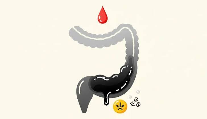
What Makes Stool Black — Melena Versus Benign Causes
Normal stool gets its brown color from stercobilin, a pigment produced when colonic bacteria metabolize bilirubin (derived from the breakdown of hemoglobin in old red blood cells). When blood enters the GI tract high enough — in the esophagus, stomach, or duodenum — stomach acid and digestive enzymes denature the hemoglobin, converting it to hematin. As hematin passes through the intestine, it undergoes further breakdown by bacteria, ultimately producing a jet-black or very dark brown pigment that gives melena its characteristic appearance.
Melena has three distinguishing features that separate it from benign causes of dark stool:
- Color: jet black or very dark, similar to tar or coffee grounds — not simply a dark brown
- Consistency: sticky, tarry, and semi-liquid in texture — not well-formed or dry
- Odor: distinctively foul and penetrating — noticeably different from normal stool odor; often described by patients as metallic or putrid
These features are caused by the chemistry of digested blood mixed with intestinal contents and are not reproducible by any dietary or supplement-related cause. Iron supplements and bismuth (Pepto-Bismol) can produce black or very dark stool, but the texture remains normal and the odor is not distinctively foul. If there is any genuine uncertainty, testing the stool for blood — with a fecal occult blood test — and consulting a clinician is the appropriate response.
It is also important to recognize that not all upper GI bleeds produce melena. A very rapid bleed from an upper GI source — such as a hemorrhagic peptic ulcer or ruptured esophageal varix — can produce maroon or even bright red blood in the stool if transit is fast enough that digestion does not occur. Conversely, melena can result from bleeding as distal as the right colon if transit is slow. The key is that melena always indicates at least 50 to 100 mL of blood loss — sufficient to indicate that evaluation is medically necessary.
Common Non-Pathological Causes of Black Stool
Not all black stool requires emergency evaluation. Several commonly used substances can produce stool that appears dark or black without indicating blood loss:
Iron supplements — ferrous sulfate, ferrous gluconate, and other iron salts produce black, dark green, or very dark brown stool. This is a predictable and harmless effect of supplemental iron reacting with sulfur compounds in the colon. Iron-related stool darkening does not have the tarry texture or foul smell of melena, and it resolves when iron supplements are discontinued or the dose is reduced. Iron deficiency anemia itself — the reason most people take iron supplements — can be caused by GI blood loss, which is an important consideration: if black stool developed before iron supplementation began, or if occult blood testing is positive, the anemia may have a GI bleeding cause that the supplement is masking.
Bismuth subsalicylate (Pepto-Bismol and equivalent products) reacts with trace sulfur compounds in saliva and the GI tract to form bismuth sulfide, a black compound that coats the stool. Black tongue (a harmless blackening of the tongue) is a related effect. The stool appears dark to black but is otherwise normal in consistency and odor. The effect is dose-dependent and typically resolves within one to two days of stopping bismuth. Patients are often alarmed by this effect if not forewarned about it.
Dark foods in large quantities — black licorice, dark berries (blueberries, blackberries), beets (which more often cause red/pink discoloration), and black pudding (blood sausage) can produce stool that appears darker than usual. Activated charcoal, used in some detox preparations, produces intensely black stool that is completely benign.
The clinical rule of thumb: if dark stool developed after clearly starting an iron supplement or bismuth-containing product, and if the stool has normal consistency without foul odor, and if the person feels well without lightheadedness or abdominal pain — a brief period of reassurance and observation is reasonable. If any doubt exists, or if associated symptoms are present, clinical evaluation is always appropriate.
Peptic Ulcer Disease — The Most Common Cause of Melena
Peptic ulcer disease (PUD) — erosions in the gastric or duodenal mucosa — accounts for the majority of acute upper GI bleeding episodes requiring hospitalization. Two primary causes drive most peptic ulcers: Helicobacter pylori infection and regular NSAID (non-steroidal anti-inflammatory drug) use.
Helicobacter pylori is a bacterium that colonizes the gastric mucosa and disrupts the protective mucus layer through urease activity and direct mucosal invasion. It is the most common chronic bacterial infection worldwide — present in approximately 44 percent of the global population — and is strongly associated with duodenal ulcers and a subset of gastric ulcers. H. pylori infection is often asymptomatic for years; peptic ulcer disease may be the first clinical manifestation. Testing and eradication of H. pylori is the primary treatment for H. pylori-associated ulcers and substantially reduces the risk of recurrence and gastric cancer.
NSAIDs (ibuprofen, naproxen, aspirin, diclofenac, and others) inhibit cyclooxygenase (COX) enzymes, reducing prostaglandin synthesis in the gastric mucosa. Prostaglandins are essential for maintaining the mucus-bicarbonate barrier, regulating mucosal blood flow, and stimulating epithelial cell turnover. NSAID use — particularly at high doses, in elderly patients, or combined with anticoagulants or corticosteroids — dramatically increases the risk of gastric and duodenal ulcers and GI bleeding. Selective COX-2 inhibitors (celecoxib) have a significantly lower GI risk than non-selective NSAIDs and are preferred in patients at elevated GI risk who require regular anti-inflammatory therapy.
Peptic ulcer bleeding ranges from slow, chronic oozing producing gradual melena and iron deficiency anemia — often discovered incidentally — to massive arterial hemorrhage from an eroded vessel at the ulcer base (the Dieulafoy lesion is a specific subtype). Major peptic ulcer bleeding typically presents with melena, hematemesis (vomiting blood or coffee-ground material), dizziness, and hemodynamic instability. The Rockford score and Blatchford score are validated risk stratification tools that estimate the likelihood of requiring intervention and guide decisions about inpatient versus outpatient management.
Esophageal Varices and Portal Hypertension
Esophageal varices are dilated submucosal veins in the distal esophagus that develop as a consequence of portal hypertension — the elevated pressure in the portal venous system caused by cirrhosis of the liver. When portal pressure exceeds the threshold at which the normal portosystemic shunts cannot accommodate blood flow, collateral vessels develop in the esophagus (esophageal varices), stomach (gastric varices), rectum (hemorrhoidal varices), and abdominal wall (caput medusae).
Variceal bleeding is one of the most feared complications of cirrhosis. Esophageal varices rupture under the high pressure of portal hypertension, producing sudden, massive upper GI bleeding — hematemesis of bright red blood or melena in large volume — often without prior warning. Six-week mortality from a first variceal bleed ranges from 15 to 25 percent even with modern treatment, and is substantially higher in patients with decompensated cirrhosis. Survivors of a first variceal bleed have a 60 to 70 percent risk of rebleeding within one year without prophylactic therapy.
Management of acute variceal bleeding requires immediate resuscitation, vasoactive medications (terlipressin or octreotide to reduce portal pressure), antibiotics (norfloxacin or ceftriaxone to reduce bacterial infection and improve survival), and urgent upper endoscopy with endoscopic variceal ligation (EVL) — rubber band ligation of bleeding varices. Transjugular intrahepatic portosystemic shunt (TIPS) is reserved for refractory bleeding. Long-term management includes non-selective beta-blockers (propranolol, nadolol) or carvedilol to reduce portal pressure and serial endoscopic surveillance and band ligation of large varices before they bleed.
Adults with known cirrhosis who develop black tarry stool or vomit blood should call emergency services immediately — this is one of the few GI presentations where the speed of response directly affects survival. Our article on right upper abdominal pain and liver health covers cirrhosis and its complications in greater detail.
Mallory-Weiss Tears and Other Upper GI Causes
A Mallory-Weiss tear is a longitudinal mucosal laceration at the gastroesophageal junction, typically caused by a sudden increase in intra-abdominal pressure from forceful vomiting, retching, or coughing. The classic presentation is painless hematemesis (vomiting red blood) that follows a bout of non-bloody vomiting — the initial vomiting is from a benign cause (alcohol intoxication, viral gastroenteritis, or pregnancy morning sickness) and then blood appears in subsequent vomits. Mallory-Weiss tears can also produce melena when the blood volume is insufficient to cause hematemesis. Most tears heal spontaneously without intervention; endoscopic therapy (injection, hemoclipping, or band ligation) is reserved for active arterial bleeding.
Other causes of upper GI bleeding that can present with melena include gastric cancer (most often a slowly bleeding lesion presenting with anemia rather than acute melena), GAVE (gastric antral vascular ectasia — “watermelon stomach” — a pattern of dilated mucosal blood vessels in the gastric antrum), and aortoenteric fistula (a rare but catastrophic complication of aortic aneurysm repair in which the prosthetic graft erodes into the duodenum, causing massive GI bleeding).
When to Seek Emergency Care Versus Scheduled Evaluation
Black stool requires a different urgency response depending on accompanying symptoms and the clinical context:
- Emergency (call 999 / go to ER immediately): black stool with lightheadedness, dizziness, fainting, rapid heartbeat, severe weakness, or hypotension — these indicate hemodynamic compromise from significant blood loss; black stool with hematemesis (vomiting blood or dark brown coffee-ground material); known cirrhosis with sudden black stool or vomiting blood
- Urgent evaluation (same day to 24 hours): new black tarry stool with clearly foul odor and sticky texture, even without hemodynamic symptoms — this warrants urgent evaluation to characterize the bleeding source and stabilize if needed
- Prompt scheduled evaluation (within a few days to one week): black stool that may be iron- or bismuth-related but where uncertainty exists; black stool without the classic tarry texture but in an adult at risk for GI disease; gradual onset iron deficiency anemia with persistently dark stools not explained by supplements
- Observation and reassurance: dark stool that clearly began with iron supplementation, has normal consistency, no foul odor, and is accompanied by no symptoms — particularly in a younger adult without GI risk factors. Confirm with fecal occult blood test if uncertain.
Diagnosis — Upper Endoscopy and Risk Stratification
When melena is suspected, upper endoscopy (esophagogastroduodenoscopy, EGD) is the primary diagnostic and therapeutic tool. Performed while the patient is sedated, EGD allows direct visualization of the esophagus, stomach, and duodenum — the most common sites of upper GI bleeding. Active bleeding, a visible vessel, or adherent clot at an ulcer base indicates high risk for rebleeding and prompts endoscopic treatment (injection with epinephrine, thermal coagulation, or hemostatic clipping).
Risk stratification before or at the time of endoscopy helps determine who can be safely managed as an outpatient and who requires intensive care monitoring. The Glasgow-Blatchford Score (GBS) uses hemoglobin, blood urea nitrogen, systolic blood pressure, heart rate, and clinical features to estimate the need for intervention. Patients with a GBS of 0 or 1 have a very low risk of complications and may be candidates for outpatient management; those with high scores require urgent endoscopy and close monitoring.
Blood testing at presentation includes a complete blood count (hemoglobin and hematocrit to assess the severity of blood loss), blood urea nitrogen (BUN, which is elevated in upper GI bleeding due to digested blood protein absorption — a BUN:creatinine ratio above 20 suggests upper GI bleeding), type and crossmatch, coagulation studies, and liver function tests (to identify portal hypertension). IV access and resuscitation with crystalloid and blood products is initiated simultaneously with diagnostic evaluation in hemodynamically unstable patients.
Frequently Asked Questions About Black Stool
My stool was black after taking Pepto-Bismol — is this normal?
Yes. Bismuth subsalicylate reacts with trace sulfur compounds in the digestive tract to form bismuth sulfide, which is harmless but turns the stool (and sometimes the tongue) black. This is completely expected and resolves within one to two days after stopping the medication. If you are taking bismuth for an active GI illness and develop new symptoms — particularly dizziness, significant abdominal pain, or stool that appears tarry and foul-smelling in a way that seems different from bismuth — a clinical assessment is appropriate.
Can iron deficiency anemia cause black stool even without taking iron supplements?
No — iron deficiency anemia itself does not cause black stool. However, the cause of the iron deficiency (such as a bleeding ulcer or colorectal cancer causing slow GI blood loss) may cause black or dark stool. If you have iron deficiency anemia and notice dark or black stool — particularly if not on iron supplements — this warrants evaluation for an occult GI bleeding source. See also our article on blood in the stool: possible causes and warning signs.
How much blood does it take to produce melena?
Approximately 50 to 100 mL of blood (about 3 to 6 tablespoons) entering the upper GI tract is the threshold needed to produce visible melena. This represents a meaningful but not always life-threatening volume of blood loss. However, bleeding sources capable of producing melena (peptic ulcers, varices) can escalate quickly, which is why evaluation should not wait to see whether symptoms progress.
I had black stool once but it has resolved — do I still need to see a doctor?
A single episode of black stool that resolved without clear explanation warrants evaluation, particularly in an adult over 45, in anyone taking NSAIDs or anticoagulants, or in someone with known liver disease, prior peptic ulcer disease, or GI symptoms. Bleeding sources that cause melena can stop temporarily and then rebleed. A fecal occult blood test can be performed to check for persistent blood loss even if the stool has returned to a normal color.
What does black stool from a peptic ulcer feel like?
Peptic ulcer bleeding does not always cause abdominal pain — many patients with bleeding ulcers, particularly those on NSAIDs that blunt pain perception, have no significant epigastric pain and present primarily with melena, lightheadedness, or anemia. When pain is present, it is typically epigastric — dull or burning, often worse before meals (duodenal ulcer) or after meals (gastric ulcer). The absence of pain does not exclude a peptic ulcer as the bleeding source. Our broader guide to changes in bowel habits provides additional context for interpreting GI symptoms in combination.
Sources: National Institute of Diabetes and Digestive and Kidney Diseases — Peptic Ulcers (Stomach Ulcers) | American College of Gastroenterology — Gastrointestinal Bleeding | Mayo Clinic — Black Tarry Stools: Causes
Stress Ulcers, NSAID Gastropathy, and Black Stool in Hospitalized Patients
Black stool from upper gastrointestinal bleeding does not occur exclusively in ambulatory patients presenting to primary care. A significant subset of melena cases develops in hospitalized individuals, particularly those in intensive care units, and the mechanisms differ meaningfully from the peptic ulcer disease and esophageal varices that dominate outpatient presentations. Understanding these hospital-acquired causes is important because the clinical context changes how rapidly the bleeding is recognized, how aggressively it is treated, and what prophylaxis strategies can prevent it.
Stress-Related Mucosal Disease
Critically ill patients in the ICU are at elevated risk for stress-related mucosal disease (SRMD), a spectrum that ranges from superficial erosions to full-thickness ulcers that bleed. The mechanism involves impaired mucosal blood flow during physiological stress — mechanical ventilation, sepsis, multi-organ failure, major surgery, burns, and traumatic brain injury all trigger splanchnic vasoconstriction that compromises the stomach’s protective barrier. Acid accumulates in an environment where bicarbonate secretion, mucus production, and prostaglandin synthesis are suppressed, leading to mucosal injury.
Clinically significant bleeding from SRMD — defined as overt bleeding causing hemodynamic instability or requiring transfusion — occurs in roughly one to four percent of mechanically ventilated ICU patients. Black tarry stool or coffee-ground material via nasogastric tube is often the first sign. Because these patients are already ill, the additional hemodynamic burden of bleeding worsens outcomes substantially. Proton pump inhibitor prophylaxis is standard of care for ventilated patients and those with coagulopathy, and it has substantially reduced the incidence of clinically significant SRMD over the past two decades.
NSAID Gastropathy and Anticoagulant-Associated Bleeding
Nonsteroidal anti-inflammatory drugs are among the most widely used medications worldwide, and their contribution to upper gastrointestinal bleeding is substantial. NSAIDs inhibit cyclooxygenase enzymes, reducing prostaglandin synthesis throughout the gastric mucosa. This impairs mucus secretion, reduces mucosal blood flow, and allows acid to damage the epithelium. Chronic NSAID use — particularly at higher doses or in combination with aspirin — increases the risk of peptic ulcer and gastrointestinal bleeding by three to five times compared to non-users. When NSAID-related ulcers bleed, the presentation is identical to H. pylori-associated peptic ulcer bleeding: melena, coffee-ground emesis, and a drop in hemoglobin.
Anticoagulant medications including warfarin, direct oral anticoagulants (DOACs), and heparin do not directly cause ulcers but amplify bleeding from pre-existing mucosal lesions. A patient on anticoagulation who develops melena may have had a small subclinical ulcer that would have self-limited without treatment but becomes a significant hemorrhage when clotting is impaired. Reversal of anticoagulation is often part of the acute management alongside endoscopy. For patients who genuinely need chronic anticoagulation, co-prescription of a proton pump inhibitor substantially reduces the risk of gastrointestinal bleeding events, and this combination is recommended in guidelines for high-risk individuals.
Dieulafoy Lesions — A Rare but Important Cause
Dieulafoy lesions are an uncommon but clinically important cause of upper gastrointestinal bleeding that can produce massive melena without the characteristic mucosal ulceration seen in peptic ulcer disease. The lesion consists of an abnormally large submucosal artery — typically two to three millimeters in diameter, compared to the normal 0.1 millimeter caliber of mucosal vessels — that erodes through an otherwise normal overlying mucosa. Because there is no surrounding ulcer or inflammatory tissue, the lesion is easily missed on endoscopy unless the scope is positioned precisely over the minute erosion at the moment of active bleeding.
Dieulafoy lesions occur most commonly in the proximal stomach within six centimeters of the gastroesophageal junction, though they can appear anywhere in the gastrointestinal tract. Patients typically present with recurrent episodes of sudden-onset melena or hematemesis, often massive, alternating with spontaneous cessation. Because the lesion is not visible between bleeding episodes, multiple endoscopies are sometimes required before the diagnosis is established. When identified, endoscopic treatment with hemoclips, band ligation, or coagulation is highly effective, with recurrence rates below five percent after successful therapy.