
Pale, clay-colored, or chalky white stools are unusual enough to notice but unfamiliar enough that many people don’t know what to make of them. While occasional slight variation in stool color is normal, consistently pale stools — often described as the color of putty, clay, or light tan — point to a specific physiological problem: reduced or absent bile reaching the intestine. Because bile is produced by the liver and delivered to the small intestine through the bile ducts, pale stool almost always reflects a disruption somewhere along that pathway.
The clinical significance of pale stool depends heavily on context. A single pale bowel movement may reflect a brief dietary change or unusually fast intestinal transit. But stools that are persistently pale — especially when accompanied by dark urine, yellowing of the skin or eyes, upper abdominal pain, or generalized itching — represent a pattern that warrants medical evaluation, sometimes urgently. This combination, often called cholestatic syndrome, points to bile duct obstruction or significant hepatic dysfunction and is one of the clearer visual signals that the liver or biliary system requires attention.

What Gives Stool Its Normal Brown Color
Normal stool color comes from a cascade of biochemical events that begins in the liver. Liver cells (hepatocytes) continuously produce bile, a greenish-yellow digestive fluid that contains bile salts, cholesterol, phospholipids, and bile pigments. The primary pigment is bilirubin — a yellow-orange compound formed from the breakdown of heme, the iron-containing component of hemoglobin. When red blood cells reach the end of their approximately 120-day lifespan, the spleen and liver break them down. The heme is converted to biliverdin and then to bilirubin, which is taken up by hepatocytes, conjugated to make it water-soluble, and excreted into bile.
Bile flows from the liver through the hepatic ducts into the common bile duct, which joins the pancreatic duct at the ampulla of Vater before emptying into the duodenum. In the intestine, bile aids in the digestion and absorption of dietary fats and fat-soluble vitamins. As stool moves through the colon, intestinal bacteria convert bilirubin into a series of compounds collectively called urobilinoids. Urobilinogen is reabsorbed in small amounts and either recycled through the liver or excreted in urine (giving urine its pale yellow color). The remainder is oxidized to stercobilin — the brown pigment that gives stool its characteristic color.
This entire process depends on bile flowing unimpeded from the liver into the intestine. Any condition that reduces bile production, blocks bile flow, or prevents bile from reaching the intestine will reduce stercobilin formation. As stercobilin levels fall, stool transitions from its normal brown toward tan, then clay-colored, then gray or chalky white in severe cases. At the same time, bilirubin that cannot be excreted accumulates in the blood and begins appearing in urine, darkening it to the color of tea or cola — a clinical finding that often accompanies pale stool and helps confirm that the cause is biliary or hepatic rather than dietary.
Biliary Obstruction — Gallstones and Bile Duct Blockage
The most common cause of pale stool in adults is obstruction of the bile ducts — the network of tubes that carry bile from the liver and gallbladder to the small intestine. When a bile duct becomes blocked, bile cannot reach the intestine, bilirubin backs up into the bloodstream, and stools lose their normal color. Gallstones are the leading single cause of bile duct obstruction. While most gallstones remain in the gallbladder without causing symptoms, stones that migrate into the common bile duct (choledocholithiasis) can partially or completely obstruct bile flow. The presentation typically includes right upper quadrant or epigastric pain — often severe and colicky — along with pale stool, dark urine, and sometimes jaundice and fever.
Bile duct strictures represent another important cause of obstruction. These narrowings of the bile duct can be benign — caused by inflammation following gallstone passage, previous biliary surgery, or chronic pancreatitis — or malignant, caused by cholangiocarcinoma (bile duct cancer). Cholangiocarcinoma most commonly occurs in the perihilar region where the right and left hepatic ducts join (Klatskin tumor), and it tends to present insidiously with progressively worsening obstructive jaundice, pale stool, weight loss, and fatigue. Because symptoms develop gradually and are often attributed to other causes, cholangiocarcinoma is frequently diagnosed at an advanced stage.
Pancreatic cancer that involves the head of the pancreas is another major cause of bile duct obstruction and pale stool. The pancreatic head lies adjacent to the common bile duct and ampulla of Vater, and tumors in this region can compress or invade the duct as they grow. Painless jaundice with pale stool and progressive weight loss in a patient over fifty should raise immediate concern for pancreatic head cancer. Ampullary carcinoma — arising from the ampulla of Vater itself — is rarer but produces similar obstructive features and is often detected earlier because of the prominent symptoms it causes even at small tumor sizes. In all these malignant causes, the absence of pain initially (particularly in pancreatic cancer) makes them especially important not to dismiss.
Liver Disease as a Cause of Pale Stool
Liver disease causes pale stool through a different mechanism than bile duct obstruction. Rather than blocking the physical passage of bile, hepatic conditions impair the liver’s ability to produce or excrete bile in the first place. In acute hepatitis — whether from viral infection (hepatitis A, B, C, or E), alcohol, medications, or autoimmune triggers — widespread inflammation damages hepatocytes, reducing their capacity to conjugate and excrete bilirubin. The result is intrahepatic cholestasis: bilirubin accumulates in the blood, jaundice develops, and bile reaching the intestine decreases, causing stools to lighten.
Alcoholic hepatitis is a particularly important cause. In heavy drinkers, acute alcoholic hepatitis can develop rapidly after a period of especially high intake, presenting with jaundice, pale stool, right upper quadrant tenderness, fever, and elevated liver enzymes. Severe alcoholic hepatitis carries a substantial short-term mortality risk and requires urgent medical assessment. Drug-induced liver injury (DILI) can mimic this presentation — many prescription medications, herbal supplements, and over-the-counter products can cause cholestatic liver injury with pale stool and jaundice as prominent features. Amoxicillin-clavulanate, anabolic steroids, and certain antifungal agents are among the more common offenders.
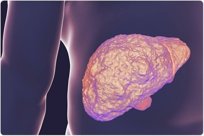
Advanced cirrhosis — the end stage of chronic liver disease from any cause — impairs bile synthesis and excretion across the entire hepatic parenchyma. As functional liver mass decreases, bilirubin excretion falls and bile production becomes insufficient for normal stool pigmentation. In cirrhotic patients, pale stool is one of several signs of decompensation, typically appearing alongside jaundice, ascites, and coagulopathy. Unlike acute obstructive causes, cirrhosis-related pale stool develops gradually as the disease progresses, often alongside other well-recognized features of liver failure. Related symptoms of right upper abdominal pain and liver health often accompany this presentation.
Primary Biliary Cholangitis and Primary Sclerosing Cholangitis
Primary biliary cholangitis (PBC) is a chronic autoimmune liver disease that specifically targets the small intrahepatic bile ducts. The immune system attacks and progressively destroys the epithelial cells lining these ducts, causing inflammation and fibrosis that impairs bile flow. Over time, impaired bile excretion leads to cholestasis with characteristic symptoms: fatigue, severe pruritus (itching caused by bile salt accumulation in the skin), and eventually jaundice with pale stool. PBC affects women more than men (roughly 9:1) and most commonly presents in middle age. The diagnosis is supported by elevated alkaline phosphatase and gamma-glutamyl transferase (GGT), positive anti-mitochondrial antibodies (AMA) in over 90% of cases, and characteristic liver biopsy findings.
Primary sclerosing cholangitis (PSC) is a distinct chronic disease that causes inflammation, fibrosis, and progressive stricturing of both intrahepatic and extrahepatic bile ducts. Unlike PBC, PSC affects more men than women and is strongly associated with inflammatory bowel disease — approximately 70–80% of PSC patients have concurrent ulcerative colitis. As bile duct strictures develop and progress, bile flow becomes increasingly impaired, leading to recurrent cholangitis (bile duct infection), progressively severe cholestasis, and eventual biliary cirrhosis. Pale stool in PSC reflects the intermittent and progressive nature of ductal obstruction. PSC also carries a significantly elevated risk of cholangiocarcinoma, making regular surveillance an important part of management.
Both PBC and PSC are diagnosed through a combination of serological markers, imaging (MRCP showing characteristic biliary tree abnormalities in PSC), and sometimes liver biopsy. Ursodeoxycholic acid is the standard treatment for PBC and improves biochemical markers and liver outcomes in the majority of patients. For PSC, obeticholic acid and several investigational agents are being studied, but endoscopic dilation of dominant strictures remains an important management tool for symptom relief and prevention of cholangitis. Both conditions carry a risk of progression to cirrhosis and liver failure, making early diagnosis and specialist follow-up important for long-term outcomes.
Associated Symptoms That Point Toward the Cause
Pale stool rarely appears in isolation. The accompanying symptoms — or their absence — are often as diagnostically informative as the pale stool itself. The most significant associated finding is dark urine: tea-colored or cola-colored urine in the context of pale stool strongly indicates that bilirubin is being excreted in the urine because it cannot reach the intestine through the bile duct. This combination (pale stool plus dark urine) is called acholic stool with bilirubinuria and is virtually diagnostic of biliary obstruction or significant intrahepatic cholestasis. When jaundice is also present — visible yellowing of the sclera (whites of the eyes) or skin — the clinical picture is complete and evaluation is urgent regardless of whether the cause is ultimately benign or malignant.
Pruritus (itching) without a rash is another hallmark of cholestasis. Bile salts that accumulate in the skin when bile cannot flow normally activate itch receptors, producing generalized itching that is often worst at night, on the palms, and on the soles of the feet. This symptom can precede jaundice and pale stool by weeks in slowly progressive biliary obstruction. Abdominal pain, when present, helps localize the cause: episodic right upper quadrant pain radiating to the shoulder is typical of gallstone-related biliary colic; dull epigastric pain radiating to the back is more characteristic of pancreatic disease; and diffuse upper abdominal tenderness with fever suggests cholangitis — infection within an obstructed bile duct that represents a medical emergency requiring immediate antibiotics and biliary decompression. Patients with changes in bowel habits alongside pale stool should be evaluated promptly.
The absence of pain can be clinically significant. Painless jaundice with progressive pale stool and weight loss — particularly in a patient over fifty — is a pattern that raises immediate concern for malignant obstruction (pancreatic cancer, cholangiocarcinoma) and warrants urgent imaging and specialist referral. Fatigue and unintentional weight loss accompanying pale stool also increase the likelihood of a serious underlying condition, distinguishing this picture from the benign dietary causes of transient stool color changes that resolve without intervention.
When Pale Stool Requires Urgent Evaluation
The urgency of evaluation depends on the full clinical picture rather than the pale stool alone. Seek same-day or emergency evaluation for pale stool accompanied by any of the following: high fever with chills and jaundice (Charcot’s triad of cholangitis — a biliary infection requiring emergency decompression); severe abdominal pain; visible jaundice that has developed rapidly over days; altered mental status or confusion in a jaundiced patient (a sign of acute liver failure or advanced cholangitis). These presentations can deteriorate rapidly and require hospital-level assessment.
Prompt evaluation within days — rather than emergency care — is appropriate for pale stool with any of the following: dark urine appearing alongside the pale stool; new jaundice without pain; unexplained weight loss; progressive itching without rash; or pale stool persisting for more than a week without an obvious benign explanation. Patients with known liver disease who develop pale stool should contact their gastroenterologist or hepatologist promptly, as this may represent a change in disease status. Pale stool in a newborn or infant — never normal — requires same-day pediatric evaluation, as biliary atresia (absence or underdevelopment of the bile ducts) can progress to liver failure if not treated within the first few months of life.
Observation without immediate evaluation may be reasonable for a single pale stool with no other symptoms in an otherwise healthy adult who recently changed diet (for example, significantly reduced fat intake, which decreases bile stimulation) or took bismuth-containing medications. However, if pale stools persist beyond two or three days or any accompanying symptoms develop, medical evaluation should not be delayed further. The approach to evaluating concerning stool changes follows a similar risk-stratification framework: benign causes are common, but systematic evaluation rules out the serious ones that require timely intervention.
Diagnosis — Blood Tests, Imaging, and Endoscopic Evaluation
The initial evaluation of pale stool centers on distinguishing between two broad categories: obstructive (extrahepatic) cholestasis, where bile cannot pass through the bile ducts, and intrahepatic cholestasis, where the liver itself is failing to produce or excrete bile properly. Liver function tests provide the first diagnostic layer. An elevated alkaline phosphatase (ALP) and GGT with relatively preserved transaminases (AST, ALT) suggests obstructive pathology. Markedly elevated transaminases with moderate ALP elevation points toward hepatocellular disease — hepatitis, drug injury, or ischemic hepatitis. Total and direct bilirubin help quantify the severity of cholestasis and distinguish conjugated from unconjugated hyperbilirubinemia. Coagulation studies (INR) and albumin assess synthetic liver function, which falls in severe hepatic disease but is typically preserved in early or purely obstructive cholestasis.
Abdominal ultrasound is the first-line imaging study for suspected biliary obstruction. It is widely available, inexpensive, and effectively identifies dilated bile ducts (a key sign of downstream obstruction), gallstones in the gallbladder, liver texture abnormalities, and mass lesions in the liver or pancreatic head. Its limitation is poor visualization of the distal common bile duct and ampullary region due to overlying bowel gas. When ultrasound is inconclusive or biliary obstruction is strongly suspected, MRCP (magnetic resonance cholangiopancreatography) provides non-invasive, high-resolution imaging of the entire biliary tree without requiring contrast injection into the ducts, making it the preferred follow-up study in most cases.
ERCP (endoscopic retrograde cholangiopancreatography) combines diagnostic imaging with therapeutic capability — it can directly visualize the ampulla, inject contrast into the bile ducts, obtain brushings for cytology, and immediately treat obstruction by removing stones, placing stents, or dilating strictures. Because ERCP carries procedure-related risks (post-procedure pancreatitis in three to five percent of cases, bleeding, perforation), it is typically reserved for patients in whom therapeutic intervention is anticipated rather than used as a first-line diagnostic study. Endoscopic ultrasound (EUS) has emerged as an important complementary tool for evaluating the distal bile duct and pancreatic head with high resolution, and it allows fine-needle aspiration of suspicious masses for tissue diagnosis when malignancy is suspected. Together, these diagnostic approaches allow precise identification of the cause of pale stool in nearly all patients.
Frequently Asked Questions About Pale Stool
Can a pale stool be caused by something I ate?
Yes, in some cases. A diet very low in fat reduces the stimulus for bile release into the intestine, which can result in slightly lighter stool. Bismuth-containing medications (such as Pepto-Bismol) usually cause black or very dark stool rather than pale stool, but the color effect depends on dose and transit time. Calcium supplements and antacids with calcium carbonate can lighten stool color in some individuals. However, these dietary effects typically produce tan or light brown stool rather than true clay-colored or white stool. Consistently pale, clay-colored stool is not explained by diet alone and warrants evaluation.
How is pale stool different from changes in bowel habits like diarrhea?
Pale stool refers specifically to the color of the stool rather than its consistency or frequency. A person can have normal-frequency, well-formed stool that is pale in color due to reduced bile, or they can have diarrhea that is also pale. The two problems are not mutually exclusive and can coexist — for example, in fat malabsorption syndromes, stools may be pale, bulky, and oily (steatorrhea) as well as loose. The presence of pale-colored diarrhea together with weight loss and abdominal discomfort warrants evaluation for gastrointestinal conditions affecting nutrient absorption and bowel pigmentation.
Is pale stool in a baby or infant a medical emergency?
Pale or acholic (colorless) stool in a newborn or young infant is a potential emergency. Biliary atresia — a condition in which the bile ducts fail to develop normally or are destroyed in early infancy — causes progressively acholic stools and jaundice. If untreated, biliary atresia leads to liver cirrhosis and failure within the first two years of life. The Kasai procedure (hepatoportoenterostomy), performed ideally before eight weeks of age, can restore bile flow and delay or prevent the need for liver transplantation. Pale stool in a newborn, even without obvious jaundice, should prompt same-day pediatric evaluation.
What blood tests confirm that pale stool is from a liver problem?
A liver function panel including AST, ALT, ALP, GGT, total and direct bilirubin, albumin, and INR provides a comprehensive initial picture. Elevated direct (conjugated) bilirubin confirms that bilirubin is being produced and conjugated but not excreted — consistent with obstruction or intrahepatic cholestasis. An ALP more than three times the upper limit of normal alongside elevated GGT (which confirms the ALP elevation is of hepatic rather than bone origin) is the biochemical hallmark of cholestasis. These tests, combined with imaging, usually establish the category of disease before any invasive evaluation is necessary.
Can pale stool resolve on its own?
If the cause is a temporary, self-limited condition — such as a brief episode of viral hepatitis A, a dietary change, or a medication that has since been stopped — stool color will typically normalize as the underlying cause resolves. However, pale stool from a bile duct stone, malignant obstruction, or progressive liver disease will not resolve without treatment of the underlying condition. Because the causes range from benign and self-limited to serious and requiring intervention, persistent pale stool should not be waited out without at least a basic laboratory and imaging evaluation to identify or exclude a treatable cause.
Sources: NIDDK — Liver Disease · Mayo Clinic — Clay-Colored Stools · ACG — Gallstones
Pale Stool During Pregnancy and in Older Adults
Pale stool in pregnancy requires the same evaluation as in any adult, though the list of common causes shifts slightly. Intrahepatic cholestasis of pregnancy (ICP) is a liver condition specific to pregnancy in which bile acid transport in the liver becomes impaired, leading to accumulation of bile acids in the blood and reduced bile excretion. Symptoms include severe generalized itching — often beginning on the palms and soles and worsening at night — followed by pale stool and sometimes mild jaundice. ICP carries a risk to the fetus and is managed with ursodeoxycholic acid and careful obstetric monitoring, with delivery typically planned around 36–37 weeks to reduce the risk of stillbirth associated with elevated bile acids.
In older adults, the threshold for evaluating pale stool should be lower than in younger individuals. Age-related changes in gallstone prevalence, higher rates of malignancy, and more frequent use of medications that can cause cholestatic liver injury all increase the likelihood that pale stool in someone over sixty reflects a condition requiring treatment. Painless jaundice with pale stool and weight loss in an older adult should be considered a pancreatic or biliary malignancy until proven otherwise, and a CT scan of the abdomen and pelvis with contrast — or at minimum an abdominal ultrasound — should be arranged promptly rather than deferred. Early detection significantly improves outcomes in pancreatic and biliary cancers where surgical resection remains possible.