
Helicobacter pylori (H. pylori) is a spiral-shaped bacterium that colonises the gastric mucosa — the lining of the stomach — and is responsible for the majority of peptic ulcer disease worldwide, the most common cause of gastric cancer after Epstein-Barr virus, and a significant contributor to functional dyspepsia. Understanding what the H. pylori test measures, which type of H. pylori test to use in different clinical situations, how to interpret results, and what happens after a positive diagnosis is essential information for the millions of people each year who undergo testing for this highly prevalent pathogen. H. pylori testing spans non-invasive methods — the urea breath test (UBT), the stool antigen test (HpSA), and serology — and invasive biopsy-based methods including rapid urease test (RUT), histology, and culture. Each has specific indications, strengths, and limitations. This article explains each H. pylori test in detail so that patients receiving testing or results can understand what each test actually detects, why their clinician chose one method over another, and what a positive or negative result means for their digestive health.
H. pylori infection affects approximately forty-four percent of the global population — over three billion people — with highest prevalence in low- and middle-income countries, older age groups, and populations with childhood exposure to crowded living conditions, contaminated water, or poor sanitation. In developed countries, prevalence is approximately thirty to forty percent in adults over fifty, falling to fifteen to twenty percent in younger age groups reflecting improved sanitation over recent decades. Transmission is primarily oral-oral or faecal-oral, typically acquired in childhood and persisting lifelong unless treated. The majority of people with H. pylori infection never develop symptomatic disease — the bacterium persists in a state of chronic low-grade gastritis in most hosts, with the minority developing ulcers, gastric cancer, or severe symptoms depending on bacterial virulence factors, host immune responses, and environmental cofactors. Testing is therefore targeted rather than universal — directed at patients with dyspepsia, peptic ulcer disease, early gastric cancer, first-degree relatives of gastric cancer patients, and patients before long-term NSAID or aspirin therapy.
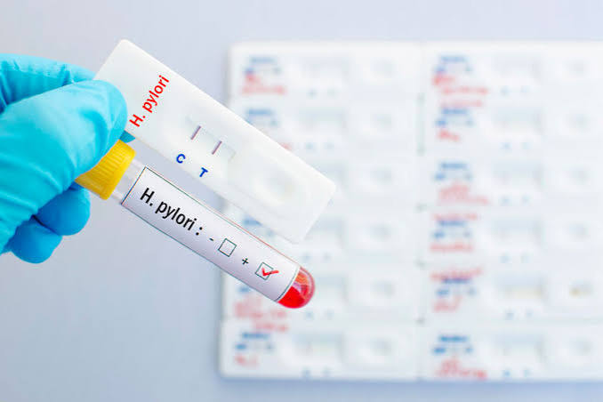
Urea Breath Test — The Gold Standard Non-Invasive Test
The urea breath test (UBT) is the most accurate non-invasive test for active H. pylori infection and is considered the gold standard for both initial diagnosis and post-treatment eradication confirmation. The test exploits H. pylori’s unique ability to produce large quantities of urease — an enzyme that breaks down urea into ammonia and carbon dioxide. The patient swallows a capsule or solution containing carbon-13 labelled urea (a stable, non-radioactive isotope used in clinical testing). If H. pylori is present in the stomach, its urease enzyme breaks down the labelled urea, and carbon-13 labelled CO₂ is absorbed into the bloodstream and exhaled. The patient breathes into a collection bag before and after swallowing the labelled urea, and the breath samples are sent to a laboratory where mass spectrometry measures the ratio of carbon-13 to carbon-12 in the exhaled CO₂. An elevated ratio compared to baseline confirms active H. pylori infection. Sensitivity and specificity both exceed ninety-five percent, making the UBT highly accurate. The test requires specific preparation to avoid false-negative results: proton pump inhibitors (PPIs like omeprazole, lansoprazole, pantoprazole) must be stopped at least two weeks before the test, bismuth compounds and antibiotics must be stopped at least four weeks before testing. PPIs suppress H. pylori activity without eradicating it, leading to false-negative results if the test is performed while the patient is taking them. The UBT is the preferred method for confirming successful eradication after antibiotic therapy, tested at least four weeks after completing treatment. It is appropriate across all age groups, requires no endoscopy, and produces a reliable real-time answer about active infection status. The article on stool tests for digestive health covers the stool antigen test as the alternative non-invasive option for settings where UBT is not available.
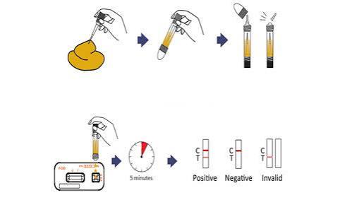
Stool Antigen Test and Serology for H. pylori
The H. pylori stool antigen test (HpSA) detects H. pylori-specific proteins in a faecal sample using monoclonal antibody-based immunoassay, with sensitivity and specificity comparable to the UBT (both exceeding ninety percent). The HpSA test requires the same preparation as the UBT — PPIs stopped two weeks before, antibiotics four weeks before. It is used for initial diagnosis of active infection and for post-treatment eradication confirmation (tested at least four weeks after completing antibiotic therapy). The stool antigen test is often preferred over the UBT in paediatric populations and in settings where UBT facilities are limited. Like the UBT, it detects active, current infection rather than past exposure, making it appropriate for both initial testing and eradication confirmation. H. pylori serology detects IgG antibodies against H. pylori antigens in the blood — a fundamentally different approach with important practical consequences. Because IgG antibodies persist for months to years after successful H. pylori eradication, a positive serology result does not confirm current active infection — it confirms past or current exposure. This makes serology inappropriate for post-treatment eradication confirmation: even after successful antibiotic cure, serology remains positive for six to twelve months or longer. Serology is used in specific circumstances: epidemiological research (population prevalence studies), initial diagnosis in patients who cannot stop PPIs (serology is not affected by PPI use), and assessment in patients with active gastrointestinal bleeding (where the acidity and protein from bleeding can temporarily suppress H. pylori activity, causing UBT and stool antigen false negatives). In clinical practice, serology has largely been replaced by the UBT and stool antigen test for initial diagnosis and has no role in post-treatment confirmation. The full liver and digestive biochemistry context that complements H. pylori testing is covered in the overview of liver function tests and the role of ALT and AST in detecting gastrointestinal-associated liver disease.
Biopsy-Based H. pylori Tests — Rapid Urease, Histology, Culture
When a patient undergoes upper gastrointestinal endoscopy for investigation of dyspepsia, peptic ulcer, or gastric pathology, the endoscopist can take gastric mucosal biopsies specifically for H. pylori testing. Three biopsy-based H. pylori tests are used: Rapid urease test (RUT, CLO test) — a gastric biopsy is placed in a urea-containing gel with a pH indicator; if H. pylori urease is present, it converts urea to ammonia, raising the pH and changing the gel colour from yellow to pink within one to two hours. The RUT is fast, inexpensive, and has good sensitivity (greater than ninety percent) when two biopsies from both antrum and corpus are tested. Like the UBT, it requires two weeks off PPIs for accurate results. Histology — biopsy sections are stained with Giemsa or haematoxylin and eosin and examined microscopically for H. pylori organisms. Histology additionally characterises the degree of gastritis, identifies intestinal metaplasia (a precancerous gastric mucosal change that increases gastric cancer risk), and can detect dysplasia or early cancer — making it the most informative biopsy-based test when gastric cancer risk assessment or mucosal pathology characterisation is the clinical priority. Culture and sensitivity testing — H. pylori is grown from gastric biopsy specimens on selective media, allowing identification of the specific antibiotic susceptibility profile of the infecting strain. Culture is the definitive method for guiding antibiotic selection in patients who have failed prior eradication therapy and are suspected to carry antibiotic-resistant H. pylori strains. Clarithromycin resistance rates are increasing globally (exceeding fifteen to twenty percent in many European and Asian countries), making susceptibility-guided therapy increasingly important in patients who fail first-line treatment. Culture is technically demanding, requires specialist laboratory capability, and takes several days — it is not routine initial testing but is valuable in the treatment-failure scenario.
H. pylori Eradication Therapy and Post-Treatment Testing
A positive H. pylori test result in a symptomatic patient is an indication for eradication therapy — antibiotic treatment aimed at eliminating the infection. Standard first-line regimens include triple therapy (PPI + clarithromycin + amoxicillin for fourteen days, achieving eradication in approximately eighty-five to ninety percent of cases in low-resistance populations) and quadruple therapy (PPI + bismuth + metronidazole + tetracycline — the preferred regimen in areas with high clarithromycin resistance, achieving eradication in approximately eighty-five to ninety-five percent). The fourteen-day regimen has higher eradication rates than seven-day regimens and is preferred in current guidelines. Bismuth-based quadruple therapy is increasingly used as first-line treatment in regions where clarithromycin resistance exceeds fifteen percent. After completing eradication therapy, post-treatment confirmation of eradication is strongly recommended for all patients by both the British Society of Gastroenterology and the American College of Gastroenterology: the UBT or stool antigen test should be performed at least four weeks after finishing antibiotics and two weeks after stopping PPIs. Confirming eradication matters for several reasons: patients with healed peptic ulcers have dramatically reduced ulcer recurrence risk after eradication (from approximately eighty percent five-year recurrence to less than ten percent); patients with MALT lymphoma (a gastric lymphoma driven by H. pylori) may achieve complete lymphoma remission after eradication alone; and patients with chronic active gastritis or intestinal metaplasia have reduced progression risk toward gastric cancer after eradication, particularly when treated before significant mucosal damage has occurred. Failure to confirm eradication leaves patients with persistent infection, continued peptic ulcer risk, and ongoing mucosal inflammation without appropriate management.
Frequently Asked Questions About H. pylori Testing
Do I need to be tested for H. pylori if I have no symptoms?
Routine testing of asymptomatic individuals is not currently recommended in most countries outside of specific high-risk populations. Testing is recommended for people with active peptic ulcer disease or a history of peptic ulcer, uninvestigated dyspepsia (the “test and treat” strategy endorsed by NICE and ACG guidelines for patients under fifty-five without alarm symptoms), first-degree relatives of gastric cancer patients (particularly where H. pylori prevalence is high), patients requiring long-term low-dose aspirin or NSAID therapy (H. pylori eradication before starting NSAIDs reduces ulcer risk), patients with unexplained iron deficiency anaemia unresponsive to iron supplementation, and patients with idiopathic thrombocytopenic purpura (ITP — H. pylori eradication can improve platelet counts). Screening of asymptomatic individuals in high-prevalence communities is under active investigation in countries with high gastric cancer incidence (Japan, South Korea, China) where the cancer burden justifies population-level eradication programmes. If you are concerned about H. pylori risk based on family history, previous symptoms, or living in a high-prevalence region, discussion with your GP about the appropriateness of testing is worthwhile. The article on when digestive symptoms need medical attention covers the symptom thresholds that trigger H. pylori testing in the uninvestigated dyspepsia pathway.
I tested positive for H. pylori. Does this mean I will get stomach cancer?
A positive H. pylori test does not mean you will develop gastric cancer. While H. pylori is classified as a Group 1 carcinogen by the WHO and is the most important modifiable risk factor for non-cardia gastric cancer, the vast majority of people infected with H. pylori — estimated at over ninety-five percent — never develop gastric cancer. Gastric cancer risk depends on multiple factors: bacterial virulence (the CagA-positive strains carrying the cytotoxin-associated gene A virulence factor are associated with higher gastric cancer risk), host genetic factors, the degree of gastric mucosal damage already present, and environmental cofactors including smoking, high-salt diet, and low fruit and vegetable intake. The greatest individual benefit from H. pylori eradication in terms of gastric cancer prevention occurs when treatment is given early — before intestinal metaplasia (a pre-cancerous change) has developed. Eradication after intestinal metaplasia is established reduces but does not eliminate gastric cancer risk — surveillance endoscopy is recommended for these patients. The appropriate response to a positive H. pylori test is eradication therapy and confirmation of cure — not anxiety about inevitable cancer. Early eradication of H. pylori is one of the most effective single-pathogen cancer prevention interventions available in medicine.
Sources: ACG — H. pylori and Peptic Ulcer Disease · NIDDK — Peptic Ulcer Diagnosis · WHO — H. pylori Antibiotic Resistance
H. pylori and Peptic Ulcer Disease
Before the discovery of H. pylori by Barry Marshall and Robin Warren in 1982 — for which they received the Nobel Prize in Physiology or Medicine in 2005 — peptic ulcer disease was considered a chronic condition driven by lifestyle factors (stress, spicy food, excess acid production) requiring lifelong acid suppression and frequently relapsing. The discovery that H. pylori infection is responsible for approximately seventy to ninety percent of duodenal ulcers and sixty to eighty-five percent of gastric ulcers transformed peptic ulcer disease from a chronic incurable condition into a curable infection in the majority of patients. H. pylori causes ulcers through multiple mechanisms: the bacterium’s urease enzyme produces ammonia that damages the mucous layer protecting the gastric epithelium; cytotoxins (particularly VacA and CagA virulence factors) induce direct mucosal cell damage and chronic inflammation; and H. pylori disrupts the normal balance of acid-stimulating and acid-inhibiting mechanisms in the stomach, increasing acid secretion in patients with duodenal ulcers. The net result is a chronically inflamed, poorly protected gastric and duodenal mucosa susceptible to acid-induced erosion and ulceration. H. pylori testing is indicated in all patients with confirmed peptic ulcer disease — both at initial diagnosis (to guide eradication therapy) and after treatment (to confirm eradication and explain healed ulcers). Patients with H. pylori-negative peptic ulcers should be evaluated for NSAID/aspirin use (the second most common cause of peptic ulcers), Zollinger-Ellison syndrome (gastrinoma-driven acid hypersecretion causing multiple, refractory peptic ulcers), and rare causes. Successful H. pylori eradication heals most duodenal ulcers within four to eight weeks and reduces ulcer relapse rates from approximately eighty percent at five years (without eradication) to under ten percent — a treatment benefit so dramatic that it redefined the entire field of peptic ulcer management. Understanding the biochemical changes associated with peptic ulcer disease — including the relationship to gastric acid and the markers covered in the liver function test overview — provides the full clinical context for H. pylori’s role in digestive pathology.
H. pylori, Gastric Cancer, and MALT Lymphoma
H. pylori is classified as a definite Group 1 carcinogen by the International Agency for Research on Cancer (IARC) and the WHO — one of the few infections formally classified as a cause of human cancer. H. pylori causes non-cardia gastric adenocarcinoma (the predominant gastric cancer type) through a multi-step carcinogenic cascade known as the Correa cascade: H. pylori infection leads to chronic active gastritis, which progresses over years to decades through atrophic gastritis (loss of normal gastric glands), intestinal metaplasia (replacement of gastric epithelium with intestinal-type epithelium), dysplasia, and ultimately invasive adenocarcinoma. This cascade is accelerated by the CagA-positive strains of H. pylori, smoking, high-salt diet, and genetic susceptibility factors. Approximately one to two percent of infected individuals develop gastric cancer over a lifetime — a low individual risk that translates to enormous population burden given the global scale of H. pylori infection: H. pylori is estimated to be responsible for approximately eighty-nine percent of non-cardia gastric cancers and approximately seventy-five percent of all gastric cancers worldwide. H. pylori eradication before the development of intestinal metaplasia significantly reduces gastric cancer incidence. H. pylori is also the primary cause of gastric MALT lymphoma (mucosa-associated lymphoid tissue lymphoma) — a low-grade B-cell lymphoma driven by chronic H. pylori-induced gastric mucosal immune stimulation. Remarkably, in localised gastric MALT lymphoma, H. pylori eradication alone (without chemotherapy or radiation) achieves complete lymphoma remission in approximately seventy-five to eighty percent of patients — making it one of the only oncological conditions where antibiotic therapy is the primary anti-cancer treatment. This striking therapeutic response reinforces the importance of identifying and treating H. pylori infection in all appropriate clinical settings. Patients with H. pylori and a family history of gastric cancer, those with high-grade intestinal metaplasia found on endoscopy, and those from high gastric cancer prevalence populations warrant discussion of long-term gastric cancer surveillance with their gastroenterologist.
What to Expect When Your H. pylori Test Is Positive
A positive H. pylori test result (by any validated method — UBT, stool antigen, or biopsy-based testing) in a patient with appropriate clinical indications is an indication to offer eradication therapy. The management pathway after a positive result typically proceeds as follows: the clinician discusses the test result and its implications with the patient, prescribes a first-line eradication regimen (most commonly fourteen-day triple or quadruple therapy as above), advises on the importance of completing the full course (antibiotic non-completion is a leading cause of treatment failure and antibiotic resistance), and schedules post-treatment testing at four to eight weeks after completing antibiotics — always at least two weeks after stopping PPIs to avoid false-negative confirmation results. The majority of patients experience no significant side effects from eradication therapy, though nausea, taste disturbance, and loose stools are common with metronidazole-containing regimens, and amoxicillin can cause diarrhoea. Patients should be counselled to complete the full course even if they feel well and to contact their prescriber if they develop significant adverse effects. If post-treatment testing confirms eradication success, no further H. pylori-specific management is typically needed for patients with uncomplicated dyspepsia or healed peptic ulcer — though patients with complicated peptic ulcer history (bleeding, perforation) and those with gastric intestinal metaplasia or gastric atrophy may require ongoing surveillance endoscopy as discussed with their gastroenterologist. If post-treatment testing shows persistent H. pylori infection, second-line therapy using a different antibiotic combination is prescribed, ideally guided by susceptibility testing from biopsy culture if available. Understanding the full landscape of digestive health testing — from stool tests to blood-based hepatitis markers covered in hepatitis blood tests explained — allows patients to understand how H. pylori investigation fits within the broader framework of gastrointestinal diagnostic workup.
H. pylori testing and eradication represent one of medicine’s clearest examples of a single intervention — identifying and treating an infection — preventing multiple serious outcomes including peptic ulcer disease, gastric cancer, and MALT lymphoma. For patients receiving a positive H. pylori result, the appropriate response is to complete eradication therapy, confirm success with post-treatment testing, and follow any additional surveillance recommendations from their gastroenterologist based on findings at endoscopy. For patients with negative H. pylori testing whose dyspepsia persists, investigation continues along the functional dyspepsia pathway — the article on GGT and liver health addresses the broader liver and biliary context that sometimes underlies upper abdominal symptoms attributed to dyspepsia. The increasingly global problem of H. pylori antibiotic resistance makes susceptibility-guided eradication an important tool for clinicians managing patients who have failed initial therapy, and culture-based sensitivity testing from endoscopic biopsy remains the definitive approach to selecting an effective regimen for treatment-resistant cases.
I had a positive urea breath test three months ago, completed the 14-day triple therapy, and just did my follow-up breath test — negative, H. pylori eradicated. My GP explained I needed to wait at least 4 weeks after finishing antibiotics before testing again, which I didn’t realise at first. The difference in my symptoms is remarkable — the bloating and burning pain I’d had for two years has almost completely resolved. I wish I’d been tested sooner.
Rachel, your experience illustrates the treatment pathway perfectly — the 4-week post-treatment wait before breath testing is critical, and many patients don’t realise the test will be falsely negative if done too soon after antibiotics or if PPIs haven’t been stopped beforehand. Symptom improvement after eradication can be dramatic, particularly in patients whose dyspepsia was driven by active H. pylori-related gastritis. A confirmed negative post-treatment UBT gives genuine peace of mind that the infection has been eliminated.
The section on MALT lymphoma is something most patients don’t know about — that a lymphoma can go into complete remission from antibiotics alone is genuinely remarkable and illustrates how profoundly H. pylori drives disease beyond simple gastritis and ulcers. I’m a retired pharmacist and still find this one of the most compelling examples of infection-driven oncology in medicine.