
Colonoscopy is a procedure in which a gastroenterologist or colorectal surgeon passes a long, flexible video endoscope through the rectum and colon to directly visualise the lining of the large intestine from the rectum to the caecum and, in most cases, the terminal ileum. It is the most accurate diagnostic tool for colonic pathology, enabling the detection and removal of polyps, the diagnosis of colorectal cancer, inflammatory bowel disease, and a range of other colonic conditions, and the investigation of symptoms including rectal bleeding, iron-deficiency anaemia, a change in bowel habits, and lower abdominal pain. For adults who have been referred for a colonoscopy — whether for investigation of symptoms, screening for colorectal cancer, or surveillance after previous polyp removal — understanding the preparation, the procedure itself, what the findings mean, and what to expect during recovery makes the experience significantly more manageable and the results more meaningful.
Colorectal cancer is one of the most common cancers in adults over fifty, and colonoscopy occupies a central role in its prevention as well as its diagnosis: the removal of adenomatous polyps (precancerous growths) at colonoscopy prevents a substantial proportion of colorectal cancers from ever developing. This dual role — diagnosis and prevention in a single procedure — makes colonoscopy one of the most valuable investigations in adult medicine. The stool-based colorectal cancer screening tests that may precede a colonoscopy referral are covered in the stool tests for digestive health article; the full range of diagnostic imaging that complements colonoscopy is discussed in the CT scan and abdominal ultrasound articles in this series.
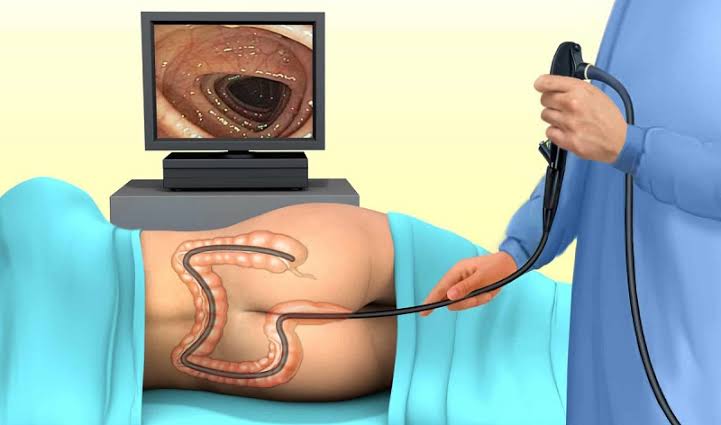
What Colonoscopy Examines and Why It Matters
The colonoscope is a flexible instrument approximately 160–180 cm in length and 11–13 mm in diameter, containing a CCD camera at the tip, an air/CO2 insufflation channel, a water jet, a suction channel, and a working channel for therapeutic instruments. The procedure visualises the entire colon: rectum (0–15 cm from the anal verge), sigmoid colon, descending colon, splenic flexure, transverse colon, hepatic flexure, ascending colon, caecum (identified by the appendiceal orifice and ileocaecal valve), and in most cases the terminal ileum (last 10–30 cm of the small intestine, intubated through the ileocaecal valve). Total colonoscopy — reaching the caecum — is the technical standard; incomplete colonoscopy (failure to reach the caecum) occurs in approximately five percent of procedures and may require alternative investigations such as CT colonography.
The clinical value of colonoscopy derives from three capabilities that no other investigation provides simultaneously: direct mucosal visualisation with high-definition imaging (detecting lesions as small as 1–2 mm); tissue sampling (biopsy forceps through the working channel); and therapeutic intervention (polypectomy, haemostasis, dilation). Imaging investigations — CT colonography, CT abdomen, MRI — can detect large colonic lesions and extra-colonic pathology but cannot obtain tissue or treat polyps. Stool-based tests detect evidence of colonic pathology indirectly but cannot localise or characterise the source. Colonoscopy integrates these capabilities, which is why it remains the investigation of choice for symptomatic colonic disease and is the gold standard for colorectal cancer surveillance in high-risk patients.
When Colonoscopy Is Recommended
Symptomatic investigation: Rectal bleeding (bright red blood per rectum or mixed with stool), particularly in adults over fifty or with associated change in bowel habit, weight loss, or anaemia — these combinations represent alarm features that mandate urgent colonoscopy. Iron-deficiency anaemia in adults, particularly post-menopausal women and men of any age, requires investigation of both the upper (endoscopy) and lower GI tract (colonoscopy) to exclude a colorectal source. Change in bowel habit (new onset constipation, diarrhoea, or alternating pattern) persisting for six or more weeks, particularly in adults over forty-five to fifty. Lower abdominal pain or discomfort without clear non-colonic explanation. Unexplained weight loss with lower GI symptoms. Mucus in stool combined with other alarm features.
Colorectal cancer screening: In patients with a positive stool-based colorectal cancer screening test — faecal immunochemical test (FIT) or faecal occult blood test (FOBT) — colonoscopy is performed to determine the source of occult blood and identify or exclude colorectal neoplasia. In the UK, the NHS Bowel Cancer Screening Programme offers FIT testing to adults aged fifty to seventy-four; positive results trigger a colonoscopy referral. Patients in higher-risk categories — those with a confirmed family history of colorectal cancer or adenomatous polyposis syndromes — may be offered primary colonoscopy screening at an earlier age and more frequently than the general population. The role of stool-based tests in colorectal cancer detection and when they trigger colonoscopy referral is covered in detail in the stool tests article.
Surveillance after previous pathology: Patients who have had adenomatous polyps removed at a previous colonoscopy require surveillance colonoscopy at intervals determined by the number, size, and histology of polyps found (as per BSG and ESGE post-polypectomy surveillance guidelines). Patients with a personal history of colorectal cancer require colonoscopy surveillance post-resection. Patients with long-standing ulcerative colitis or Crohn’s colitis require surveillance colonoscopy for dysplasia monitoring, with intervals based on disease extent and duration. Patients with hereditary colorectal cancer syndromes (Lynch syndrome, familial adenomatous polyposis) have intensive ongoing surveillance protocols.
Bowel Preparation: The Critical Step
The quality of bowel preparation is the single most important determinant of colonoscopy quality: an inadequately prepared colon obscures mucosal detail, increases procedure time and discomfort, reduces polyp detection rates, and frequently necessitates repeat colonoscopy. Understanding why preparation matters — and exactly how to follow the instructions — is the most important thing a patient can do before a colonoscopy.
Modern bowel preparations are high-volume or low-volume osmotic laxative solutions. High-volume preparations (PEG-based solutions such as MoviPrep or Klean-Prep, 2–4 litres total) work by osmotic action — drawing water into the bowel lumen to flush contents. Low-volume preparations (such as PLENVU, Picolight, or CitraFleet — 1 litre or sachet-based) use a combination of osmotic and stimulant laxative mechanisms and are preferred by many patients for their lower volume requirement. Split-dose preparation — half the preparation taken the evening before the procedure and half on the morning of the procedure (for afternoon colonoscopies) or the entire preparation the afternoon and evening before (for morning procedures) — consistently produces better bowel cleansing than single-dose preparation and is the current recommended standard for most patients.
During preparation: patients follow a low-residue or clear liquid diet for one to two days before the procedure; dietary restriction reduces the faecal load and significantly improves preparation quality. High-fibre foods (vegetables, fruit, nuts, seeds, high-fibre cereals) and red or purple foods (which can mimic blood endoscopically) are avoided. Adequate hydration during the preparation process prevents dehydration — patients should drink clear fluids liberally (water, clear broths, clear juice without pulp, black tea or coffee) throughout. Medications that affect bowel motility (iron supplements, constipating opiates) are typically adjusted before preparation. Patients should be near a toilet during the active preparation phase — the onset of action is typically within one to three hours of beginning the preparation, and the preparation phase may last four to six hours.
The Colonoscopy Procedure: What to Expect
On arrival at the endoscopy unit, patients complete a pre-procedure checklist and change into a hospital gown. A cannula is inserted, monitoring leads are attached, and sedation or Entonox (nitrous oxide inhalation) is administered — sedation choices for colonoscopy typically include intravenous midazolam with or without fentanyl. General anaesthesia is not routinely used for colonoscopy in most centres (though it is available in specific circumstances). The patient lies in the left lateral position; the endoscopist introduces the scope through the anus and advances it carefully around the colon. Carbon dioxide is used for insufflation in preference to air at most modern endoscopy units — CO2 is more rapidly absorbed, resulting in significantly less post-procedure bloating and discomfort than air.
Colonoscopy requires skill in scope manipulation to navigate the natural curves and angulations of the colon (sigmoid, splenic flexure, hepatic flexure) without causing excessive patient discomfort. The insertion phase (advancing to the caecum) and the withdrawal phase (careful inspection of all mucosal surfaces) both contribute to the quality of the examination. The withdrawal phase — typically taking at minimum six minutes for an adequate examination in a clean colon — is where the majority of polyps and lesions are detected. The adenoma detection rate (ADR) — the proportion of patients in whom at least one adenoma is found — is a key quality indicator for colonoscopists, with a minimum threshold of twenty-five percent (and higher benchmarks for experienced endoscopists) mandated by quality assurance programmes.
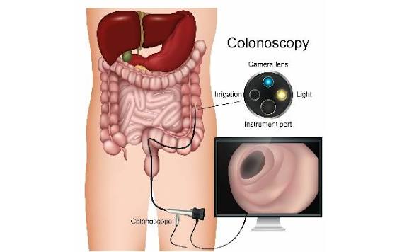
What Colonoscopy Can Find: Key Findings Explained
Polyps: Colonic polyps are protrusions from the mucosal surface that are found in approximately thirty to forty percent of adults over fifty at screening colonoscopy. They are classified by morphology (pedunculated on a stalk; sessile sitting flat; flat/depressed — the latter often more challenging to detect) and by histology: Hyperplastic polyps (typically small, located in the rectum and sigmoid, very low malignant potential); Adenomatous polyps (tubular, tubulovillous, or villous — these are the principal precursor to colorectal cancer; risk of malignant transformation correlates with size, villous component, and degree of dysplasia); Sessile serrated lesions (a more recently characterised polyp type, often flat and pale, particularly in the right colon, associated with an accelerated pathway to colorectal cancer via microsatellite instability); Traditional serrated adenomas (rare, high malignant potential). Polyps are removed endoscopically at the time of detection: small polyps (≤5 mm) by cold biopsy or cold snare; medium polyps (6–19 mm) by cold snare polypectomy; large polyps (≥20 mm) by endoscopic mucosal resection (EMR) or endoscopic submucosal dissection (ESD) or referral to a tertiary centre for complex resection.
Colorectal cancer: Colorectal cancer appears endoscopically as a mass, stricture, ulcerated lesion, or fungating growth — most commonly in the sigmoid colon or rectum, though right-sided cancers are increasingly recognised. Biopsy confirms the diagnosis. CT staging (covered in the CT scan article) follows to assess local and distant disease extent. Early-stage cancers (T1 confined to the submucosa, lymph node negative) detected through surveillance may be amenable to endoscopic resection.
Inflammatory bowel disease: Ulcerative colitis appears as continuous mucosal inflammation extending from the rectum proximally — endoscopic features include mucosal erythema, granularity, loss of vascular pattern, superficial ulceration, and in severe disease, spontaneous bleeding and deep ulceration. Crohn’s disease of the colon typically shows skip lesions (discontinuous involvement), deep longitudinal ulcers, cobblestone mucosa, and relative rectal sparing. Biopsies from multiple sites (including uninvolved mucosa) are essential for histological confirmation and disease activity assessment. The terminal ileum is particularly important to visualise in suspected Crohn’s disease, as ileocaecal involvement is present in the majority of patients. Colitis is also assessed and graded endoscopically using validated scoring systems (Mayo score for ulcerative colitis, Simple Endoscopic Score for Crohn’s) to guide treatment decisions and assess mucosal healing after therapy.
Diverticular disease: Diverticula — outpouchings of the colonic mucosa through the muscle wall — appear as multiple round openings in the colonic lumen, particularly in the sigmoid colon, in approximately forty to sixty percent of adults over sixty. Uncomplicated diverticulosis requires no treatment. Active diverticulitis (inflammation/infection) is diagnosed clinically and on CT; colonoscopy during an acute episode is avoided due to perforation risk but may be performed after recovery to exclude underlying malignancy mimicking diverticulitis. Haemorrhoids: Internal haemorrhoids are visible as mucosal protrusions at the dentate line on retroflexion in the rectum — they are a common cause of bright red rectal bleeding in adults of all ages but are a diagnosis of exclusion in those over fifty, in whom colonoscopy is required to exclude a proximal colonic source of bleeding.
Recovery and Results After Colonoscopy
Recovery after colonoscopy is typically rapid. Bloating from gas insufflation during the procedure resolves within a few hours — the use of carbon dioxide instead of air significantly reduces this. Mild cramping or discomfort is common and self-limiting. Patients who received intravenous sedation require thirty to sixty minutes of observation in the recovery bay before discharge. A responsible adult must escort sedated patients home; driving is prohibited for twenty-four hours after sedation. Patients who received Entonox only may recover more quickly.
The endoscopist will usually provide a verbal summary of findings before discharge. A written report is provided with the discharge documentation or sent to the referring clinician. Biopsy results typically take one to two weeks and are communicated by letter or at a follow-up appointment. After polypectomy, patients are advised to monitor for post-polypectomy bleeding (bright red rectal bleeding, which occasionally occurs up to two weeks after cold snare polypectomy) and to seek emergency medical attention if significant bleeding or severe abdominal pain develop. Diet is unrestricted after the procedure (assuming no major complications). Patients on anticoagulants may require specific instructions about when to restart their medication after polypectomy — the endoscopy unit will advise on this.
Risks and Complications of Colonoscopy
Colonoscopy carries a slightly higher complication risk than upper GI endoscopy, reflecting both the longer procedural duration and the therapeutic interventions commonly performed. For diagnostic colonoscopy without polypectomy, the serious complication rate is approximately one in two thousand to one in three thousand. The principal complications are: Perforation: Occurs in approximately one in one thousand to one in three thousand diagnostic colonoscopies and at higher rates after therapeutic polypectomy (particularly large or complex polypectomy). Perforation may present as severe abdominal pain, peritoneal signs, and clinical deterioration during or after the procedure, though delayed presentations (twelve to twenty-four hours later) occur. Management ranges from endoscopic clip closure to emergency surgical repair depending on size, location, and clinical status. Post-polypectomy bleeding: Occurs in approximately one to two percent of polypectomies. Immediate intra-procedural bleeding is managed endoscopically. Delayed post-polypectomy haemorrhage (occurring up to fourteen days after the procedure) typically presents as painless rectal bleeding — the majority resolve spontaneously, but significant haemorrhage requires endoscopic re-evaluation and haemostasis. Cardiopulmonary events: Minor oxygen desaturation under sedation is common and self-correcting; serious sedation-related complications are rare with appropriate pre-procedure assessment and monitoring. Splenic injury: Very rare; occurs through traction on the splenocolic ligament during scope manipulation at the splenic flexure — presents with left upper quadrant pain after the procedure and requires urgent CT assessment. Patients who develop severe abdominal pain, rectal bleeding exceeding a tablespoon or two, fever above 38°C, or feeling unwell after colonoscopy should seek emergency medical assessment without delay.
Frequently Asked Questions About Colonoscopy
The preparation is the worst part — is there any way to make it easier?
This is a widely shared experience and a genuine barrier to colonoscopy uptake. The practical strategies that help: choosing a low-volume preparation if available (1 litre split-dose rather than 4 litres significantly reduces preparation burden); chilling the solution in the refrigerator before drinking (improves palatability); consuming it through a straw; following each portion with clear fluid; and keeping a clear fluid diet for the day before rather than a full clear liquid fast, which reduces preparation volume required. The most important factor is starting the preparation as scheduled and completing the full dose — underpreparation is the primary cause of inadequate bowel cleansing, repeat procedures, and missed pathology. If you have previous experience with preparation that was difficult to complete, discuss this with the colonoscopy team before the procedure — alternatives may be available. The relationship between bowel habit, fibre intake, and colonic health that underpins both preparation adherence and colonoscopy outcomes is covered in the digestive health series on this site.
Adenomatous polyps were found and removed — what does my surveillance schedule mean?
Post-polypectomy surveillance intervals are determined by the number, size, and histological type of polyps found. Under current BSG guidelines: patients with one or two small adenomas (less than 10 mm, low-grade dysplasia) are considered low-risk and do not require colonoscopy surveillance — they return to routine bowel cancer screening. Patients with three or four small adenomas, or one or two adenomas of which at least one is 10 mm or more, are intermediate-risk — surveillance colonoscopy at three years. Patients with five or more adenomas, or three or more with at least one 10 mm or more, are high-risk — surveillance at one year. Patients with adenomas showing high-grade dysplasia or with sessile serrated lesions 10 mm or more may require more intensive surveillance. Understanding your surveillance interval means understanding the risk category the pathology placed you in — if this has not been clearly explained, ask your endoscopy unit or GP for clarification. The CT imaging that may accompany colorectal cancer staging for patients in whom a cancer is detected at colonoscopy is covered in the CT scan article.
How does colonoscopy fit into the overall digestive and liver investigation picture?
Colonoscopy addresses the lower gastrointestinal tract — it does not examine the oesophagus, stomach, or small bowel, and provides no information about liver, biliary, or pancreatic pathology. For patients whose symptoms span both upper and lower GI systems, colonoscopy may be complemented by upper GI endoscopy. For patients with lower GI symptoms accompanied by abnormal liver blood tests, the liver investigation pathway — liver function tests, abdominal ultrasound — runs in parallel with colonoscopy rather than being replaced by it. The broader question of how gastroenterologists and hepatologists select and sequence investigations is covered in the article on how doctors diagnose digestive and liver problems, which provides the systematic clinical framework within which colonoscopy sits.
Sources: BSG — Post-Polypectomy Surveillance Guidelines · ASGE — Colonoscopy Quality Indicators · NCI — Colorectal Cancer
For adults considering whether colonoscopy or an alternative investigation is more appropriate for their situation, the choice between colonoscopy and CT colonography (virtual colonoscopy) is a common clinical question. CT colonography uses CT imaging to create a three-dimensional reconstruction of the colon without a scope — it requires the same bowel preparation as conventional colonoscopy, provides accurate polyp detection for lesions above 6 mm, and avoids the procedural risks of perforation and sedation. Its limitations are that biopsies cannot be taken and polyps cannot be removed — patients with positive CT colonography findings require a follow-up conventional colonoscopy for tissue sampling and polypectomy. CT colonography is preferred in patients with incomplete conventional colonoscopy, patients who are anticoagulated and cannot be bridged safely for polypectomy, or patients with comorbidities that make colonoscopy higher risk. The CT imaging principles relevant to digestive investigations are covered in the CT scan for digestive and liver problems article, and the abdominal ultrasound article covers what ultrasound can and cannot detect in the context of lower GI symptoms and suspected colonic pathology at abdominal ultrasound. Understanding how these investigations complement colonoscopy — and when one is preferred over another — is the foundation of evidence-based lower gastrointestinal investigation.
Just had my colonoscopy following a positive FIT test — three adenomas found and removed, all tubular and low-grade. The post-polypectomy surveillance section is really helpful. My gastroenterologist said I’m intermediate risk and need a three-year repeat, but I didn’t fully understand the risk categorisation at the time. The BSG guidelines breakdown makes it much clearer.
Thank you Patrick — three tubular adenomas with low-grade dysplasia and no adenoma 10 mm or larger puts you squarely in the intermediate-risk category under current BSG guidelines, where three-year surveillance is the appropriate interval. The rationale is that the colonoscopy has cleared the existing polyp burden, and three years allows adequate time for any new polyps to develop to a detectable size while not leaving them long enough to progress significantly. Adhering to the three-year interval is the most important thing — the risk of colorectal cancer developing from low-grade tubular adenomas over three years in a clean colon is very low, so the surveillance programme is designed to maintain that protection. Glad you found the article helpful.
The section on bowel preparation is the most realistic description I’ve read. The advice to use a straw and chill the solution genuinely helped me get through my second colonoscopy more easily than my first. One thing I’d add: asking your unit about low-volume options before assuming you’ll have to manage 4 litres made a real difference for me — I got a 1-litre split-dose prep and it was far more manageable.