
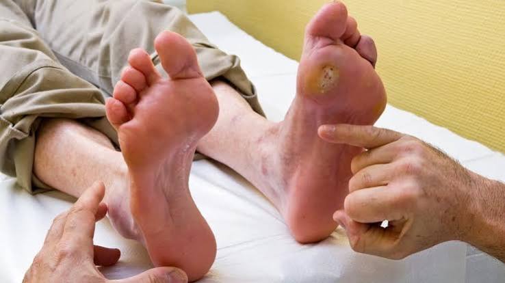
Diabetic foot problems represent one of the most devastating and yet most preventable of all diabetes complications — a fact that makes the continued high rate of diabetes-related amputations in the United States both a medical failure and an opportunity. The cascade that leads from diabetes to foot ulcer to amputation is well understood, the risk factors are identifiable, and the prevention strategies are straightforward and low-cost. Yet over 100,000 non-traumatic lower extremity amputations are performed annually in the U.S. in adults with diabetes — a rate 10 times higher than in the general population. Diabetic foot problems encompass a spectrum from minor calluses and nail deformities (which are significant risk factors in the presence of neuropathy and peripheral arterial disease) to life-threatening infections and gangrenous limbs. The unifying principle of diabetic foot care is that prevention is always superior to treatment: a daily foot inspection takes 2 minutes and prevents consequences that can take months of hospitalizations, surgeries, and rehabilitation to manage — and that sometimes cannot be prevented from culminating in amputation.
Diabetic foot disease accounts for more hospital bed-days than any other diabetes complication in the United States. Approximately 15% of adults with diabetes will develop a foot ulcer during their lifetime. Once a foot ulcer develops, the risk of amputation increases dramatically — approximately 20% of foot ulcers result in lower extremity amputation. Adults with diabetes who undergo below-knee amputation face a 5-year mortality rate of approximately 50%, which is higher than many cancers. The economic burden of diabetic foot disease — including hospitalizations, wound care, prosthetics, and rehabilitation — exceeds $9 billion annually in the United States. Yet studies consistently show that multidisciplinary foot care programs with regular foot examination, patient education, and early wound treatment reduce amputation rates by 50–85%.
Why Diabetes Makes Feet Uniquely Vulnerable
The diabetic foot is uniquely vulnerable to injury and infection because of the convergence of three simultaneous pathological processes — peripheral neuropathy (loss of protective sensation), peripheral arterial disease (reduced blood flow and healing capacity), and impaired immunity (reduced infection-fighting ability) — that act synergistically to create conditions where a minor injury becomes a life-threatening crisis:
- Peripheral neuropathy — losing the warning system that protects feet: Healthy feet are protected by an exquisitely sensitive pain system: sharp objects, excessive pressure, heat, and cold all generate pain signals that prompt immediate behavioral responses — removing the shoe, shifting weight, treating the wound. In adults with diabetic peripheral neuropathy and loss of protective sensation, this warning system is disabled. A pebble in a shoe, a poorly fitted shoe rubbing the side of a toe, a plantar callus over a bony prominence, or a nail penetrating the insole generates no warning — and the wound accumulates for hours, days, or weeks before the person removes their shoe and discovers an open wound. Neuropathy also causes motor fiber damage — loss of intrinsic foot muscle function that creates hammer toe, claw toe, and pes cavus (high-arched foot) deformities that concentrate plantar pressure at specific points, dramatically increasing callus formation and ulceration risk at those sites. Autonomic neuropathy contributes by reducing sweating in the foot — dry, cracked skin is far more susceptible to bacterial entry than healthy skin — and by causing the paradoxical vasodilation of Charcot neuroarthropathy. Our diabetic neuropathy: symptoms and prevention guide covers peripheral and autonomic neuropathy comprehensively.
- Peripheral arterial disease — impaired blood flow and healing: Peripheral arterial disease (PAD) — atherosclerosis of the arteries supplying the legs and feet — is two to four times more prevalent in adults with diabetes than in the general population, occurs at a younger age, and typically involves below-the-knee (tibial and peroneal) arteries more than the above-knee vessels that PAD primarily affects in non-diabetic adults. Tibial artery disease is particularly problematic for wound healing: reduced blood flow to the foot impairs the delivery of oxygen, nutrients, and immune cells needed for tissue repair, and reduces the concentration of antibiotics that penetrate the wound site. Wounds that would heal normally in a person with intact circulation fail to heal in the ischemic diabetic foot — bacteria multiply in the nutrient-rich but poorly perfused wound bed, causing rapidly spreading infection. The assessment of pedal pulses, ankle-brachial index (ABI), and toe pressures at each comprehensive foot examination identifies adults at highest risk for ischemia-complicated wound healing. Calcification of tibial arteries in diabetes can falsely elevate ABI, making toe-brachial index (TBI) — which measures blood pressure at the toe, where calcification is less severe — the preferred test for PAD assessment in adults with diabetes.
- Immune dysfunction — impaired infection fighting in the wound: Hyperglycemia impairs multiple components of the immune response that normally protect against skin infections: neutrophil chemotaxis (migration toward infection sites), phagocytosis (engulfment and killing of bacteria), and oxidative burst (the reactive oxygen species that destroy bacteria inside neutrophils) are all significantly reduced in poorly controlled diabetes. The result is that bacteria that would be rapidly eliminated in a person without diabetes can establish a foothold in the diabetic foot wound, progress from superficial skin infection to deep tissue infection involving tendons, fascial planes, and bone (osteomyelitis), and trigger systemic sepsis. Osteomyelitis complicates approximately 20% of moderate and severe diabetic foot ulcers and dramatically increases the likelihood of amputation — bone infection is very difficult to eradicate without surgical debridement of infected bone tissue, which may require partial or complete toe amputation, ray resection (removal of a toe and its metatarsal), or more proximal amputation if the infection has spread. Our diabetes complications: what adults should know guide covers the comprehensive complication context including immune dysfunction.
Recognizing Diabetic Foot Problems: What to Look For
Early recognition of diabetic foot problems — before a superficial wound becomes a deep infection — requires both daily self-examination by the adult with diabetes and regular professional evaluation by a healthcare provider. Understanding what specific findings to look for, and which findings require urgent attention, is the practical skill that translates foot examination from a recommendation to a life-saving behavior:
- Calluses and corns — the precursors to ulceration: Calluses — areas of thickened skin caused by chronic, repetitive pressure or friction — are not simply a cosmetic concern in adults with diabetic neuropathy: they are direct precursors to foot ulceration. As a callus grows thicker, the concentrated pressure it transmits to the underlying dermis and subcutaneous tissue eventually causes tissue breakdown from the inside out — creating an “inside-out” ulcer that begins in the deep tissue layers and breaches the callus surface. Adults with diabetes who have any callus — particularly plantar calluses under the metatarsal heads or heel — should have them professionally debrided by a podiatrist on a regular schedule (typically every 6–8 weeks) and should use prescribed accommodative insoles or custom orthotics to redistribute plantar pressure away from the callus site. Attempting to remove calluses with over-the-counter chemical agents (salicylic acid plasters) or sharp instruments at home is dangerous in the setting of neuropathy and reduced healing capacity.
- Nail abnormalities — infection risk and ingrown nail hazard: Nail pathology is a significant but underappreciated risk factor for diabetic foot infection. Onychomycosis (fungal nail infection) — which causes thickened, discolored, crumbly nails — is two to three times more common in adults with diabetes. Thickened nails are more prone to trauma, particularly when they are compressed in a shoe, and create a reservoir for bacteria and fungi that can seed adjacent skin injuries. Ingrown toenails — when the nail plate grows into the lateral nail fold — cause tissue trauma and open portals for bacterial infection that can progress to soft tissue infection and potentially osteomyelitis of the distal phalanx. Adults with diabetes should never cut their nails too short or round the corners — nails should be cut straight across with a nail clipper, and any ingrown nail should be evaluated by a podiatrist rather than treated at home. Nail care should be performed by a podiatrist for adults who cannot see their nails clearly, who have reduced sensation in their hands, or who have very thickened nails.
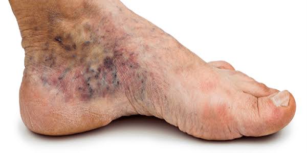
- Skin changes that signal vascular compromise: Several skin findings indicate reduced blood flow to the foot and signal elevated ulceration and poor-healing risk: dependent rubor (the foot turns dark red when lowered below heart level and blanches when elevated, due to passive capillary filling in the setting of arterial insufficiency), hair loss on the dorsum of the foot and toes (from reduced vascular delivery of nutrients to hair follicles), atrophic shiny skin (thin, tight skin without normal subcutaneous tissue padding), and slow capillary refill time (when the hallux is compressed and released, the nail bed should return to pink within 2 seconds — delayed refill indicates impaired perfusion). Cold feet compared with normal skin temperature are an additional indicator of reduced arterial flow. Any of these findings warrant assessment with ABI or TBI testing and potential vascular surgery referral for revascularization evaluation.
Diabetic Foot Ulcer: Treatment and When to Seek Urgent Care
When a diabetic foot ulcer develops — despite prevention efforts, or in an adult who presents with an established ulcer — the treatment goals are: relieving pressure on the wound (offloading), treating infection aggressively, restoring blood flow if ischemia is present, and providing appropriate wound care. The severity of the ulcer determines the urgency and setting of treatment:
- Offloading — removing pressure from the ulcer site: Offloading — reducing or eliminating the mechanical forces that caused the ulcer and prevent its healing — is the single most important wound care intervention for neuropathic plantar ulcers. Without offloading, the healing process is continuously disrupted by the pressure and shear forces of walking. Total contact casting (TCC) — a custom-molded, non-removable cast that distributes weight across the entire plantar foot surface — is the gold-standard offloading device for neuropathic plantar ulcers, with healing rates of 65–90% within 6–12 weeks in studies. Despite its superior efficacy, TCC is underused because of the skill required to apply it, the need for weekly cast changes for wound inspection, and the perception (often unfounded) that patients will not comply. Removable cast walkers — air-cast boots that can be made non-removable with an overwrap — offer a practical alternative with healing rates intermediate between TCC and standard footwear.
- Infection assessment and treatment — what requires hospitalization: The Infectious Diseases Society of America (IDSA) classifies diabetic foot infections by severity: mild infections (localized cellulitis less than 2 cm around the ulcer, no systemic signs) can be managed with oral antibiotics on an outpatient basis. Moderate infections (cellulitis beyond 2 cm, deeper tissue involvement, or lymphangitis) typically require intravenous antibiotics. Severe infections (systemic signs of infection — fever, elevated white count, hypotension — or deep space infection with necrotizing fasciitis, myositis, or osteomyelitis) require immediate hospitalization and surgical consultation. Osteomyelitis is suspected when the ulcer is deep enough that the bone can be probed (the “probe-to-bone” test, which has a positive predictive value of approximately 89% for osteomyelitis), or when imaging (MRI — the most sensitive test) shows bone marrow edema. Osteomyelitis often requires surgical debridement and prolonged antibiotic courses (4–6 weeks of IV antibiotics if not surgically debrided, or 4–6 weeks of combined IV/oral therapy for surgically treated disease).
- Advanced wound care modalities: For ulcers that do not respond to standard wound care within 4 weeks, advanced therapies are available with varying levels of evidence: negative pressure wound therapy (NPWT/VAC therapy) — which applies sub-atmospheric pressure through a sealed wound dressing to remove exudate, reduce edema, stimulate granulation tissue formation, and improve blood flow — is well-supported by evidence for accelerating diabetic foot wound closure. Bioengineered skin substitutes (acellular dermal matrix products, cellular skin grafts) provide extracellular matrix scaffolding for wound healing and growth factors that stimulate tissue regeneration. Hyperbaric oxygen therapy (HBO) — breathing 100% oxygen at increased atmospheric pressure — increases tissue oxygen delivery in ischemic wounds and has shown benefit in randomized trials for limb salvage in patients with diabetic foot infections and ischemia who are not candidates for vascular intervention. Our diabetes complications overview covers all foot complication management in the broader context of diabetes care. The American Podiatric Medical Association’s diabetic foot resources, the NIDDK’s diabetic foot problems information, and the CDC’s diabetic foot problems overview provide authoritative guidance on prevention and treatment.
Daily Foot Care Practices That Prevent Diabetic Foot Problems
The following daily foot care practices, recommended by the ADA, the NIDDK, and major podiatric organizations, have the strongest evidence for preventing diabetic foot ulcers and amputations in adults with diabetes, peripheral neuropathy, or peripheral arterial disease:
- Daily foot inspection — the most important prevention practice: Every adult with diabetes and neuropathy or PAD should inspect both feet completely every day — examining the plantar surface (using a mirror or asking a family member), between the toes, the heels, and the nail beds for any redness, blistering, callus formation, crack, cut, swelling, or discoloration. This inspection should take 2–3 minutes and should be done at a consistent time each day (after bathing is ideal, as the skin is clean and visible). Any new finding that was not present the previous day should be evaluated — either by the person themselves if clearly minor (small superficial abrasion, minor redness resolving within 24 hours) or by a healthcare provider if the finding is larger, not improving, or involves broken skin, swelling, or warmth. Adults who discover they have been walking on an open wound without knowing it require immediate professional evaluation.
- Proper footwear — the physical barrier between the foot and injury: Footwear selection is the most modifiable structural risk factor for diabetic foot ulcers. Key principles: shoes must be professionally fitted (foot size and width change with age, and the person cannot rely on past shoe size); there must be at least one thumb’s width of space between the longest toe and the shoe toebox; the shoe must be wide enough to accommodate the forefoot without compressing the fifth metatarsal head; the internal shoe surfaces must be smooth (no internal seams that create pressure points); and the shoe should have a firm heel counter for stability. Adults with significant foot deformities (hammer toes, claw toes, Charcot deformity, prominent metatarsal heads), neuropathy with loss of protective sensation, or a prior foot ulcer should be evaluated for prescription therapeutic footwear — including custom-molded shoes and accommodative or custom orthotic insoles — which are covered by Medicare and most insurance plans when prescribed by a physician and provided by a certified pedorthist or podiatrist. Never purchase shoes without trying them on and walking in them, and always inspect the shoe interior before putting it on each time — foreign objects, torn linings, and internal seams are common undetected injury sources in adults with neuropathy.
- Skin care, nail care, and temperature precautions: Keeping the skin of the feet well-moisturized — using unscented lotion applied to the plantar surface and heels (but not between the toes, where moisture promotes fungal infection) — prevents the dry skin cracking that creates bacterial entry portals. Water temperature should always be tested with an elbow or thermometer before foot bathing — adults with sensory neuropathy cannot feel scalds that would be immediately obvious to a person with normal sensation. Never use a heating pad, hot water bottle, or electric blanket on the feet — burns from these devices are a significant cause of serious foot injuries in adults with neuropathy. Never walk barefoot — indoors or outdoors — including on the beach (hot sand burns are a common cause of serious foot injuries in adults with neuropathy who are unaware of the danger). Trimming nails straight across (not into the corners) reduces ingrown nail risk. The complete A1C monitoring guide — because glucose control is the upstream prevention strategy for all foot complications — is in our A1C testing schedule guide. The comprehensive annual monitoring that coordinates foot examinations with all other diabetes care is in our annual diabetes care checklist.
Charcot Neuroarthropathy: The Most Misdiagnosed Diabetic Foot Condition
Charcot neuroarthropathy — also called Charcot foot — is one of the most severe and most frequently misdiagnosed diabetic foot conditions. It occurs when peripheral neuropathy leads to fractures and dislocations in the bones and joints of the foot and ankle that the person cannot feel, resulting in progressive bone destruction and foot deformity that dramatically elevates ulceration and amputation risk:
- Presentation and misdiagnosis: The acute Charcot foot presents as a hot, swollen, red foot — findings that are commonly misdiagnosed as cellulitis, gout, deep vein thrombosis, or osteomyelitis. The key distinguishing feature is that the acute Charcot foot is typically non-tender despite appearing severely inflamed — because the underlying fractures and dislocations are painless due to neuropathy. Skin temperature is often markedly elevated (3–7°C above the contralateral foot) from the inflammatory hyperemia. Delaying diagnosis and treatment — including any weight-bearing — allows progressive bone collapse into the pathognomonic rocker-bottom deformity: a collapsed midfoot arch that creates a new bony plantar prominence that is at extreme risk for ulceration. Misdiagnosis as cellulitis and treatment with antibiotics while continuing to weight-bear is the most common error and can be catastrophic. Any hot, swollen, red foot in an adult with diabetes and neuropathy must be evaluated for Charcot neuroarthropathy before any other diagnosis is assumed.
- Diagnosis and treatment: Plain X-rays may show fractures and dislocations in Charcot foot, but are often normal in the acute phase before collapse is visible — MRI is more sensitive for detecting the bone marrow edema that precedes structural disruption. Treatment is immediate total non-weight-bearing (wheelchair or crutches — no walking even with a cast) and referral to a multidisciplinary diabetic foot center with experience managing Charcot neuroarthropathy. Total contact casting in the non-weight-bearing phase protects the foot from collapse while the acute phase resolves. The active Charcot phase can last 6–12 months — defined by persistent warmth, swelling, and active bone remodeling visible on imaging. Bisphosphonates (pamidronate, zoledronic acid) reduce bone turnover in the acute phase and may shorten the active Charcot phase duration. Surgical stabilization (internal fixation with screws, plates, or external fixators) is considered for severe deformity or instability that cannot be managed non-operatively. Our diabetic neuropathy guide covers the autonomic neuropathy mechanisms that contribute to Charcot development. The comprehensive annual monitoring that includes foot examination is in our annual diabetes care checklist.
Sources: American Diabetes Association — Standards of Medical Care in Diabetes, diabetic foot care and comprehensive foot examination; NIDDK — diabetic foot problems prevention and treatment; CDC — diabetes-related lower extremity amputation statistics; American Podiatric Medical Association — diabetic foot care clinical practice guidelines; IDSA (Infectious Diseases Society of America) — diabetic foot infection classification and antibiotic guidelines; Charcot neuroarthropathy — diagnosis, acute management, and total contact casting; osteomyelitis probe-to-bone test positive predictive value (89%); total contact casting efficacy for neuropathic plantar ulcers (65–90% healing within 12 weeks); NPWT/vacuum-assisted closure for diabetic foot wound healing; hyperbaric oxygen therapy randomized trial evidence for limb salvage; peripheral arterial disease tibial artery involvement and below-knee ABI limitations in diabetes; toe-brachial index as preferred PAD test in calcified tibial arteries; amputation rates in adults with diabetes (10x general population); 5-year mortality after below-knee amputation (approximately 50%); multidisciplinary foot care programs reducing amputation rates by 50–85%; onychomycosis prevalence in diabetes; motor neuropathy, intrinsic foot muscle wasting, and hammer toe/claw toe deformity; Medicare coverage for therapeutic footwear in adults with diabetes; negative pressure wound therapy mechanism of action and evidence; bioengineered skin substitutes for chronic wound healing.
This breakdown of diabetic foot problems: prevention and care is exactly what patients need before a specialist appointment. I especially valued the explanation of why these recommendations exist, not just what they are. Will definitely be coming back to this site for more health information.
Finally a resource that explains diabetic foot problems: prevention and care in plain language. This is the kind of evidence-based writing that actually changes how people approach their health. Forwarding this to others in my support group who are dealing with similar issues.
This is one of the clearest explanations of diabetic foot problems: prevention and care I have found. The section on managing this condition day-to-day was especially useful for planning. I wish I had found this article earlier — would have saved a lot of confusion.