
Bruising more easily than usual — noticing bruises that appear from minor contact, develop in unusual locations, or take an unusually long time to fade — can be one of the earliest visible signs of impaired liver function. The liver plays a central role in hemostasis, the physiological process by which bleeding is controlled and wounds are sealed. It synthesizes the majority of clotting factors, produces thrombopoietin (which stimulates platelet production), and clears activated coagulation factors from the circulation. When liver function declines — whether from chronic hepatitis, cirrhosis, alcohol-related liver disease, or other hepatic conditions — these hemostatic processes are impaired in multiple simultaneous ways, producing a bleeding tendency that manifests clinically as easy bruising, prolonged bleeding from minor cuts, and, in advanced liver disease, life-threatening hemorrhage from varices or coagulopathy.
Easy bruising is often dismissed by patients and clinicians as a minor issue — attributed to aging, aspirin use, or simply being “fair-skinned” — without consideration of underlying liver disease. In reality, easy bruising in a patient with risk factors for liver disease (alcohol use, viral hepatitis exposure, metabolic syndrome, or unexplained fatigue) warrants liver function testing as part of a systematic evaluation. Understanding the hepatic mechanisms of easy bruising and the conditions that cause it provides a framework for recognizing when this common symptom has a clinically significant cause.
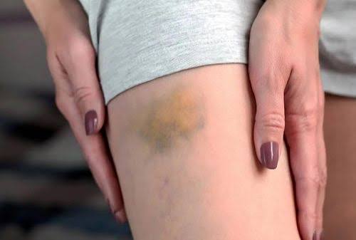
How the Liver Controls Bleeding — Clotting Factors and Hemostasis
Normal hemostasis requires the coordinated action of platelets, coagulation factors, and vascular endothelium — and the liver contributes to all three. The coagulation cascade — the sequence of enzymatic reactions that culminates in fibrin clot formation — depends on factors I (fibrinogen), II (prothrombin), V, VII, VIII, IX, X, XI, XII, and XIII, most of which are synthesized exclusively or predominantly in hepatocytes. Factors II, VII, IX, and X are vitamin K-dependent — they require vitamin K for a post-translational modification (gamma-carboxylation) that is essential for their activation. Because the liver both synthesizes these factors and processes the fat-soluble vitamin K absorbed from bile (and bile production is impaired in cholestatic liver disease), liver disease disrupts vitamin K-dependent factor production through two overlapping mechanisms: reduced hepatocyte synthetic capacity and reduced bile availability for vitamin K absorption.
The prothrombin time (PT) and its derived measure the INR (International Normalized Ratio) measure the extrinsic coagulation pathway — which includes factor VII, the shortest-lived clotting factor — and are the most sensitive standard coagulation tests for detecting early hepatic synthetic dysfunction. Factor VII has a half-life of only four to six hours, meaning that even brief periods of impaired hepatic synthesis produce measurable PT/INR prolongation. In clinical practice, an elevated INR in a patient without anticoagulant therapy is a reliable indicator of reduced hepatic clotting factor production. In the MELD score used to prioritize liver transplantation, INR is one of three components — alongside creatinine and bilirubin — reflecting its prognostic importance in end-stage liver disease.
Beyond clotting factors, the liver also synthesizes anticoagulant proteins including protein C, protein S, and antithrombin. The simultaneous deficiency of both pro- and anticoagulant factors in cirrhosis produces what hepatologists call “rebalanced hemostasis” — a state that appears relatively stable under standard coagulation testing (PT/INR, APTT) but is actually fragile and prone to tipping into either excessive bleeding or thrombosis depending on the clinical context. Standard coagulation tests in cirrhosis therefore overestimate bleeding risk and do not fully capture the complexity of hemostatic balance in liver disease — a clinically important point when managing these patients perioperatively or when interpreting coagulation results.
Thrombocytopenia — Low Platelet Count in Liver Disease
Thrombocytopenia — a platelet count below 150,000 per microliter — is common in chronic liver disease and contributes to easy bruising alongside clotting factor deficiency. Multiple mechanisms converge to reduce platelet counts in liver disease. Portal hypertension from cirrhosis causes splenomegaly (an enlarged spleen from venous congestion), and the enlarged spleen sequesters and destroys platelets at an accelerated rate — a phenomenon called hypersplenism. Because the spleen normally sequesters approximately one-third of the body’s platelets, even modest splenomegaly can substantially reduce the circulating platelet count. Platelet counts of 50,000–100,000 per microliter are common in compensated cirrhosis; counts below 50,000 produce clinically significant bleeding risk and are associated with advanced portal hypertension.
Thrombopoietin (TPO), the primary hormone stimulating platelet production by the bone marrow, is predominantly produced by the liver. Reduced TPO production in cirrhosis decreases the bone marrow’s platelet output, compounding the hypersplenism-related platelet destruction. Viral infections — particularly hepatitis C — can directly suppress bone marrow platelet production through immune-mediated mechanisms. Alcohol — which directly suppresses bone marrow hematopoiesis including platelet production — is an additional contributor in patients with alcoholic liver disease. The combined effect of reduced TPO, hypersplenism, direct bone marrow suppression (in relevant causes), and, in some patients, immune-mediated platelet destruction produces the multi-factorial thrombocytopenia of advanced liver disease. Avatrombopag and lusutrombopag — oral TPO receptor agonists — are now available for temporary platelet count increase before invasive procedures in patients with chronic liver disease and severe thrombocytopenia, replacing the need for platelet transfusions in many circumstances.
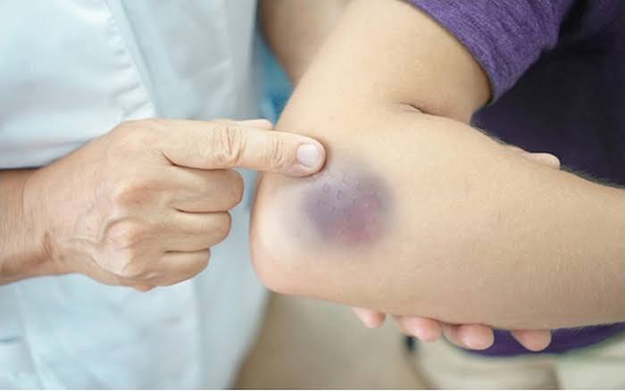
Causes of Easy Bruising Related to Liver Disease
Any condition that significantly reduces hepatic synthetic function can produce the coagulopathy that causes easy bruising. Cirrhosis from any cause — alcoholic liver disease, chronic viral hepatitis B or C, MASLD with progression to MASH and fibrosis, autoimmune hepatitis, or primary biliary cholangitis — reduces clotting factor synthesis proportional to the degree of hepatocellular loss. The coagulopathy of cirrhosis is progressive: patients with Child-Pugh A (compensated) cirrhosis may have mild PT prolongation and mild thrombocytopenia with minimal clinical impact, while those with Child-Pugh C (decompensated) cirrhosis may have severe coagulopathy requiring careful management before any invasive procedure.
Acute liver failure — from acetaminophen overdose, acute viral hepatitis, drug-induced liver injury, or Wilson’s disease crisis — produces particularly severe and rapidly evolving coagulopathy because all hepatic clotting factor synthesis is abruptly lost. The development of INR above 1.5 in the absence of anticoagulant therapy in a patient with acute hepatocellular injury is one of the criteria for diagnosing acute liver failure — a medical emergency requiring immediate transfer to a liver transplant center. In acute liver failure, the coagulopathy reflects not just factor deficiency but also disseminated intravascular coagulation (DIC), platelet consumption, and fibrinolysis — producing a complex hemostatic failure that is difficult to manage and carries high mortality without transplantation.
Cholestatic liver disease — including primary biliary cholangitis, primary sclerosing cholangitis, and biliary obstruction — impairs vitamin K absorption by reducing bile salt availability in the intestine. This vitamin K deficiency specifically affects the vitamin K-dependent clotting factors (II, VII, IX, X) and anticoagulant proteins (C and S), producing a coagulopathy that is initially reversible with parenteral (injected) vitamin K supplementation. A therapeutic trial of intravenous or intramuscular vitamin K is a useful diagnostic and therapeutic maneuver in a patient with elevated PT/INR and cholestatic liver disease — correction of the INR toward normal confirms vitamin K deficiency rather than true synthetic failure, while failure to correct confirms significant hepatocellular dysfunction. Associated symptoms of liver disease including jaundice, dark urine, and pruritus frequently accompany easy bruising in cholestatic conditions.
Non-Hepatic Causes of Easy Bruising to Differentiate
Easy bruising has many causes beyond liver disease. Medications are among the most common: aspirin and non-steroidal anti-inflammatory drugs (NSAIDs) impair platelet function and increase bruising tendency; anticoagulants (warfarin, direct oral anticoagulants such as rivaroxaban and apixaban) directly impair coagulation; long-term corticosteroids cause skin fragility and perivascular tissue atrophy that increases susceptibility to bruising from minor trauma; and certain antidepressants and antiepileptic drugs can affect platelet function or coagulation. A comprehensive medication review is essential in any patient presenting with easy bruising.
Hematological conditions — including thrombocytopenic purpura (ITP), von Willebrand disease, hemophilia, and hematological malignancies (leukemia, lymphoma, multiple myeloma) — produce bleeding tendencies and easy bruising through mechanisms independent of liver disease. Cushing’s syndrome from endogenous cortisol excess or exogenous corticosteroid use produces characteristic bruising patterns (typically on the trunk, arms, and thighs) from cutaneous fragility. Vitamin C deficiency (scurvy) — underrecognized in malnourished adults, the elderly, and those with dietary restrictions — causes perifollicular hemorrhage and bruising from impaired collagen synthesis. Simple senile purpura — superficial bruises appearing on the dorsum of the hands and forearms in older adults from age-related skin and vascular changes without any hemostatic defect — is the most common cause of easy bruising in elderly patients and is entirely benign. The clinical history, distribution of bruising, medication review, and targeted laboratory testing (complete blood count, coagulation panel, liver function tests) efficiently differentiate these possibilities.
Frequently Asked Questions About Easy Bruising and Liver Function
What liver tests detect bleeding risk from liver disease?
The most clinically relevant tests are PT/INR (measuring clotting factor activity in the extrinsic pathway, primarily factor VII), APTT (measuring the intrinsic pathway factors), platelet count, and fibrinogen level. Liver function tests — ALT, AST, ALP, GGT, bilirubin, and albumin — assess the degree of hepatocellular injury and synthetic function. In practice, the combination of elevated INR, reduced albumin, and thrombocytopenia in a patient with known or suspected liver disease is a reliable indicator of significant hepatic dysfunction with clinical bleeding risk. Viscoelastic testing methods such as thromboelastography (TEG) and rotational thromboelastometry (ROTEM) provide a more comprehensive picture of the whole-blood coagulation process in cirrhosis and are increasingly used to guide hemostatic management during procedures and bleeding episodes.
Does easy bruising from liver disease always mean cirrhosis?
No. Significant clotting factor deficiency can occur in acute hepatitis — including acute alcoholic hepatitis and acute drug-induced liver injury — before cirrhosis develops. Cholestatic liver disease impairing vitamin K absorption produces coagulopathy independently of hepatocellular fibrosis. However, cirrhosis is the most common context for clinically significant liver-related coagulopathy because it represents years of cumulative hepatocellular loss. Early-stage liver disease with preserved synthetic function rarely produces clinically apparent easy bruising — the symptom typically indicates that a substantial proportion of hepatic synthetic capacity has been lost.
Is it safe to take aspirin or NSAIDs if you have liver disease?
With caution. Aspirin and NSAIDs impair platelet function and increase bleeding risk — risks that are amplified in patients with underlying liver disease and thrombocytopenia or coagulopathy. In patients with cirrhosis, particularly those with portal hypertension and varices, NSAIDs also carry a risk of precipitating gastrointestinal bleeding and acute kidney injury. Acetaminophen (paracetamol) at recommended doses is generally preferred over NSAIDs for analgesia in patients with liver disease when anti-inflammatory effects are not specifically needed, but chronic high-dose acetaminophen should also be used cautiously in cirrhosis. Discussing all over-the-counter and prescription analgesics with a hepatologist or gastroenterologist is advisable for patients with known liver disease and easy bruising.
Sources: NIDDK — Cirrhosis · ACG — Coagulation in Liver Disease · Mayo Clinic — Easy Bruising
Managing Coagulopathy Before Procedures in Liver Disease
One of the most important clinical contexts in which liver-related coagulopathy matters is procedural management. Patients with cirrhosis and liver disease frequently require invasive procedures — diagnostic paracentesis, liver biopsy, endoscopy, dental procedures, or surgery — and the decision about how to manage their coagulopathy before and during these procedures is clinically complex. Traditional approaches involved prophylactic fresh frozen plasma (FFP) to correct an elevated INR before invasive procedures, but this practice has been substantially reconsidered as evidence accumulated showing that FFP does not reliably prevent procedural bleeding in cirrhosis and carries its own risks including volume overload, allergic reactions, and transfusion-related acute lung injury.
Current guidelines from major hepatology societies take a more nuanced approach. For procedures with low bleeding risk — including diagnostic paracentesis, thoracentesis, and standard gastroscopy — no specific prophylactic hemostatic correction is required regardless of the INR or platelet count, because the procedural bleeding risk is low and the evidence for benefit from FFP or platelet transfusions is absent. For procedures with moderate to high bleeding risk — liver biopsy, therapeutic endoscopy with band ligation or polypectomy, and major surgery — platelet transfusions are considered when the platelet count is below 50,000 per microliter, and TPO receptor agonists (avatrombopag, lusutrombopag) provide a pharmacological alternative that avoids transfusion-related risks. Prophylactic vitamin K can be given if cholestatic vitamin K deficiency is suspected as a contributing factor. The decision to use FFP should be individualized, with awareness that it provides only temporary and incomplete factor correction and does not normalize the INR in patients with true hepatic synthetic failure.
Thromboelastography (TEG) and rotational thromboelastometry (ROTEM) — point-of-care viscoelastic tests that measure whole-blood clot formation, strength, and fibrinolysis in real time — provide a more accurate assessment of the actual coagulation state in cirrhosis than the PT/INR and platelet count alone. They are particularly useful in guiding hemostatic management during active bleeding episodes and major surgery in patients with liver disease, allowing targeted correction of specific hemostatic deficits (platelet dysfunction, clotting factor deficiency, or hyperfibrinolysis) rather than empirical blanket treatment. These technologies are increasingly available in liver transplant centers and major hospitals with active hepatology programs and represent the standard of care for complex hemostatic management in advanced liver disease.
Variceal Bleeding — The Most Dangerous Consequence of Liver-Related Coagulopathy
While easy bruising represents the mild end of the liver disease coagulopathy spectrum, variceal bleeding represents the most dangerous end. Esophageal and gastric varices — dilated venous collaterals that develop in the esophagus and stomach as a consequence of portal hypertension — are at risk of rupture and bleeding, and the combination of portal hypertension and impaired hemostasis from liver disease makes variceal hemorrhage extremely difficult to control. Variceal bleeding presents as vomiting of bright red blood or large volumes of “coffee ground” material, or as passage of large quantities of black, tarry stool (melena) — it is a medical emergency with an in-hospital mortality of fifteen to twenty percent even with optimal treatment.
Endoscopic band ligation (EBL) — placing small rubber bands around variceal columns to obliterate them — is the primary endoscopic treatment for acute esophageal variceal bleeding. Vasoactive drugs including terlipressin (or octreotide where terlipressin is unavailable) reduce portal pressure and control bleeding before and during endoscopy. Prophylactic antibiotics — typically ceftriaxone — are given at presentation because bacterial infections complicate variceal bleeding in up to fifty percent of cases and worsen outcomes. In patients with variceal bleeding that cannot be controlled with endoscopy and pharmacotherapy, TIPS (transjugular intrahepatic portosystemic shunt) is the rescue procedure of choice, providing immediate portal pressure reduction and hemostasis. All patients who survive a variceal bleeding episode require secondary prophylaxis with non-selective beta-blockers (propranolol, carvedilol, or nadolol) plus regular endoscopic band ligation sessions until varices are obliterated, to prevent recurrence. The risk of a first variceal bleed in patients with known cirrhosis and portal hypertension is managed with primary prophylaxis — non-selective beta-blockers or band ligation — based on the size and characteristics of varices found on screening endoscopy.
For patients with easy bruising from liver disease, understanding the spectrum from minor bruising to variceal hemorrhage — and the role of regular hepatological monitoring in preventing progression to the most dangerous end of that spectrum — is an important part of illness literacy. Routine screening endoscopy for varices, regular liver function testing, and optimization of the underlying liver disease (including alcohol cessation, antiviral therapy for viral hepatitis, and metabolic risk factor management) represent the primary prevention strategy for the severe bleeding complications of cirrhosis. A patient who acts on early warning signs — including easy bruising, abdominal swelling, and persistent fatigue — by seeking hepatological evaluation creates an opportunity for intervention before decompensation and life-threatening complications develop.
Thrombosis Risk in Liver Disease — The Other Side of Rebalanced Hemostasis
While easy bruising and bleeding dominate the clinical picture of liver disease coagulopathy, the concept of “rebalanced hemostasis” means that patients with cirrhosis also carry a paradoxically elevated thrombosis risk in certain vascular beds — particularly the portal venous system. Portal vein thrombosis (PVT) occurs in approximately ten to twenty-five percent of patients with cirrhosis and can acutely worsen portal hypertension, precipitate liver decompensation, and complicate liver transplantation by narrowing the available vascular anatomy for anastomosis. The reduced portal blood flow velocity in cirrhosis (from increased hepatic resistance and splanchnic vasodilation), combined with deficiencies of natural anticoagulants such as protein C, protein S, and antithrombin, creates a prothrombotic microenvironment in the portal system even as the patient appears coagulopathic on standard laboratory testing.
This counterintuitive thrombosis risk has significant clinical implications. Anticoagulation with low-molecular-weight heparin or direct oral anticoagulants is increasingly used to treat PVT in cirrhotic patients and may also slow the progression of liver fibrosis by reducing sinusoidal thrombotic events — an area of active research. The safety of anticoagulation in cirrhotic patients with thrombocytopenia and elevated INR requires careful individualized assessment, but the historical reflex to avoid anticoagulation in all patients with liver disease coagulopathy is no longer evidence-based. The key clinical principle is that easy bruising and elevated INR in cirrhosis do not automatically imply a safe hemostatic state — they reflect a fragile balance that can fail in either direction depending on the clinical trigger. Patients with newly diagnosed PVT or known thrombotic risk factors alongside liver disease should have their anticoagulation decision made by a hepatologist experienced in this area rather than by reflex avoidance based on the INR alone.
For patients who have been told they have abnormal clotting tests or easy bruising related to liver disease, several practical steps reduce bleeding risk in daily life while awaiting further evaluation or treatment. Avoiding aspirin and NSAIDs (unless prescribed for a specific indication that outweighs the bleeding risk, as discussed with the prescribing physician) removes a controllable platelet function impairment on top of the existing hepatic defect. Informing all healthcare providers — including dentists and surgeons — of the liver disease and abnormal coagulation status before any procedure ensures appropriate pre-procedural planning. Wearing protective clothing in situations where falls or trauma might occur, using electric razors rather than blades, and handling sharp objects carefully reduce the frequency of bruising incidents. Reporting any bruise that is unusually large, rapidly expanding, accompanied by a hard mass, or appearing in a site suggesting deep-tissue bleeding (rather than superficial skin bruising) to a clinician promptly is important — deep hematomas can cause compartment syndrome or compressive injury requiring specific treatment. The combination of vigilance about bleeding risk, avoidance of precipitating medications, and regular hepatological follow-up to monitor and optimize liver function represents the practical framework for managing daily life with liver disease coagulopathy.
Early identification of liver-related coagulopathy through routine blood tests — a complete blood count revealing thrombocytopenia, liver function tests showing elevated INR or low albumin — allows timely referral for hepatological evaluation before complications such as variceal hemorrhage develop. For many patients, the discovery of easy bruising and abnormal clotting tests is the first indication that liver disease requiring active management is present, prompting the diagnostic cascade that identifies the underlying condition and opens the door to disease-modifying treatment.